흑대두․감초 혼합추출물이 생쥐의 비장세포 및 대식세포의 활성에 미치는 영향
서승용․방금엽․이일화․권 진1․안문생2․은재순*
우석대학교 약학대학, 1 : 한국재활복지대학, 2 : 한국약선연구소
Effects of the Combined Extracts of Glycine Max Merr. and Glycyrrhiza Uralensis on the Activity of Murine Splenocytes and Macrophages
Seung Yong Seo, Jinye Pang, Rihua Li, Jin Kwon1, Mun Saeng Ahn2, Jae Soon Eun*
Department of Pharmacy, Woosuk University, 1 : Korea National College of Rehabilitation & Welfare, 2 : Institute of Korea Medicated-Diets
The purpose of this research was to investigate the effects of the extracts of Glycyrrhiza uralensis (GE) and the combined extracts of Glycine max Merr. and Glycyrrhiza uralensis (GGE) on the activity of murine splenocytes and macrophages. GE and GGE were administered orally twice a day for 7 days at the dose of 500 mg/kg. GE decreased the viability of T- and B-lymphocytes in splenocytes, but GGE increased the viability of B-lymphocytes in splenocytes.
GE increased the population of B-lymphocytes in splenocytes, but decreased the population of T-lymphocytes and splenic CD4+ cells. Also, GGE decreased the population of B-lymphocytes in splenocytes, but increased the population of T-lymphocytes and splenic CD4+ cells. Furthermore, GE and GGE enhanced the phagocytic activity of peritoneal macrophages and the production of nitric oxide. These results suggest that the regulative action of immune response of GGE is more potent than their of GE.
Key words : Glycine max Merr., Glycyrrhiza uralensis, splenocytes, macrophages
* 교신저자 : 은재순, 전북 완주군 삼례읍 후정리 490, 우석대학교 약학대학
․E-mail : [email protected], ․Tel : 063-290-1569
․접수 : 2009/10/28 ․수정 : 2009/11/15, ․채택 : 2009/11/30
서 론
흑두 (黑豆)에는 흑태, 서리태, 서목태 3종이 있으며, 흑태는 흑대두라고도 하며 살이 노랑색이고, 서리태는 속살이 연두색이 고, 서목태는 일명 쥐눈이콩이라고도 한다. 흑대두의 성질은 따 뜻하고, 맛은 달며, 독이 없는 것으로 알려져 있다. 신장병이 있 을 때 먹으면 좋고, 삼초 중 중초를 고르게 하고, 기를 내려서 모 든 풍열 (風熱)을 억제하며, 맥이 막힌 것을 통하게 하고, 광물성 약재의 독을 비롯한 모든 독을 풀며, 혈액순환을 활발하게 해 준 다. 법제하는 데 따라 적용하는 증세가 달라, 달인 물은 성질이 몹시 차갑기 때문에 몸에 열이 몹시 나고 가슴속이 답답하며 괴 로운 증세에 효과가 있고, 모든 약의 독을 푸는데 반해, 볶아서 먹으면 몸이 더워지고, 술에 담갔다가 먹으면 풍증 (風症)에 효과 가 있는 것으로 알려져 있다1,2).
흑두는 다량의 단백질 외에도 lecithin, phytoestrogen, 불포 화지방산, anthocyanins 등이 함유되어 있으며3-5), 항암작용6-8), 항비만작용9,10), estrogenic 활성11), 항산화작용12), 항바이러스작용
13), 면역조절작용14,15) 등 다양한 약리작용이 보고되었다.
한편, 감초는 human B cell의 증식을 촉진하고16), 대식세포 의 면역기능을 촉진하며17), splenocyte의 증식을 촉진하는 등18) 다양한 면역조절작용이 있음이 보고되었다.
면역이란 생체가 자기 성분 이외의 물질 등이 생체의 항상 성을 깨뜨리거나 자기를 위협하는 것을 배제하기 위해 일어나는 일련의 생체방어반응을 의미하며, T- 및 B-lymphocyte가 관련된 특이적면역과 대식세포가 관련된 비특이적면역으로 분류할 수 있다19).
본 연구에서는 면역능을 조절할 수 있는 기능성식품을 개발 하고자, 흑대두 추출물과 흑대두와 감초를 1 : 1 비율로 혼합하여 추출한 혼합추출물을 생쥐에 경구투여한 후, 비장세포에 의한 특 이적면역 및 대식세포에 의한 비특이적면역에 미치는 영향을 측 정한 결과 약간의 지견을 얻었기에 이에 보고하고자 한다.
재료 및 방법
1. 실험동물
본 실험에 사용한 생쥐는 ICR계 수컷 20 ± 2 g을 대한실험 동물(주)에서 구입하여, 온도 20 ± 3℃, 습도 50 ± 5%, dark/light 12시간의 조건하에서 1 주일 이상 실험실에 적응시킨 후 사용하 였으며, 고형사료와 물을 자유스럽게 섭취하도록 하였다.
2. 시약
실험에 사용한 시약은 RPMI 1640 medium, fetal bovine serum (FBS)은 Gibco Co.에서, lucigenin, zymosan, MTT, sulfanilamide, concanavalin A (Con A), lipopolysaccharide (LPS), N-naphthylethylenediamine․2HCl, γ-interferon (γ-IFN) 은 Sigma Co.에서, PE-conjugated anti-CD4, FITC-conjugated anti-CD8 antibody, PE-conjugated anti-B220, FITC-conjugated anti-Thy1 mAbs는 Dainippon seiyaku Co.에서, FITC- conjugated E. coli particle은 Molecular Probes Co.에서 구입하 여 사용하였으며, 기타 시약은 cell culture용 및 1급 시약을 사용 하였다. 사용기구는 multi-well plate (96-well, 24-well)는 Costar Co.에서, white multi-well plate (96-well)는 Nunc Co.에서 구입 하여 사용하였으며, microplate-reader (Molecular Devices, SoftMax Pro5), CO2 incubator (Vision scientific Co.), flow cytometer (Coulter EPICS-XL), inverted fluoromicroscope (Zeiss Co.) Infinite F200 (TECAN Co.) 등을 사용하였다.
3. 검액의 조제
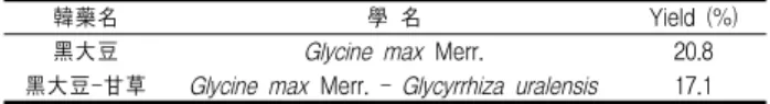
흑대두는 부곡농산에서 판매하는 것을 구매하여 사용하였으 며, 감초는 시중건재상에서 구입하여 사용하였고, 흑대두 50 g 또는 흑대두 25 g과 감초 25 g을 혼합하여, 각각 증류수 1,000 ml 로 2회 가열 추출한 후, 여과하여 여액을 농축한 다음, freeze dryer로 동결건조하여 (이하 흑대두 추출물을 GE라 하고, 흑대 두와 감초 혼합추출물을 GGE라 함.), 동물실험 시에는 생리식염 수에 용해시켜 사용하였으며, 세포실험시에는 membrane filter (0.45 μM)로 여과멸균하여 사용하였다(Table 1).
Table 1. Yield of Samples
韓藥名 學 名 Yield (%)
黑大豆 Glycine max Merr. 20.8
黑大豆-甘草 Glycine max Merr. - Glycyrrhiza uralensis 17.1
4. 비장세포의 분리
생쥐의 비장세포 분리는 Wysocki20) 및 Mizel21) 등의 방법을 이용하였다. 생쥐 5마리를 1군으로 하여, 각 검액 500 mg/kg을 1일 2회씩 7일간 경구투여한 다음 생쥐를 CO2 gas로 질식시켰 다. 적출한 비장을 DPBS를 넣은 petri dish에서 잘게 분쇄하고 멸균된 stainless mesh로 여과하여 세포부유액을 얻은 후, 10%
FBS와 penicillin-streptomycin (100 units/ml, 100 ㎍/ml)을 첨가 한 RPMI 배지로 2회 세척 후 (1,500 rpm에서 10 분간 원심분리), 비장세포 부유액으로 하였다. In vitro 실험에서는 각 검액을 100,
10 및 1 μg/ml를 처리한 후 48 시간 배양 후 동일한 실험을 실시 하였다.
5. 대식세포의 분리
대식세포의 분리는 각 검액 500 mg/kg을 1일 2회씩 7일간 경구투여 하였고, 약물 투여 4일째 생쥐 복강에 3%
thioglycollate 2 ml를 주입하였다. 생쥐를 CO2 gas로 질식시켜, 복강에 DPBS 10 ml를 넣어 복강세포를 수집하였다. 수집한 세포 를 4℃에서 1,300 rpm으로 10 분간 원심분리하고, 10% FBS와 penicillin-streptomycin (100 units/ml, 100 ㎍/ml)을 첨가한 RPMI 배지로 2회 세척한 후, 직경 120 mm petri dish에 분주하 여 CO2 incubator에서 2시간 동안 배양하였다. 부착하지 않은 세 포를 제거한 다음, 부착한 세포를 복강대식세포로 사용하였다.
6. 비장세포의 세포생존율 측정
세포생존율 측정은 Mosmann22)이 개발하여 Kotnik 등23)이 변형시킨 MTT 방법으로 측정하였다. 분리한 비장세포를 96-well plate의 각 well에 100 ㎕ (1.2 × 10⁷ cells/ml)씩 분주하고, T-lymphocytes를 관찰할 때는 concanavalin A (Con A) 5 ㎍/ml 를 B-lymphocytes를 관찰할 때는 lipopolysaccharide (LPS) 10
㎍/ml를 첨가하거나 첨가하지 않은 조건으로 37℃의 CO2
incubator에서 48 시간 배양하였다. 배양 종료 4 시간 전에 5 mg/ml 농도로 DPBS에 희석된 MTT용액 20 ㎕를 각 well에 첨 가하였다. 4시간 후 0.1N-HCl에 용해시킨 10%-SDS 100 ㎕를 각 well에 첨가하여, 차광 상태에서 18 시간 더 배양한 후, 발색된 각 well의 흡광도를 microplate reader로 570 nm에서 측정하여 세포생존율을 산정하였다.
7. 비장세포의 subpopulation 측정
분리한 비장세포를 RPMI 1640 배지로 희석하여, T cell의 population은 PE-conjugated anti-CD4 및 FITC-conjugated anti-CD8 monoclonal antibody로, T 및 B cell의 subpopulation 은 PE-conjugated anti-B220 및 FITC-conjugated anti-Thy1 monoclonal antibody로 이중 염색하여 4 ℃에서 30 분간 반응시 킨 후 flow cytometer로 subpopulation을 측정하였다24).
8. 복강 대식세포로부터 phagocytosis 측정
분리한 복강대식세포를 2 × 10⁶ cells/ml가 되도록 HBS buffer에 부유시켜, 96-well microplate (white)의 각 well에 세포 부유액 50 ㎕, lucigenin 용액 50 ㎕ 및 zymosan 용액 30 ㎕를 첨 가하여, 최종 volume이 200 ㎕가 되도록한 후, 37℃에서 15분간 전처리한 다음, 30분 동안 lucigenin chemiluminescence 양을 luminometer를 이용하여 측정하였다25,26).
9. 대식세포의 engulfment 측정
FITC-conjugated E. coli particle을 HBSS에 1 mg/ml 농도로 현탁시켜 sonification한 후 사용하였으며, trypan blue는 citrate buffer (pH 4.4)에 250 ㎍/ml 농도로 용해하여 사용하였다. 분리
한 macrophage를 RPMI1640 배지로 1 x 10⁵ cells/ml 되도록 조 정한 후, 100 ㎕를 96 well에 분주하고 E. coli 현탁액 25 ㎕를 가 하여 1 시간 동안 배양한 다음 배양액을 제거하고 extracellular fluorescence를 억제하기 위해 trypan blue 100 ㎕를 첨가하여 inverted fluoromicroscope로 관찰하였다27).
10. 대식세포로부터 nitric oxide 생성량 측정
대식세포로 부터 생성되는 nitric oxide (NO)의 양은 Griess 법28)으로 측정하였다. 분리한 대식세포를 2 × 10⁶ cells/ml로 조 제하여, 24 well plate에 분주한 후, 각 well에 LPS 1 ㎍/ml와 γ -IFN 25 units/ml를 첨가하여 48 시간 배양하였다. 배양액 100
㎕와 Griess 시약 100 ㎕를 혼합하여 96 well plate에 넣고, 37℃
에서 10 분간 방치한 후 570 nm에서 microplate reader로 흡광도 를 측정하여 미리 작성한 NaNO2의 검량선에 의해 NO2-
의 농도 를 환산하였다.
11. 통계처리
모든 실험 결과들은 mean ± S.E.로 나타내었고 통계처리는 Student's t-test를 실시하여 p<0.05를 기준으로 유의성 여부를 판 정하였다.
결 과
1. 비장세포의 증식능에 미치는 효과
분리한 비장세포에 lymphocyte mitogen을 처리하지 않았을 때의 세포생존율을 100%로 하였을 때, Con A를 처리하였을 때 세포생존율은 168.5 ± 2.9%로 증가하였으며, LPS를 처리하였을 때 세포생존율은 161.1 ± 2.2%로 증가하였다. GE를 투여하고 분 리한 비장세포에 lymphocyte mitogen을 처리하지 않았을 때의 세포생존율은 95.6 ± 1.5%로, Con A를 처리하였을 때의 세포생 존율은 158.6 ± 2.8%로, LPS를 처리하였을 때의 세포생존율은 146.5 ± 2.7%로 대조군에 비해 감소하였으며, GGE를 투여하고 분리한 비장세포에 lymphocyte mitogen을 처리하지 않았을 때 의 세포생존율은 100.5 ± 1.6%로 GE 투여군에 비해 증가하였으 며, Con A를 처리하였을 때의 세포생존율은 156.6 ± 1.7%로 GE 투여군과 별 차이가 없었으나, LPS를 처리하였을 때의 세포생존 율은 159.8 ± 2.5%로 GE 투여군에 비해 증가하였다(Fig. 1).
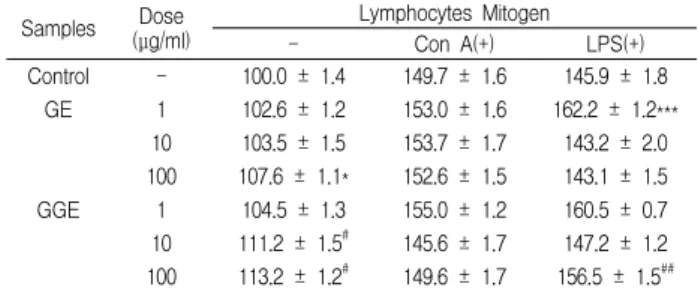
In vitro 실험에서 lymphocyte mitogen을 처리하지 않은 대 조군의 세포생존율은 100.0%로 하였을 때 Con A를 처리하였을 때는 149.7 ± 1.6%로, LPS를 처리하였을 때는 145.9 ± 1.8%로 증 가하였다. GE 1, 10 및 100 μg/ml을 처리하고 lymphocyte mitogen을 처리하지 않았을 때의 세포생존율은 102.6 ± 1.2, 103.5 ± 1.5 및 107.6 ± 1.1%로, 100 μg/ml 농도에서 대조군에 비 해 증가하였으며, Con A를 처리하였을 때는 153.0 ± 1.6, 153.7 ± 1.7 및 152.6 ± 1.5%로 대조군과 별 차이가 없었으나, LPS를 처리 하였을 때는 162.2 ± 1.2, 143.2 ± 2.0 및 143.1 ± 1.5%로 대조군에 비해 증가하였다. GGE 1, 10 및 100 μg/ml을 처리하고 lymphocyte mitogen을 처리하지 않았을 때의 세포생존율은
104.5 ± 1.3, 111.2 ± 1.5 및 113.2 ± 1.2%로, 10 μg/ml 이상의 농 도에서 대조군에 비해 증가하였으며, Con A를 처리하였을 때는 155.0 ± 1.2, 145.6 ± 1.7 및 149.6 ± 1.7%로 대조군과 별 차이가 없었으나, LPS를 처리하였을 때는 160.5 ± 0.7, 147.2 ± 1.2 및 156.5 ± 1.5%로 GE 투여군에 비해 증가하였다(Table 2).
Fig. 1. Effects of the administration of water extracts of Glycine max Merr. (GE) and the combined water extracts of Glycine max Merr.
and Glycyrrhiza uralensis (GGE) on the cell viability of mitogen treated-splenocytes in mice. The samples (500 mg/kg) were administered p.o. twice a day for 7 days, and the separated splenocytes (1.2 × 10⁷ cells/ml) were cultured for 48 h in RPMI1640 media mixed with an activating mitogen of concanavalin A (Con A) or lipopolysaccharide (LPS). The data represents the mean ± S.E. of 5 mice. *; Significantly different from control group (*; p<0.05, ***;
p<0.001). #; Significantly different from GE-treated group (p<0.05). Con A;
Concanavalin A, LPS; Lipopolysaccharide.
Table 2. Effects of GE and GGE on the cell viability of splenocytes in vitro system.
Samples Dose (μg/ml)
Lymphocytes Mitogen
- Con A(+) LPS(+)
Control - 100.0 ± 1.4 149.7 ± 1.6 145.9 ± 1.8 GE 1 102.6 ± 1.2 153.0 ± 1.6 162.2 ± 1.2***
10 103.5 ± 1.5 153.7 ± 1.7 143.2 ± 2.0 100 107.6 ± 1.1* 152.6 ± 1.5 143.1 ± 1.5 GGE 1 104.5 ± 1.3 155.0 ± 1.2 160.5 ± 0.7 10 111.2 ± 1.5# 145.6 ± 1.7 147.2 ± 1.2 100 113.2 ± 1.2# 149.6 ± 1.7 156.5 ± 1.5##
The various concentration of samples were treated into splenocytes and then the cells were cultured for 48 hrs. The data represents the mean ± SE of 3 experiments. *;
Significantly different from control group (*; p<0.05, ***; p<0.001). #; Significantly different from GE-treated group (#; p<0.05, ##; p<0.01).
2. 비장세포의 subpopulation에 미치는 효과
비장세포 중 대조군의 B220 positive 세포 (B220+) 세포는 38.1 ± 1.5% 이었으며, Thy1 positive 세포 (Thy1+)는 20.8 ± 1.7%
이었다. GE를 투여하고 분리한 비장세포 중 B220+ 세포는 44.5 ± 1.4%로, Thy1+ 세포는 14.5 ± 1.1%로 대조군에 비해 B220+ 세포 는 증가하고, Thy1+ 세포는 감소하였다. GGE를 투여하고 분리한 비장세포 중 B220+ 세포는 37.2 ± 1.8%로, Thy1+ 세포는 21.7 ± 1.8%로 GE 투여군에 비해 B220+ 세포는 감소하고, Thy1+ 세포는 증가하였다. Splenic T-lymphocytes 중 대조군의 CD4+ 세포는 16.2 ± 0.4% 이었고, CD8+ 세포는 7.4 ± 0.3% 이었으며, GE를 투 여하고 분리한 splenic T-lymphocytes 중 CD4+ 세포는 13.1 ± 0.5%로, CD8+ 세포는 7.2 ± 0.3%로 CD4+ 세포가 대조군에 비해 감소하였으며, GGE를 투여하고 분리한 splenic T-lymphocytes
중 CD4+ 세포는 20.7 ± 0.5% 로 CD8+ 세포는 7.7 ± 0.3%로 GE 투여군에 비해 CD4+ 세포가 증가하였다(Fig. 2).
Fig. 2. Effects of the administration of GE and GGE on the subpopulation of murine splenocytes. The samples (500 mg/kg) were administered p.o. twice a day for 7 days, and the separated splenocytes were stained with PE-conjugated anti-B220 and FITC-conjugated anti-Thy1 monoclonal antibody or PE-conjugated anti-CD4 and FITC-conjugated anti-CD8 monoclonal antibody for 30 minutes at 4℃. The subpopulation was determined with a flow cytometer. The data represents the mean ± S.E. of 5 mice. *; Significantly different from control group (*; p<0.05, ***; p<0.001). #; Significantly different from control group (#; p<0.05, ##; p<0.01, ###; p<0.001).
3. 대식세포의 탐식능에 미치는 효과
Chemiluminescence은 대식세포가 탐식을 하는 동안 생성되 는 oxygen radical에 의해 발생되며, lucigenin에 의해 증가되는 것으로 알려져 있다29). 대조군의 대식세포로부터 생성되는 lucigenin chemiluminescence에 비해, GE를 투여하고 분리한 대 식세포에서 생성되는 lucigenin chemiluminescence가 증가하였 으며, GGE를 투여하고 분리한 대식세포에서 생성되는 lucigenin chemiluminescence는 GE 투여군에 비해 더욱 증가하였다(Fig.
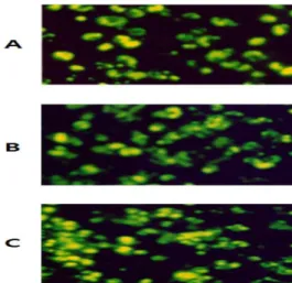
3). 또한 FITC-conjugated E. coli particle의 탐식능을 측정하였을 때도 GE를 투여하고 분리한 대식세포의 engulfment가 대조군에 비해 증가하였으며, GGE를 투여하고 분리한 대식세포의 engulfment가 GE 투여군에 비해 증가하였다(Fig. 4).
Fig. 3. Effects of the administration of GE and GGE on the level of lucigenin chemiluminescence in murine peritoneal macrophages. The samples (500 mg/kg) was administered orally twice a day for 7 days and the separated peritoneal macrophages (2 × 10⁶ cells/ml) were cultured in HBS mixed with opsonized zymosan. The level of lucigenin chemiluminescence was measured for 30 min with a luminometer. Each bar represents the mean ± S.E. of 5 mice.
*; Significantly different from control group (p<0.05). #; Significantly different from GE-administered group (p<0.05).
Fig. 4. Photomicrographs of the engulfment of FITC-conjugated E.
coli particles in peritoneal macrophages obtained from GE- or GGE-administered mice. Photographs (taken at 200X magnification) showing the uptake of FITC-conjugated E. coli particles in control (A), GE-administered mice (B) and GGE-administered mice (C). The macrophages were observed with an inverted fluoromicroscope.
4. 복강 대식세포로 부터 nitric oxide의 생성에 미치는 효과 대조군의 대식세포에 LPS와 γ-IFN을 처리하지 않고 48 시 간 배양하였을 때 nitric oxide(NO) 생성양은 1.6 ± 0.2 μM 이었 으며, GE를 투여하고 분리한 대식세포에서 NO 생성양 및 GGE 를 투여하고 분리한 대식세포에서 NO 생성양은 대조군과 별 차 이가 없었으나, LPS와 γ-IFN을 처리하였을 때 대조군의 NO 생 성양은 16.5 ± 0.4 μM로 증가하였으며, GE를 투여하고 분리한 대식세포에서 NO 생성양은 22.7 ± 0.5 μM로 대조군에 비해 증 가하였으며, GGE를 투여하고 분리한 대식세포에서 NO 생성양 은 28.3 ± 0.7 μM로 GE 투여군에 비해 증가하였다(Fig. 5).
Fig. 5. Effects of the administration of GE and GGE on the production of nitric oxide from murine peritoneal macrophages. The samples (500 mg/kg) was administered p.o. twice a day for 7 days, and then 3%
thioglycollate was injected i.p. at the 4th day. Peritoneal macrophages (2 x 10⁶ cells/ml) obtained after 2 hr adherence period were cultured for 48 hrs. in the presence or in the absence of LPS and γ-IFN. The production of nitric oxide was determined with a Griess reagent. The data represents the mean ± S.E. of 5 mice.
*; Significantly different from control group (p<0.001). #; Significantly different from RE group (p<0.05).
고 찰
흉선세포는 thymus의 피질 및 수질에서 분화과정을 거쳐
helper T lymphocyte (CD4+) 및 cytotoxic T lymphocyte (CD8+) 로 분화되며, 분화된 helper T (Th) 세포 중 Th1 세포에서는 γ -IFN 및 IL-2가, Th2 세포에서는 IL-4, IL-6 및 IL-10 등의 cytokine이 분비되어, 다른 T, B 임파구 및 macrophage의 증식과 분화를 촉진하는 것으로 알려져 있고, cytotoxic T cell은 tumor cell의 lysis를 일으키며 macrophage를 활성화시키는 것으로 알 려져 있다. 한편 B 임파구는 항체의 생성을 촉진하는 것으로 알 려져 있다30).
GE를 투여하고 분리한 비장세포의 생존율은 대조군에 비해 감소하였으나, GGE를 투여하고 분리한 비장세포의 생존율은 대 조군과 비슷한 정도로 회복되었다. 또한, GE를 투여하고 분리한 비장세포의 생존율은 T lymphocytes mitogen인 Con A를 처리 하였을 때나, B lymphocytes mitogen인 LPS를 처리하였을 때 모 두 대조군에 비해 감소하였으나, GGE를 투여하고 분리한 비장 세포의 생존율은 Con A를 처리하였을 때는 GE 투여군에 비해 별 차이가 없었으나, LPS를 처리하였을 때는 GE 투여군에 비해 증가하였다. 이러한 결과는 GE 투여에 의해 T 및 B lymphocytes 의 생존율이 감소되나, GGE 투여에 의해서는 T lymphocytes의 생존율만 감소됨을 의미하는 것이다. 이러한 생존율 감소가 GE 및 GGE를 고용량 투여한 결과인지에 대해서는, 향후 다양한 용 량을 투여하여 관찰하여야 할 과제이다.
GE 및 GGE에 의한 비장세포의 생존율을 저하시키는 작용 이 GE 및 GGE의 직접작용에 의한 것인지를 관찰하기 위해 in vitro 실험을 실시하였다. GE 1, 10 및 100 μg/ml를 처리하였을 때, 100 μg/ml 농도에서 세포생존율이 증가하였으며, GGE를 처 리하였을 때는 GE 투여군에 비해 증가하였다. T lymphocytes mitogen인 Con A를 처리하였을 때는 GE 및 GGE 처리하였을 때 대조군과 별 차이가 없었으나, B lymphocytes mitogen인 LPS 를 처리하였을 때는 GE를 처리하였을 때 대조군에 비해 증가하 였으며, GGE를 처리하였을 때 GE 투여군에 비해 증가하였다.
이는 GE 및 GGE를 in vitro 처리하였을 때 주로 B lymphocytes 의 생존율이 증가하고 있음을 의미하는 것이다. In vivo 실험에서 GE 투여에 의해 세포생존율이 감소하였는데, in vitro 처리하였 을 때는 세포생존율이 증가하였다는 것은, GE 및 GGE를 경구투 여하였을 때, 이들 물질이 lymphocytes에 간접적으로 작용하여 세포생존율을 감소시킴을 시사하는 결과라 할 수 있다.
GE를 투여하였을 때 비장세포의 B220+ 세포의 population은 대조군에 비해 증가하였으나, Thy1+ 세포의 population은 감소하 였으며, GGE를 투여하였을 때 비장세포의 B220+ 세포의 population은 GE 투여군에 비해 감소하였으나, Thy1+ 세포의 population은 GE 투여군에 비해 증가하고, GE를 투여하였을 때 splenic CD4+ 세포의 population은 대조군에 비해 감소하고, splenic CD8+ 세포의 population은 대조군과 별 차이가 없었으며, GGE를 투여하였을 때 splenic CD4+ 세포의 population은 GE 투 여군에 비해 증가하고, splenic CD8+ 세포의 population은 대조군 과 별 차이가 없었다. 이는 GE 투여에 의해 변화되는 비장세포의 population을 GGE가 회복시킬 수 있음을 의미하는 것이다.
Lymphocytes에서 분비되는 cytokines들은 대식세포를 조절
하는 것으로 알려져 있기 때문에31), 복강 대식세포에서 분비되는 NO 및 phagocytic activity를 측정하였다. 외부로부터 이물질이 침입하게 되면 생체는 자기방어를 위해 대식세포가 활성화되어 탐식작용이 촉진된다. 이러한 탐식능은 polymorphonuclear leukocytes에서도 일어난다. 본 실험에서 대식세포의 phagocytic activity를 측정하는데 chemiluminescence를 측정하는 방법을 이 용하였다. 이 방법의 원리는 대식세포가 particle을 phagocyte하 는 동안 oxygen radical을 생성하는데, 이때 생성된 oxygen radical과 lucigenin이 반응하여 lucigenin chemiluminescence를 발생하는 것을 측정함으로써 phagocytic activity가 진행되는 것 을 확인하는 것이다32). GE를 투여하고 분리한 복강 대식세포로 부터 lucigenin chemiluminescence을 측정한 결과 대조군에 비 해 증가하였으며, GGE를 투여하고 분리한 복강 대식세포로부터 lucigenin chemiluminescence을 측정한 결과 GE 투여군에 비해 증가하였다. 또한, FITC-conjugated E. coli를 이용하여 실험하였 을 때도, 동일한 실험결과를 얻었다. 이러한 결과는 GE를 투여하 면 대식세포의 탐식능을 증가시키고, GGE를 투여하면 GE 보다 더 강하게 탐식능을 증가시킴을 의미하는 것이다.
LPS와 γ-IFN을 처리하지 않은 대조군에서 분비되는 NO양 은 LPS와 γ-IFN을 처리하였을 때 현저히 증가하였으며, GE를 투여하고 분리한 대식세포로부터 분비되는 NO양은 LPS와 γ -IFN을 처리하였을 때 대조군에 비해 증가하였으며, GGE를 투 여하고 분리한 대식세포로부터 분비되는 NO양은 GE 투여군에 비해 증가하였다. NO는 활성화된 대식세포의 pseudopodia 형성 을 억제하는 것으로 알려져 있다33). GE 및 GGE가 탐식능 및 NO 생성을 동시에 증가시켰다는 결과는, 이들의 탐식능 증가작 용이 NO와는 다른 경로를 통하여 나타나고 있음을 시사하는 것 이라 할 수 있다.
결 론
흑대두 추출물 (GE)과 흑대두·감초 혼합추출물 (GGE)을 생쥐에 경구투여 하였을 때 비장세포와 대식세포의 활성을 측정 한 결과 다음과 같은 결론을 얻었다.
GE 경구투여는 비장세포의 T- 및 B-lymphocytes 생존율을 감소시키나, GGE 경구투여는 비장세포의 B-lymphocytes 생존율 을 증가시켰다. GE 및 GGE를 비장세포에 in vitro 처리시 B-lymphocytes의 생존율을 증가시켰다. GE 경구투여는 B-lymphocytes의 population을 증가시키고, T-lymphocytes의 population 및 splenic T-lymphocytes의 CD4+ cell의 population 을 감소시켰으나, GGE 경구투여는 B-lymphocytes의 population 을 감소시키고, T-lymphocytes의 population 및 splenic T-lymphocytes의 CD4+ cell의 population을 증가시켰다. GE 및 GGE 경구투여는 복강 대식세포의 탐식능을 증가시켰다. GE 및 GGE 경구투여는 복강 대식세포로부터 nitric oxide 생성을 촉진 시켰다.
이상의 실험결과 GGE가 GE 보다 비장세포 및 대식세포의 활성을 더욱 강하게 증가시켰다.
감사의 글
이 논문은 2009년도 교육과학기술부(지역거점연구단육성사 업/헬스케어기술개발사업단) 및 우석대학교 교내학술연구비 지 원에 의하여 연구되었음.
참고문헌
1. 李時珍, 本草綱目, 北京, 人民衛生出版社, p 1501, 1982.
2. 許浚, 東醫寶鑑, 서울, 南山堂, p 589, 1987.
3. Yoshida, K., Sato, Y., Okuno, R., Kameda, K., Isobe, M., and Kondo, T. Structural analysis and measurement of anthocyanins from colored seed coats of Vigna, Phaseolus, and Glycine legumes. Biosci. Biotechnol. Biochem. 60:
589-593, 1996.
4. Sakai, T. and Kogiso, M. Soy isoflavones and immunity. J.
Med. Invest., 55: 167-173, 2008.
5. Wu, D., Meydani, M., Leka, L.S., Nightingale, Z., Handelman, G.J., Blumberg, J.B., Meydani, S.N. Effect of dietary supplementation with black currant seed oil on the immune response of healthy elderly subjects. Am. J. Clin.
Nutr., 70(4):536-543, 1999.
6. Cassileth, B.R., Vickers, A.J. Soy: an anticancer agent in wide use despite some troubling data. Cancer Invest., 21(5):817-818, 2003.
7. Maeda, H., Katsuki, T., Akaike, T., Yasutake, R. High correlation between lipid peroxide radical and tumor-promoter effect: suppression of tumor promotion in the Epstein-Barr virus/B-lymphocyte system and scavenging of alkyl peroxide radicals by various vegetable extracts. Jpn J. Cancer Res., 83(9):923-928, 1992.
8. Liao, H.F., Chou, C.J., Wu, S.H., Khoo, K.H., Chen, C.F., Wang, S.Y. Isolation and characterization of an active compound from black soybean [Glycine max (L.) Merr.]
and its effect on proliferation and differentiation of human leukemic U937 cells. Anticancer Drugs, 12(10):841-846, 2001.
9. Jang, E.H., Moon, J.S., Ko, J.H., Ahn, C.W., Lee, H.H., Shin, J.K., Park, C.S., Kang, J.H. Novel black soy peptides with antiobesity effects: activation of leptin-like signaling and AMP-activated protein kinase. Int. J. Obes. (Lond)., 32(7):1161-1170, 2008.
10. Kim, H.J., Bae, I.Y., Ahn, C.W., Lee, S., Lee, H.G.
Purification and identification of adipogenesis inhibitory peptide from black soybean protein hydrolysate. Peptides, 28(11):2098-2103, 2007.
11. Zhao, Q.W., Lou, Y.J. Estrogenic activity and its mechanism of ethanol extract from black soybean. Zhongguo Zhong Yao Za Zhi, 31(10):825-828, 2006.
12. Takahashi, R., Ohmori, R., Kiyose, C., Momiyama, Y., Ohsuzu, F., Kondo, K. Antioxidant activities of black and yellow soybeans against low density lipoprotein oxidation.
J. Agric. Food Chem., 53(11):4578-4582, 2005.
13. Yamai, M., Tsumura, K., Kimura, M., Fukuda, S., Murakami, T., Kimura, Y. Antiviral activity of a hot water extract of black soybean against a human respiratory illness virus. Biosci. Biotechnol. Biochem., 67(5):1071-1079, 2003.
14. Sakai, T., Kogiso, M. Soy isoflavones and immunity. J.
Med. Invest., 55(3-4):167-173, 2008.
15. Chan, Y.C., Wu, C.C., Chan, K.C., Lin, Y.G., Liao, J.W., Wang, M.F., Chang, Y.H., Jeng, K.C. Nanonized black soybean enhances immune response in senescence- accelerated mice. Int. J. Nanomedicine, 4: 27-35, 2009.
16. Chung, W.T., Lee, S.H., Kim, J.D., Sung, N.S., Hwang, B., Lee, S.Y., Yu, C.Y., Lee, H.Y. Effect of the extracts from Glycyrrhiza uralensis Fisch on the growth characteristics of human cell lines: Anti-tumor and immune activation activities. Cytotechnology, 37(1):55-64, 2001.
17. Cheng, A., Wan, F., Wang, J., Jin, Z., Xu, X. Macrophage immunomodulatory activity of polysaccharides isolated from Glycyrrhiza uralensis Fish. Int Immunopharmacol.
8(1):43-50, 2008.
18. Sun, H.X. and Pan, H.J. Immunological adjuvant effect of Glycyrrhiza uralensis saponins on the immune responses to ovalbumin in mice. Vaccine, 24(11):1914-1920, 2006.
19. Abbas, A.K., Lichtman, A.H. and Pober, J.S. Cellular and Molecular Immunology. 2ed. Saunders, p 5, 1994.
20. Wysocki, L.J. and Sato, V.L. Planning for lymphocytes: A method for cell selection. Proc. Nat1. Acad. Sci. USA., 75:
2844, 1978.
21. Mizel, S.B., Openheim, J.J. and Rosensteich, D.L.
Characterization of lymphocyte-activating factor(LAF) produced by the macrophage cell line P388D1. J. Immunol.
120: 1497, 1979.
22. Mosmann, T. Rapid colorimetric assay for cellular growth and survival: application to proliferation and cytotoxic assays. J. Immunol. methods, 65: 55, 1983.
23. Kotnic, V. and Fleischmann, W.R.Jr. A simple and rapid method to determine hematopoietic growth factor activity.
J. Immunol. methods, 129: 23, 1990.
24. Suda, T. and Nagata, S. Purification and characterization of the Fas-ligand that induces apoptosis. J. Exp. Med., 179:
873-879, 1994.
25. Boudard, F., Vallot, N., Cabaner, C. and Bastide, M.
Chemiluminenscence and nitrite determinations by the MALU macrophage cell line. J. Immunol. Methods, 174:
259, 1994.
26. Blair, A.L., Cree, I.A., Beck, J.S. and Hating, M.J.G.
Measurement of phagocyte chemiluminenscence in a microtiter plate format. J. Immunol. Methods, 112: 163, 1988.
27. Chok, P.W., Choon, S.P. and Benjamin, H.S. A rapid and simple microfluorometric phagocytosis assay. J. Immuno.
Methods, 162: 1, 1993.
28. Rockett, K.A., Awburn, M.M., Cowden, W.B. and Clark, I.A. Killing of Plasmodium faciparum in vitro by nitric oxide derivatives. Infec. Immunity, 59(9):3280, 1991.
29. Breiheim, G., Stendahl, O. and Dahlgren, C. Intra- and extracellular evevts in luminol-dependent chemilulinescence of polymorphonuclear leukocytes. Infect. Immun., 45: 1, 1984.
30. Miceli, M.C. and Parnes, J.R. The role of CD4 and CD8 in T cell activation and differentiation. Advances in Immunology, 53: 59, 1993.
31. Charles, A.J., Paul, T., Mark, W. The immune system in health and disease. 4ed, Garland Pub. p 463, 2000.
32. Channon, J.Y., Leslie, C.C. and Johnston, Jr.R.B.
Zymosan-stimulated production of phosphatidic acid by macrophages: relationship to release of superoxide anion and inhibition by agents that increase intracellular cyclic AMP. J. Leucocyte Biol., 41: 450-455, 1987.
33. Jun, C.D., Park, S.K., Kim, J.M., Kim, J.D. and Kim, S.H.
Nitric oxide inhibits macrophage pseudopodia formation in the activated macrophages. Kor. J. Immunol., 18: 635-644, 1996.