• 검색 결과가 없습니다.

Radiotherapy as a First-Line Modality for Tongue Base Adenoid Cystic Carcinoma : Report of 3 Cases

N/A
N/A
Protected

Academic year: 2021

Share "Radiotherapy as a First-Line Modality for Tongue Base Adenoid Cystic Carcinoma : Report of 3 Cases"

Copied!
5
0
0

로드 중.... (전체 텍스트 보기)

전체 글

(1)

Introduction

Adenoid cystic carcinoma(ACC) is a rare malignancy usu- ally presenting in the minor salivary glands.

1)

Because it can occur wherever minor salivary glands exist, 40% of cases arise in other head and neck sites such as the oral cavity, prin- cipally, other than major salivary glands.

1)

The relative indo- lent course of the disease and the propensity for perineural extension are well known characteristics, however, exact bi- ological properties are poorly understood.

1-4)

In the manage-

ment of ACC, surgical excision is generally recommended first. Radiotherapy has been applied postoperatively or to the patients with inoperable disease state while chemotherapy is reserved for the palliative setting in general.

5-8)

However, treat- ment options should be diversely considered according to the disease site, functional outcome, and especially the long sur- vival time of such patients.

We report 3 consecutive cases of adenoid cystic carcinoma occurring in the base of tongue treated with radiation thera- py alone.

Case Report

1. Patient A

A 79 year old women presented with a protruding tongue base mass that nearly obstructed the oropharynx. With inci-

대한 두경부 종양 학회지

제 30 권 제 2 호 2014

Radiotherapy as a First-Line Modality for Tongue Base Adenoid Cystic Carcinoma : Report of 3 Cases

Kyung Su Kim, MD

1

, Hong-Gyun Wu, MD, PhD

1,4,5

, Myung-Whun Sung, MD

2

, J. Hun Hah, MD

2

, Tae Min Kim, MD, PhD

3

Department of Radiation Oncology

1

and Otorhinolaryngology-Head and Neck Surgery

2

and Internal Medicine

3

and Cancer Research Institute,

4

Seoul National University College of Medicine, Seoul, Korea

Institute of Radiation Medicine,

5

Medical Research Center, Seoul National University, Seoul, Korea

설근부에 발생한 샘낭암종의 방사선치료 : 증례보고 3예

서울대학교 의과대학 방사선종양학교실,

1

이비인후과학교실,

2

내과학교실,

3

암연구소,

4

서울대학교 의학연구원 방사선의학연구소

5

김경수1·우홍균1,4,5·성명훈2·하정훈2·김태민3

= 국 문 초 록 =

주로 작은 침샘에서 발생하는 샘낭암종의 치료 방침은 수술과 수술 후 보조적 방사선치료가 주로 행해져 왔다. 그 러나 설근부에 발생한 샘낭암종에 대해서는 수술적 치료가 가져오는 삶의 질의 저하가 크기 때문에 수술적 치료를 적 용하기 쉽지 않다. 또한 샘낭암종의 치료에 있어서 항암제의 역할이 거의 없는 상황에서 방사선치료가 중요한 역할을 할 수 있겠다. 이에 본 저자들은 설근부에 발생한 샘낭암종 세 증례의 방사선치료 결과를 보고 하며 샘낭암종의 치료 에 대해서 문헌고찰을 통해 논의하고자 한다.

중심 단어

:샘낭암종ㆍ방사선 치료.

Received : September 30, 2014 / Revised : October 10, 2014 Accepted : October 13, 2014

교신저자 : 우홍균, 110-744 서울 종로구 대학로 101 서울대학교 의과대학 방사선종양학교실

전화 : (02) 2072-3177 ・ 전송 : (02) 742-2073 E-mail : [email protected]

online©MLComm
(2)

sional biopsy, the mass was diagnosed as ACC with a pre- dominantly cribriform pattern(Fig. 1). On computed tomog- raphy(CT) and magnetic resonance(MR) images, a 3×4.5 cm-sized well circumscribed enhancing mass was located in the tongue base. On the initial fluorine-18 fluorodeoxy- glucose(F-18 FDG) positron emission tomography(PET)/CT images, intense FDG uptake[standardized uptake value(SUV) 9.38] was measured in the tongue base mass and no signifi- cantly enlarged lymph node was observed. Several factors were discussed at our multidisciplinary tumor board for treat- ment decision-making, including the patient’s old age, which may cause difficulty in enduring the surgical procedure and result in poor functional outcomes, and the large mass filling the oropharynx which may stand as an obstacle during intu- bation. With these considerations, the patient was planned for radiation therapy alone.

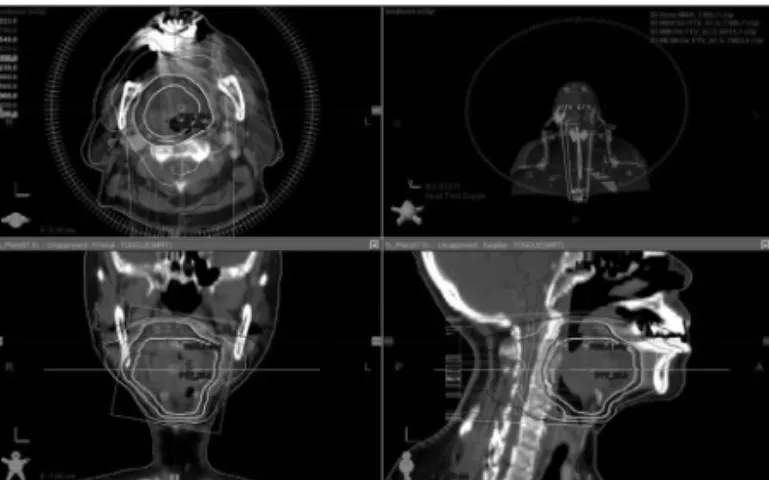
Radiotherapy was delivered with 67.5 Gy to the primary mass. 54 Gy was delivered to the primary mass+1.0 cm mar- gin for suspicious microscopic disease by 30 fractions with volumetric arc radiation therapy technique(VMRT)(Fig. 2).

During the radiotherapy the patients developed grade 3 mu- cositis however ; planned treatment was delivered. After treatment, regular follow up based on the clinical examina- tion and image modalities were performed. At 11 months af- ter completion of radiotherapy, PET/MR imaging revealed that the mass was reduced to 1.5×2 cm with a SUV uptake reduced to 3.1 and no other metastatic lesion was detected.

At the last follow up of 20 months, clinical examination re- vealed no mucosal lesion and MR image showed further de- creased mass to 1.0×1.9 cm, resulting in a state of partial re- sponse(PR)(Fig. 3). The patients complained of nothing but decreased taste.

2. Patient B

A 57-year old previously healthy women presented with

tongue bleeding and dysarthria. Symptoms persisted for about 9 months before visiting the hospital. On laryngoscope exami- nation, the tongue was deviated to the left with atrophy and an indurated mass in the tongue bass. Punch biopsy was per- formed. Pathologic examination reported that the mass was ACC with predominantly tubular pattern. On CT and MR im- ages, a 3.7 cm-sized, infiltrating, and enhancing mass involv- ing the tongue with invasion of extrinsic muscles was observed with no abnormal cervical LN enlargement. PET/CT revealed that beside the hypermetabolic lesion in the tongue, both pal- atine tonsils and posterior buccal mucosa also had hypermet- abolic activity, suggesting infiltration of cancer. No other met- astatic lesion was suggested. Considering the infiltrative cha- racteristic of the disease, the potential for systemic dissemina- tion, and the patient’s reluctance to total laryngectomy, con- current chemoradiation was decided on at our multidisciplinary tumor board discussion.

Six cycles of weekly cisplatin(35 mg/m

2

) were delivered concurrently with radiation. The total tongue, both tonsils, and posterior buccal mucosa showing hypermetabolism on PET/

CT images received 67.5 Gy. Considering the advanced state

A B

Fig. 1. Pathologic images of patients. A : Pathology of the patient A-Cribriform growth pattern of ACC, H&E staining ×200. B : Pathol- ogy of the patient B-Tubular growth pattern of ACC, H&E staining ×200 ; Pathology image of patient C was not available.

Fig. 2. Dose distribution of radiotherapy of patient A. Radiothera-

py was delivered with 67.5 Gy to the primary mass. 54 Gy was de-

livered to the primary mass+1.0 cm margin for suspicious micro-

scopic disease by 30 fractions with volumetric arc radiation therapy

technique(VMRT).

(3)

of the disease, 54 Gy was delivered to the bilateral parapha- ryngeal spaces, lymph node level IB, II and III. Using VMAT, radiation dose was delivered through 30 fractions. The pa- tient received all planned radiation and chemotherapy doses, but developed grade 3 mucositis at the end of treatment.

Complete response was observed by neck MRI after 6 months of treatments. The patient was in a state of complete response(CR) at the last follow up of 11 months by PET/MR image, showing no abnormal hypermetabolic activity except for a focal hypermetabolism on the left floor of mouth that most likely was caused by inflammation from a dental metal crown(Fig. 4).

3. Patient C

A 68 year old man complaining of dry mouth presented with a tongue base mass with soft palate immobilization. The mass was diagnosed as ACC with predominantly cribriform growth. On CT and MR images, the mass was characterized as a 3.8 cm-sized mass involving the genioglossus. Indeter- minately small lymph nodes in the left level I area was detect- ed. On PET/CT scans, no other FDG uptakes were noticed except at the tongue base mass with a SUV of 4.8. The patient was suffering from cervical spondylosis with limited neck extension that could cause intra-oral surgical procedures to be difficult, and preferred radiotherapy than surgery. Consid- ering these factors, radiation therapy was decided at our mul- A

E F G H

B C D

Fig. 3. Laryngoscopic, MR, PET/CT and PET/MR images of patient A. Laryngoscopic image(A),T1 contrast axial(B) and coronal image (C) and PET/CT image(D) before treatment showed 3×4.5 cm sized tongue base mass with hypermetabolism(SUV 9.38). Laryngoscop- ic image at 11 months after treatment(E) showed no mucosal lesion. T1 contrast axial(F) and coronal image(G) at 20 months after ra- diotherapy demonstrated reduced size of tongue base mass to 1.0×1.9 cm. PET/MR image(H) at 11months after radiotherapy showed reduced hypermetaboism(SUV 3.1).

A

E F G H

B C D

Fig. 4. Laryngoscopic, MR, PET/CT and PET/MR images of patient B. Laryngoscopic image(A), T1 axial/coronal contrast-enhanced

image(B and C) and PET/CT(D) image before treatment showed 3.7 cm-sized tongue base mass with hypermetabolism. Laryngo-

scopic image(E) at 9 months after treatment showed no abnormal lesion. T1 axial/coronal contrast-enhanced image(F and G) and

PET/MR(H) image at 11 months after radiotherapy showed no abnormal or hypermetabolic lesion.

(4)

tidisciplinary institutional tumor board.

A total of 72 Gy/30 Fx to the tongue bass mass and consid- ering indeterminate small lymph nodes detected on the imag- ing study, 54 Gy to the bilateral level I and II lymph node ar- eas including the skull base, were delivered. Grade 2 mucositis developed at the end of radiotherapy. On PET/MR imaging taken 5 months after the treatment, the mass was reduced to 2.3 cm and SUV value was 4.0 and no other metastatic lesion, making it a PR. At the last follow up of 10 months, the patient complained of minimal dry mouth with no palpable lesion on the neck, but residual mass on the tongue base.MR images re- vealed a sustained reduced mass(Fig. 5).

Discussion

Here, we report 3 patients with ACC in the tongue base treat- ed with radiation therapy. All three patients showed partial or complete response and maintained its efficacy until 10 to 20 months.

The mainstay of treatment for minor salivary gland cancer is surgery with or without postoperative radiation ; however, radiation alone as a primary treatment has been done for in- operable diseases.

5,9-11)

Considering the delayed occurrence of distant metastasis and a relatively long survival time even after distant metastasis occurs for patients with ACC

4,12)

treat- ments causing deterioration of functional outcome should be carefully applied.

Disappointingly, radiation therapy(photon) alone resulted in low local control rates(5-year local control rates<50%) in general

5-7)

and the patients treated without surgery had poor

prognosis.

1)

However, these reports reflected inoperable ad- vanced stage of the disease. Parsons et al. reported good lo- cal control especially for early T stage lesions.

5)

Moreover, there exists a subgroup with favorable prognostic factors.

3,12-15)

It is well known that cribriform or tubular growth pattern had good prognosis than solid growth pattern.

3,12,13)

Involvement of the base of skull and lymph node were poor prognostic factors for cause-specific survival

14)

and the involvement of named nerves were also associated with risk of local relapse.

15)

All three patients presented in this report had low-risk crib- riform or tubular growth pattern, no involvement of the base of skull, negative lymph nodes, and no involvement of named nerves. Recently, prognostic factors based on genetic and mo- lecular study are actively under investigation.

16-18)

However, in the clinical application of these studies, further investiga- tion is encouraged.

To enhance locoregional control rates, trials for the effec- tive treatment to inoperable patients had been conducted. Sev- eral studies reported the role of concurrent chemoradiation therapy(CCRT) for salivary gland cancer including ACC.

19)

Although a small retrospective studies, ACC patients treated with CCRT showed good responses.

20,21)

In our study, we re- port complete response of the patient B treated with radiation concurrently with cisplatin ; however, the role of adding che- motherapy in the radiation treatment of ACC should be clari- fied in the large-scale studies.

In conclusion, we report three cases showing partial or com- plete response to the radiotherapy of ACC arising in the tongue base and sustaining its efficacy to 10 to 20 months. In cases when surgical treatment could deteriorate functional outcome A

E

B

F

C

G

D

H

Fig. 5. Laryngoscopic, MR, PET/CT and PET/MR images of patient C. Laryngoscopic image(A), T1 contrast-enhance axial/coronal im-

ages(B and C) and PET/CT image(D) before treatment showed 3.8 cm-sized tongue base mass with hypermetabolism(SUV 4.8). Laryn-

goscopic image(E) at 10 month after treatment showed residual mass. T1 contrast-enhance axial/coronal images(F and G) after 10 month

of treatment showed reduced mass to 2.3 cm. PET/CT image(H) after 5 months of treatment showing reduced hypermetabolism(SUV

4.0).

(5)

due to the location of the disease, radiotherapy could be a rea- sonable option as a first-line modality for the treatment of ACC without high-risk features, leaving open the possibility of sal- vage surgery in case of local progression.

Acknowledgments

This research was supported by a grant from the National Research Foundation of Korea(NRF), which is funded by the Korea govern- ment(MEST, Grant No. 2014M2A2A7055063).

References

1) Ellington CL, Goodman M, Kono SA, Grist W, Wadsworth T, Chen AY, et al. Adenoid cystic carcinoma of the head and neck.

Cancer. 2012;118(18):4444-4451.

2) Vrielinck LJ, Ostyn F, van Damme B, van den Bogaert W, Fos- sion E. The significance of perineural spread in adenoid cystic carcinoma of the major and minor salivary glands. Int J Oral Maxillofac Surg. 1988;17(3):190-193.

3) Huang M, Ma D, Sun K, Yu G, Guo C, Gao F. Factors influenc- ing survival rate in adenoid cystic carcinoma of the salivary glands. Int J Oral Maxillofac Surg. 1997;26(6):435-439.

4) Terhaard CH, Lubsen H, Van der Tweel I, Hilgers FJ, Eijkenboom WM, Marres HA, et al. Salivary gland carcinoma: Independent prognostic factors for locoregional control, distant metastases, and overall survival: Results of the Dutch head and neck oncol- ogy cooperative group. Head Neck. 2004;26(8):681-692; discus- sion 692-683.

5) Parsons JT, Mendenhall WM, Stringer SP, Cassisi NJ, Million RR. Management of minor salivary gland carcinomas. Int J Ra- diat Oncol Biol Phys. 1996;35(3):443-454.

6) Mendenhall WM, Morris CG, Amdur RJ, Werning JW, Villar- et DB. Radiotherapy alone or combined with surgery for salivary gland carcinoma. Cancer. 2005;103(12):2544-2550.

7) Huber PE, Debus J, Latz D, Zierhut D, Bischof M, Wannenmach- er M, et al. Radiotherapy for advanced adenoid cystic carcino- ma: Neutrons, photons or mixed beam? Radiother Oncol. 2001;

59(2):161-167.

8) Laurie SA, Ho AL, Fury MG, Sherman E, Pfister DG. Systemic therapy in the management of metastatic or locally recurrent adenoid cystic carcinoma of the salivary glands: A systematic review. The Lancet Oncology. 2011;12(8):815-824.

9) Garden AS, Weber RS, Ang KK, Morrison WH, Matre J, Peters LJ. Postoperative radiation therapy for malignant tumors of minor salivary glands. Outcome and patterns of failure. Can- cer. 1994;73(10):2563-2569.

10) Kim WD, Park CI. The Role of Radiation Therapy in the Treat-

ment of Adenoid Cystic Carcinoma of the Head and Neck. Ra- diation Oncology Journal. 1992;10/1:35-42.

11) Eum KY, Wu HG. Postoperative Radiotherapy for Parotid Gland Malignancy. Radiation Oncology Journal. 2005;23/1:131-136.

12) Sung MW, Kim KH, Kim JW, Min YG, Seong WJ, Roh JL, et al. Clinicopathologic predictors and impact of distant metasta- sis from adenoid cystic carcinoma of the head and neck. Arch Otolaryngol Head Neck Surg. 2003;129(11):1193-1197.

13) Enamorado I, Lakhani R, Korkmaz H, Yoo GH, Del Mar Alon- so M, Pietraszkiewicz H, et al. Correlation of histopathological variants, cellular DNA content, and clinical outcome in adenoid cystic carcinoma of the salivary glands. Otolaryngol Head Neck Surg. 2004;131(5):646-650.

14) Douglas JG, Laramore GE, Austin-Seymour M, Koh W, Stelzer K, Griffin TW. Treatment of locally advanced adenoid cystic carcinoma of the head and neck with neutron radiotherapy. Int J Radiat Oncol Biol Phys. 2000;46(3):551-557.

15) Garden AS, Weber RS, Morrison WH, Ang KK, Peters LJ. The influence of positive margins and nerve invasion in adenoid cystic carcinoma of the head and neck treated with surgery and radiation. Int J Radiat Oncol Biol Phys. 1995;32(3):619-626.

16) Fukai J, Fujita K, Yamoto T, Sasaki T, Uematsu Y, Nakao N. In- tracranial extension of adenoid cystic carcinoma: Potential in- volvement of EphA2 expression and epithelial-mesenchymal transition in tumor metastasis: A case report. BMC Res Notes.

2014;7(1):131.

17) Tan M, Shao C, Bishop JA, Feng Z, Trock BJ, Westra WH, et al.

Aquaporin-1 Promoter Hypermethylation Is Associated with Improved Prognosis in Salivary Gland Adenoid Cystic Carci- noma. Otolaryngol Head Neck Surg; 2014.

18) Xia R, Zhou R, Tian Z, Zhang C, Wang L, Hu Y, et al. High expres- sion of H3K9me3 is a strong predictor of poor survival in pa- tients with salivary adenoid cystic carcinoma. Arch Pathol Lab Med. 2013;137(12):1761-1769.

19) Cerda T, Sun XS, Stephane V, Marcy PY, Baujat B, Baglin AC, et al. A rationale for chemoradiation(vs radiotherapy) in sali- vary gland cancers? On behalf of the REFCOR(French rare head and neck cancer network). Crit Rev Oncol Hematol; 2014.

20) Samant S, van den Brekel MW, Kies MS, Wan J, Robbins KT, Rosenthal DI, et al. Concurrent chemoradiation for adenoid cystic carcinoma of the head and neck. Head Neck. 2012;34(9):

1263-1268.

21) Haddad RI, Posner MR, Busse PM, Norris CM Jr, Goguen LA,

Wirth LJ, et al. Chemoradiotherapy for adenoid cystic carcino-

ma: Preliminary results of an organ sparing approach. Am J

Clin Oncol. 2006;29(2):153-157.

수치

Fig. 1. Pathologic images of patients. A : Pathology of the patient A-Cribriform growth pattern of ACC, H&E staining ×200
Fig. 3. Laryngoscopic, MR, PET/CT and PET/MR images of patient A. Laryngoscopic image(A),T1 contrast axial(B) and coronal image  (C) and PET/CT image(D) before treatment showed 3×4.5 cm sized tongue base mass with hypermetabolism(SUV 9.38)
Fig. 5. Laryngoscopic, MR, PET/CT and PET/MR images of patient C. Laryngoscopic image(A), T1 contrast-enhance axial/coronal im- im-ages(B and C) and PET/CT image(D) before treatment showed 3.8 cm-sized tongue base mass with hypermetabolism(SUV 4.8)

참조

관련 문서

미국은 최초 도입 시부터 내부회계관리제도 감사를 적용하였고, 그에 따라 수많은 시행착오와 어려움을 거쳐 상당한 변화와 실질적인 발전을 이뤄냈다.

contralateral extremity except 3 patients who had comminuted fracture at the last follow-up. These three cases showed an angular deformity of less than 5 degrees and

Seven patients underwent H. pyroli eradication therapy. Three patients showed complete response, but four patients failed... Conclusion

The Incomplete diffuse type PVD found primarily in patients with diabetic retinopathy and retinal detachment and complete diifuse type PVD found primarily in patients

Longitudinal study on torque transmitted from a denture base to abutment tooth of a distal extension removable partial denture with circumferential clasps..

In addition, if the patients had a high MPV level (cut-off value of 7.95 fL) without low-dose aspirin therapy, they were at risk for ischemic stroke, especially in

Polypoid lesions of the gallbladder; Report of 111 cases with surgical reference to operative indication.. Polypoid lesion of GB: Indication of carcinoma

Curves for normal freezing (liquid completely mixed at all times).. 3) Complete or partial mixing of liquid: no diffusion in solid : high cooling rate,