Various reports have suggested that hyperki- netic movement disorders are uncommon in acute stroke. Ghika Schmid et al.
1have identified 29 patients with acute or delayed movement disor- ders among 2500 acute stroke patients. Recently, we have experienced 34 post-stroke tremor patients among approximately 2000 acute stroke patients over a 5-year period. Our results sug- gested that post-stroke dyskinesia are not very rare symptoms of stroke. Furthermore, it is sus- pected that clinical patterns are also diverse. We have recently experienced a new form of post- stroke dyskinesia which was fairly rhythmic. As
far as we know, only one case of post-stroke akathisia has been reported until now.
1T h e symptoms and signs are partly compatible with akathisia. However, the rhythmic tendency and lack of any medication history of antipsychotics suggested that dyskinesia were not the typical form of akathisia. The present case may repre- sent a new clinical type of movement disorder developed after stroke. Therefore, we report the case and discuss with reference to related arti- c l e s .
Case Re po r t
A 50 year-old man was admitted to our hospital with chief complaints of altered mentality and dysarthria. Neurologic examination showed right hemiparesis and right hemifacial palsy. Brain CT showed multiple subcortical infarct(Fig. 1). Two months later, when he visited NR OPD, unvolun-
Copyright 2002 by the Korean Society for Clinical Neurophysilology 133
뇌졸중후 율동성 정좌불능증 1예
전북대학교 의과대학 신경과학교실
서만욱・오선영・성경미・신병수・김영현
A Case of Post-stroke Rhythmic Akathisia
Man-Wook Seo, M.D., Sun-Young Oh, M.D., Kyong-Mi Sung, M.D., Byoung-Soo Shin, M.D., Young-Hyun Kim, M.D.
Deparment of Neurology, College of Medicine, Jeonbuk National University, Korea
Dyskinesia can occur as a neurological abnormality due to stroke, and its incidence in stroke patients is reported to be about 1%. It is possible to classify dyskinesia into one of the morphologic types already classified clinically. However, a specific type of dyskinesia can occur; one which does not fall into the existing morphologic types. We experienced such a case of specific type dyskinesia, which couldn’t be classified into the existing classification system. A 50-year- old man visited our hospital due to rhythmic dyskinesia of the right hand, which appeared during the resting state, and had developed one month after left subcortical infarction. Flexion and extension movements of the fingers at 3Hz appeared due to the impatient impulse to move. However, this abnormal movement could be easily suppressed under the patients will. We suggested that the abnormal movement was similar to akathisia from the fact that it occurred due to the internal desire to move and that the patient could suppress dyskinesia. However, the rhythmic tendency and lack of medication history of antipsychotics suggested that the movement was not the typical form of akathisia. The present case may represent a new clinical type of movement disorder developed after stroke. Considering the clinical pattern of the present case and following a review of the literature, we believe that it can be labeled, post-stroke rhythmic akathisia.
Key Words : Rhythmicity, Akathisia, Stroke
대한임상신경생리학회지 4(2):133~136, 2002 ISSN 1229-6414
Address for correspondence Man-Wook Seo, M.D.
Department of Neurology, College of Medicine, Jeonbuk National University,
Chunju, Jeonbuk 561-712, Korea
Tel : +82-652-250-1895, Fax : +82-652-251-9363 E-mail: [email protected]
tary movements in the right hand were noticed(Fig. 2). These dyskinesia, which didn’ t seem to cause any stress to the patient, appeared during the resting state and were fairly rhythmic.
Flexion and extension movements of the fingers at 3Hz appeared due to the impatient impulse to move(Fig. 3, 4). The dyskinesia were not purpose- ful, and could be easily suppressed under the patients will. However whenever concentration was disturbed, they reappeared. There was no previous medication history of antipsychotics.
Furthermore, there were no combined extrapyra- midal signs such as rigidity, akinesia, or dysto- nia, nor any strong affective components of
서만욱・오선영・성경미・신병수・김영현
134 J Korean Society for Clinical Neurophysilology / Volume 4 / November, 2002 Figure 1. Brain CT finding. Brain CT showed multiple subcorti-
cal infarct. Right subcortical lesion was related to the dyskinesia.
Figure 2. A series of movements. Adduction of thumb was followed by subsequent movements, such as extension of index finger, extension of 3rd, 4th, 5th fingers, abduction of thumb, flexion of index finger, flexion of 3rd, 4th, 5th fingers.
fright, terror, anger, rage or anxiety. He showed additional kinetic, 10 Hz tremors in the right hand. Benztropine was prescribed, but did not prove effective.
D i s c u s s i o n
The term akathisia (not sitting) was first intro- duced by Haskovec in 1901 to describe individuals unable to remain in a seated position. In consid- eration of the combined occurrence of akathisia with extrapyramidal signs of parkinsonism, dys- tonia, and choreoathetosis, and of the improve- ment of neuroleptic akathisia with anticholinergic drugs, akathisia can be regarded as an extrapyramidal phenomenon. Patients with akathisia usually have strong subjective feelings of internal discomfort and restlessness, with a secondary need to move in order to relieve the uncomfortable sensation. We believe that the abnormal movements of our patient were similar to akathisia, given that they were present due to the internal desire to move and that the patient could suppress them. However, the dyskinesia showed strong rhythmicity, and there was no medication history of antipsychotics. Usually, strong affective components of fright, terror, anger, rage and anxiety may be included in acute akathisia. Our patient, nevertheless, had no such strong affective symptoms except for an unstable feeling of the desire to move. The movement was partly compatible with akathisia. However, some other previously described characteristics sug- gested that the movement was not the typical form of akathisia. The term, rhythmic akathisia, has not been used in the medical field yet. As far
as we know, only one case of post-stroke akathisia has been reported until now. Therefore, in consideration for the clinical patterns, the condition of our patient was termed, post-stroke rhythmic akathisia.
According to the literature, rhythmic movement is seen rarely in akathisia. Even though akathisia appears as repetitive movements, it does not per- sist. The repetitive movement usually occurs in bursts of varying duration. Periods without repetitive movements are interspersed with bursts during which the movements occur with great regularity.
2These burst-like temporal pat- terns of repetitive movements are different from the dyskinesia of our case. Our patient showed slow, persistent rhythmic movements. Braude et a l .
3identified a “coarse tremor”phenomenon in akathisia patients. It is a low frequency, large amplitude rhythmic movement in the feet of patients with akathisia. They insisted that this phenomenon could facilitate the clinical identifi- cation of akathisia. Although the body portion is different, this phenomenon is similar to the dyskinesia of our case. In accordance with their suggestion, our case could be regarded as a vari- ant of coarse tremor phenomenon. Further observations and studies are needed for the veri- fication of this “coarse tremor”p h e n o m e n o n .
The onset of dyskinesia after stroke was remarkably varied. Some patients showed dyski- nesia at stroke onset time while others exhibited delayed-onset dyskinesia 10 years after the stroke. The underlying mechanisms for the delayed onset of dyskinesia are poorly under- stood. The latency between stroke and onset of dyskinesia may reflect the time required for
뇌졸중후 율동성 정좌불능증 1예
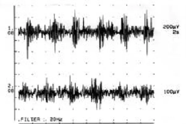
J Korean Society for Clinical Neurophysilology / Volume 4 / November, 2002 135 Figure 3. Spectral analysis of unvoluntary movements. Spectra
show angular rate RMS for the frequency components of abnormal movements plotted against frequency. 3 Hz unvolun- tary movement component was most frequent.
Figure 4. Alternating (60%) and co-contracting (40%) EMG activities at 3 Hz frequency were recorded from both finger flexor and extensor muscle groups of the right upper extremity.
occurrence of remyelinization, inflammatory changes, ephaptic transmission, oxidative reac- tions, central synaptic reorganization, trans- synaptic neuronal degeneration, and diaschisis mediated by collateral sprouting and denervation s u p e r s e n s i t i v i t y .
4It is suspected that incorrect recovery of the damaged neural circuit may be responsible for delayed onset.
K i m
5suggested that persistent failure of the proprioceptive sensory and cerebellar inputs, in addition to successful, but unbalanced, recovery of motor dysfunction, seemed to result in a pathological motor integrative system and conse- quent dyskinesia in stroke patients. His proposi- tion could be applicable to most patterns of dysk- inesia. Although the involved arm was weak in our case, dyskinesia could be suppressed com- pletely with the patient’s will. Furthermore, dyskinesia were not lessened by sensory stimuli and there were no geste antagonistique.
Therefore, it is suspected that the underlying mechanisms of our case might be different from K i m’ s suggestion. Marsden and Jenner(1980) have proposed that akathisia may be a result of post- synaptic dopamine receptor blockade in cerebral dopamine-containing regions of the brain other than the corpus striatum. Tassin et al.
7i n s i s t e d that destruction of mesocortical dopamine neu- rons is critical for the development of locomotor hyperactivity. The mesocortical dopamine system projects from the ventral tegmental area of the midbrain to the neocortex. Therefore it was sus- pected that this system might have been injured at the subcortical level in our case. Moreover, it was also suspected that underlying mechanisms of rhythmicity might have been related to the olivo-cerebellar-thalamo-cortical circuit.
Rhythmic akathisia should be differentiated from similar diseases such as pseudoakathisia, psychic agitation, tardive dyskinesia, secondary akathisia, malingering, etc. “P s e u d o a k a t h i s i a”
refers to motor manifestations of akathisia with- out subjective experiences of restlessness.
Patients with “p s e u d o a k a t h i s i a ”have a high inci- dence of orofacial and limb dyskinesia. Our
patient had no previous medication history of antipsychotics, nor any orofacial or limb dyskine- sia. Patients with psychotic agitation may exhibit excessive movement, but usually in the form of semi-purposeful movements of the upper extremities, such as handwriting and repetitive a c t i v i t i e s .
8The movement pattern of our patient was not semi-purposeful. Furthermore, our patient did not experience internal restlessness.
As previously mentioned, there was no medication history of antipsychotics. Our patient could sup- press dyskinesia completely as well. Therefore tardive dyskinesia could be differentiated. Our patient showed neither signs of Parkinsonism nor encephalitis. Secondary akathisia also could be differentiated. When our patient was disturbed, dyskinesia reappeared. So malingering was less l i k e l y .
REFERENCES
11. Ghika-Schmid F, Ghika J, Regli F, Bogousslavsky J.
Hyperkinetic movement disorders during and after acute stroke: the Lausanne Stroke Registry. J Neurol Sci 1 9 9 7 Mar 10;146(2):109-16.
12. Nemes Z, Volavka J, Bitter I, Laszlo Z. Rhythmic move- ments of chronic akathisia. Biol Psychiatr 1990;457-467.
13. Braude WM, Charles IP, Barnes TRE. Coarse, jerky foot tremor: Tremographic investigation of an objective sign of acute akathisia. Psychopharmacology 1984;82:95-101.
14. Scott BI, Jankovic J. Delayed-onset progressive movement disorders after static brain lesions. Neurology 1996;46:68- 74.
15. Kim JS. Delayed onset mixed involuntary movements after thalamic stroke.:clinical, radiological and pathophysiologi- cal findings. Brain 2001;124:299-309.
16. Marsden CD, Jenner P. The pathophysiology of extrapyra- midal side-effects of neuroleptic drugs. Psychol Med 1980;10:55-72.
17. Tassin JP, Stinus L, Simon H. Relationship between the locomotor hyperactivity induced by A 10 and the destruc- tion of the frontocortical dopaminergic innervation in the rat. Brain Res 1978;141:267-281.
18. Braude WM, Barnes TRE, Gore SM. Clinical characteris- tics of akathisia. A systemic investigation of acute psychi- atric impatient admissions. Brit J Psychiatr 1983;143:139- 152.
서만욱・오선영・성경미・신병수・김영현
136 J Korean Society for Clinical Neurophysilology / Volume 4 / November, 2002