저작자표시-비영리-변경금지 2.0 대한민국 이용자는 아래의 조건을 따르는 경우에 한하여 자유롭게 l 이 저작물을 복제, 배포, 전송, 전시, 공연 및 방송할 수 있습니다. 다음과 같은 조건을 따라야 합니다: l 귀하는, 이 저작물의 재이용이나 배포의 경우, 이 저작물에 적용된 이용허락조건 을 명확하게 나타내어야 합니다. l 저작권자로부터 별도의 허가를 받으면 이러한 조건들은 적용되지 않습니다. 저작권법에 따른 이용자의 권리는 위의 내용에 의하여 영향을 받지 않습니다. 이것은 이용허락규약(Legal Code)을 이해하기 쉽게 요약한 것입니다. Disclaimer 저작자표시. 귀하는 원저작자를 표시하여야 합니다. 비영리. 귀하는 이 저작물을 영리 목적으로 이용할 수 없습니다. 변경금지. 귀하는 이 저작물을 개작, 변형 또는 가공할 수 없습니다.
A THESIS
FOR THE DEGREE OF DOCTOR OF PHILOSOPHY
Anti-cancer effects of apigenin by enhancing tumor necrosis
factor-related apoptosis-inducing ligand-mediated death receptor 5
expression and inhibition of telomerase activity
Chang-Hee Kang
Department of Marine Life Science GRADUATE SCHOOL JEJU NATIONAL UNIVERSITY
CONTENTS
Abstract ...ⅰ
국문요약 ...ii
List of figure ...ⅳ Chapter Ⅰ. Coexistence of cell death and cell survival signals in apigenin-mediated sensitization of tumor necrosis factor-related apoptosis-inducing ligand ...1
Abstract ...2
1. Introduction ...3
2. Materials and Methods ...5
2.1. Reagents and methods ...5
2.2. Cell culture and viability ... 5
2.3. Isolation of total RAN and RT-PCR ...6
2.4. Western blot analysis ...6
2.5. Detection of apoptosis ...6
2.6. Measurement of ROS ...7
2.7. DNA fragmentation assay ...7
2.8. Flow cytometric analysis of DR5 ...7
2.9. Statistical analysis ...8
3. Results ...9
3.2. Apigenin activates caspase and antiapoptotic proteins during TRAIL sensitization ...11
3.3. Apigenin induces expression of DR5, causing to TRAIL-mediated apoptosis ...13
3.4. Apigenin-induced ROS protected TRAIL-induced apoptosis ...15
3.5. Apigenin transmits autophagy-mediated cell survival signal by generating ROS ...17
4. Discussion ...19
Chapter II. Apigenin decrease cell viability and telomerase activity in human leukemia cell lines ...25
Abstract ...26
1. Introduction ...27
2. Materials and Methods ...29
2.1. Reagents and antibodies ...29
2.2. Cell lines and culture ...29
2.3. Cell culture and viability ...29
2.4. Flow cytometric analysis ...30
2.5. Measurement of ROS ...30
2.6. DNA fragmentation assay ...30
2.7. Western blot analysis ...31
2.8. Telomerase activity assay ...31
2.9. RNA extraction and RT-PCR ...31
2.10. Electrophoretic mobility shift assay (EMSA) ...32
2.11. Statistical analysis ...33
3. Results ...34
3.1. Apigenin decreases leukemia cell viability ...34
3.2. Apigenin induces apoptosis via a caspase-dependent pathway ...36
3.3. ROS is not a main regulator in apigenin-induced apoptosis ... 38
3.4. Apigenin suppresses telomerase activity and hTERT transcriptional level ...40
3.5. Apigenin suppresses c-Myc expression in leukemia cancer cells ... 42
4. Discussion ...45
i
ABSTRACT
Apigenin is known to suppress in cancer cells proliferation, angiogenesis, motility, and induce apoptosis; however, it is little known whether apigenin triggers tumor necrosis factor (TNF)-related apoptosis-inducing ligand (TRAIL)-induced apoptosis. In addition, the underlying relationship between apoptosis and telomerase activity in response to apigenin exposure is not well understood. In this study, we found that apigenin enhances TRAIL-induced apoptosis in hepatocarcinoma cells (Hep3B and HepG2) and human leukemia cells (U937, THP-1 and HL60) by inducing caspase activation. These results were found to increase of death receptor 5 (DR5) expressions and inhibition of telomerase activity. As shown in previous studies, apigenin increased reactive oxygen species (ROS) production, however, intriguingly; ROS inhibitors sensitize combined treatment with apigenin and TRAIL-induced apoptosis, intimating that apigenin-induced ROS produces weaker cell survival signal than apoptotic one. Furthermore, we found elevated reactive oxygen species (ROS) generation following apigenin treatment. And then, apigenin-induced ROS was protection of cell death. Our results shown that apigenin-induced ROS-mediated cell protection effects was activation of autophagy. TRAIL-induced apoptosis greatly increased by specific autophagy inhibitor 3-methyladenine (3-MA). Taken together, these finding revealed that apigenin enhanced TRAIL-induced apoptosis, while on the other hand apigenin protects the apoptosis by increasing the ROS-mediated autophagy. We further showed that apigenin downregulates telomerase activity in caspase-dependent apoptosis and observed that apigenin dosing results in down regulation of telomerase activity via inhibition of c-Myc-mediated telomerase reverse transcriptase (hTERT) expression. Be sides, treatment of apigenin cells with the two ROS inhibitors did not restore telomerase activity. We conclude that apigenin enhanced TRAIL-induced apoptosis by regulation of DR 5 expression and the loss of telomerase activity.
ii 국문요약 Apigenin은 암세포의 증식, 이동능, 신생혈관생성을 억제 하고 세포사멸사를 유도 하 는 것으로 알려졌으나, TRAIL이 유도하는 암세포 사멸사와의 연관 관계에 대해서는 아직 알려진 바가 없다. 게다가 apigenin 처리에 의한 telomerase의 활성 변화와 암세포의 세 포사멸사간의 상호 작용에 대한 연구는 아직 명확 하게 밝혀지지 않았다. 이 연구에서는 Hep3B와 HepG2 암세포를 이용하여 apigenin이 TRAIL이 유도하는 세포사멸사를 강화하고, 백혈병 세포주를 이용하여 caspase 단백질 활성을 통한 세포사멸사를 확인하였다.
Chapter 1. Apigenin은 DR5의 발현을 증가 시켜 TRAIL이 유도하는 세포사멸사를 강화 시켰다. 이번 연구 결과의 흥미로운 점은 apigenin은 ROS의 생산을 증가 시켰지만 ROS억 제제와 apigenin, TRAIL을 병행 처리 하자 더욱 강한 세포사멸사를 유도 하였다. 이 결 과는 apigenin에 의해 생산된 ROS가 세포를 보호하여 TRAIL에 의한 죽음으로부터 생존할 수 있게 도와 주었음을 의미한다. 우리의 연구결과는 apigenin에 의해 생산된 ROS가 autophagy의 활성을 유도하여 세포를 보호했음을 보여 주었다. Apigenin과 TRAIL이 유도 하는 세포사멸사는 autophagy 억제제인 3-MA에 의해 더욱 강하게 증가하였다. 그리고 그 증가 폭은 apigenin과 TRAIL, ROS 억제제를 처리하여 유도된 세포사멸사의 수치와 거의 일치하였다. 이 결과는 apigenin이 DR5의 발현을 증가시켜 TRAIL이 유도하는 세포사멸사 를 강화 시키지만, 한편으로는 ROS생성을 통해 autophagy의 활성을 유도하여 세포를 보 호하기도 함을 보여 준다.
iii
Chapter 2. Apigenin의 처리는 백혈병 세포주인 THP-1, U937, HL-60 세포에서 강한 세 포사멸사를 유도하였다. Apigenin이 생성한 ROS와 세포사멸사간의 관계를 규명하고자 apigenin과 ROS 억제제인 NAC과 GSH를 병행 처리하였다. NAC과 GSH는 apigenin이 생산하 는 ROS의 생산을 억제 하였으나 세포사멸사 유도에 대한 결과에는 별다른 변화가 없었 다. 따라서 우리는 세포사멸사를 유도하는 다른 원인인 telomerase의 활성 변화를 확인 하였다. 그 결과 apigenin은 telomerase의 활성을 억제하였고, 이는 hTERT의 발현 억제 를 통한 것임을 확인 하였다. 이러한 hTERT의 발현 억제는 c-Myc의 활성 억제를 통한 것 으로서 apigenin의 처리는 c-Myc의 전사인자로서의 활성을 억제 할 뿐만 아니라 c-Myc의 발현 역시 억제 하는 것으로 확인 되었다. 하지만 ROS억제제의 처리는 apigenin이 유도 하는 c-Myc의 활성을 억제 하지 못하였으며, telomerase의 활성역시 억제 하지 못하였 다, 결론 적으로 apigenin이 유도하는 c-Myc 활성 조절에 매개한 telomerase 활성 억제 효능은 apigenin이 생산한 ROS와는 연관이 없음을 규명하였다.
종합적으로 우리는 이번 연구에서 apigenin은 DR5의 발현 증가와 c-Myc의 활성 억제를 통하여 암세포 사멸 효과를 유도 하지만, 한편으로는 ROS 생산을 유도 함으로서 세포 보 호 효과를 확인 하였다. 이러한 apigenin의 성질을 이용하여 획기적인 암세포의 특이적 항암 치료제를 개발 할 수 있을 것으로 기대된다. 따라서 이후 연구에서는 apigenin이 유도하는 ROS와 autophagy, telomerase활성에 대해 상관 관계를 규명하는 연구를 진행 하고자 한다.
iv
List of figures
Chapter Ⅰ. Coexistence of cell death and cell survival signals in apigenin-mediated sensitization of
tumor necrosis factor-related apoptosis-inducing ligand
Fig. 1. Apigenin enhanced TRAIL-induced apoptosis in hepatocarcinoma cells ...10
Fig. 2. Treatment with a combination of apigenin and/or TRAIL activates apoptotic signal ... 12
Fig. 3. Apigenin induced up-regulation of DR5 in Hep3B cells ...14
Fig 4. Apigenin-induced ROS protected TRAIL-induced apoptosis ...16
Fig. 5. Apigenin-induced autophagy protected TRAIL-induced apoptosis ...18
Chapter II. Apigenin decrease cell viability and telomerase activity in human leukemia cell lines Fig. 1. Apigenin decreases cell viability and proliferation in leukemia cell lines ...35
Fig. 2. Apigenin sensitizes apoptosis in leukemia cell line, but not in mouse spleen cells ...37
Fig. 3. ROS is not essential for apigenin-induced apoptosis ...39
Fig. 4. Apigenin inhibits telomerase activity and hTERT expression ...41
1
Chapter Ⅰ.
Coexistence of cell death and cell survival signals in apigenin-mediated
sensitization of tumor necrosis factor-related apoptosis-inducing ligand
2
Abstract
Apigenin is known to suppress cancer cells proliferation, angiogenesis, and motility, which consequently induce apoptosis; however, it is little known whether apigenin triggers tumor necrosis factor (TNF)-related apoptosis-inducing ligand (TRAIL)-induced apoptosis. In this study, we found that apigenin enhances TRAIL-induced apoptosis in hepatocarcinoma Hep3B and HepG2 cells by inducing caspase activation. Additionally, one of crucial target in TRAIL-induced therapeutics, death receptor 5 (DR5) which is a main ligand of TRAIL, was significantly expressed in response to apigenin and chimeric antibody of DR5 completely blocked combined treatment with apigenin and TRAIL-induced apoptosis. As shown in previous studies, apigenin increased reactive oxygen species (ROS) generation in a dose-dependent manner; however, intriguingly, ROS inhibitors, glutathione (GSH) and N-acetyl-L-cysteine (NAC) sensitize combined treatment with apigenin and TRAIL-induced apoptosis, suggesting that apigenin-induced ROS generates cell survival signal than apoptotic one. Our results also showed that apigenin-induced ROS-mediated cell protection effects was activation of autophagy. TRAIL-induced apoptosis greatly increased by specific autophagy inhibitor 3-methyladenine (3-MA). Taken together, these finding revealed that apigenin enhanced TRAIL-induced apoptosis, while on the other hand apigenin protects the apoptosis by increasing the ROS-mediated autophagy.
3
1. Introduction
The tumor necrosis factor (TNF)-related apoptosis-inducing ligand (TRAIL) is a member of the structurally related TNF superfamily such as Fas ligand and TNF-α. TRAIL is an encouraging anticancer agent that is evaluated in preclinical and clinical researches [1, 2]. TRAIL induces typical apoptotic signaling through complex to death receptors (DRs), which trigger the signal which death-inducing signaling complex (DISC) and caspase cascade [3]. In contrast with another TNF super family such Fas ligand and TNF-α, TRAIL has challenged on clinical trial for cancer therapy because TRAIL is cancer cell-specific without any adverse effect in normal cells [4]. However, some studies showed that development of resistance to TRAIL in cancer cells has been found and the molecular basis for TRAIL resistance is undisturbed and elusive [5, 6]. For example, mutations in apoptosis-related proteins are monitored in various types of cancer cell lines [1]. Besides, dysfunction of DRs and abnormal activation of apoptosis regulatory proteins results in TRAIL resistance in tumor cells [7]. Therefore, understanding the intrinsic mechanisms for TRAIL resistance in cancer cells will significantly improve the merit of TRAIL as an anticancer therapeutic.
Autophagy is an evolutionarily preserved mechanism for maintaining cellular homeostasis by degrading intracellular proteins and organelles through the lysosomal and proteasomal pathway [8]. During the autophagy induction, a small vesicular sac elongates and afterwards encloses a segment of the cytoplasm, shaping a double-membrane structure known as an autophagosome in which the enclosed materials are degrade [9]. Recent studies also showed that autophagy plays important and paradoxical functions in tumorigenesis and cell death/survival in cancer cells [10, 11]. Gelinas et al. reported that autophagy inhibits tumorigenesis via eliminating damaged organelles/proteins and suppressing cell growth [12]. On the other hand, induction of autophagy promoted tumor cell survival and tumorigenesis through metabolic stress from stress damage in cancer cells [13]. Many preclinical researches suggest that various stimuli-induced autophagy is superior cytoprotective in cancer cells and suppression of autophagy enhances cancer cell death through diverse anticancer therapies, although the effect of autophagy is still contradictory between cell survival and death [14, 15].
4
Reactive oxygen species (ROS), which is by-products of general metabolic processes and produced via exogenous source, are secondary signal messengers in tumorigenesis and cancer therapeutics [16]. Significant downstream mediators of ROS-induced signaling are the induction of CCAAT/enhancer-binding protein (C/EBP) homologous protein (CHOP) and mitogen-activated protein kinases (MAPKs) [16]. We previously reported that ROS are main regulators to sensitize TRAIL-mediated apoptosis by inducing CHOP expression, which enhances DR5 expression [17]. The above results mean that ROS could offer potential as anticancer agents by stimulating various apoptotic signal pathway. In addition, Scherz-Shouval et al. determined that damaged mitochondria and oxidized proteins are removed in the cells under high accumulation of ROS by leading to autophagy, which suggests that autophagy causes to defense strategies against oxidative stress [18]. However, whether relationship between ROS and autophagy influences sensitization of TRAIL-mediated cancer cell death is still not evaluated.
Apigenin (4`,5,6,-trihydroxyflavone) is a widespread dietary flavonoid in various fruits and vegetables, including oranges, teas, onions and parsley [19]. Apigenin is mostly found in hydroxylated form and suppresses cancer cell proliferation, angiogenesis, and metastasis, which consequently induces apoptosis [20-22]. Even though many studies showed that apigenin possesses anticancer effects, its molecular mechanisms underlying relationship ROS and autophagy remain unclear in sensitization of TRAIL. Therefore, we, in the current study, investigated the role of ROS and autophagy apigenin-induced TRAIL sensitization.
5
2. Materials & methods
2.1. Reagents and methods
Antibodies against Bcl-2, IAP-1, IAP-2, XIAP, DR4 and DR5, were obtained from Santa Cruz Biotechnology (Santa Cruz, CA). Antibodies against caspase-3, caspase-8, caspase-9, PARP, and β-actin were obtained from Cell Signaling (Beverly, MA). The antibodies against LC-3B, SQSTM1/p62, and Atg-7 were purchased from Thermo Scientific (Waltham, MA). Peroxidase-labeled donkey anti-rabbit immunoglobulin, and recombinant human TRAIL/Apo2 ligand (the nontagged 19-kDa protein, amino acids 114-281) were purchased from KOMA Biotechnology (Seoul, Republic of Korea). The chimeric antibody against DR5 was obtained from R&D Systems (Minneapolis, MN). 6-Carboxy-2',7'-dichlorofluorescein diacetate (DCFDA) was obtained from Molecular Probes (Eugene, OR). Glutathione (GSH) and N-acetyl-L-cysteine (NAC) were purchased from Sigma (St. Louise, MO). Apigenin was obtained from Tocris (Ellisville, MO) and dissolved in DMSO (vehicle). Rapamycin (RAPA), bafilomycin A1 (BAF), and 3-methyladenine (3-MA) were also purchased from Tocris.
2.2. Cell culture and viability
Human hepatocarcinoma Hep3B and HepG2 cells were obtained from American Type Culture Collection and cultured in RPMI 1640 (WelGENE Inc, Daegu, Republic of Korea) supplemented with 10% fetal bovine serum and antibiotics (WelGENE Inc). The cells were seeded at a density of 1 ´ 105 cells/ml, incubated for 12 h, and then treated with the indicated concentrations of apigenin in the presence and absence of TRAIL. After 24 h, 3-(4,5-dimethylthiazol-2-yl)-2,5-diphenyltetrazolium bromide (MTT) assay was performed to determine cell viability.
6
2.3. Isolation of total RNA and RT-PCR
Total RNA was extracted from Hep3B cells using the Easy-Blue reagent (iNtRON Biotechnology, Sungnam, Republic of Korea). The DR5 sense primer 5′-GTC TGC TCT GAT CAC CCA AC-3′ and the anti-sense primer 5′-CTG CAA CTG TGA CTC CTA TG-3′ were used to amplify human DR5 mRNA. For glyceraldehyde-3-phosphate dehydrogenase (GAPDH), the sense primer 5′-CGT CTT CAC CAT GGA GA-3′ and the anti-sense primer 5′-CGG CCA TCA CGC CCA CAG TTT-3′ were used. The following PCR conditions were applied: GAPDH, 25 cycles of denaturation at 94°C for 30 s, annealing at 63°C for 30 s and extended at 72°C for 30 s; DR5, 31 cycles of denaturation at 94°C for 30 s, annealing at 60°C for 30 s and extended at 72°C for 30 s. GAPDH was used as an internal control to evaluate relative expression of DR5.
2.4. Western blot analysis
Total cell extracts were prepared using PRO-PREP protein extraction solution (iNtRON Biotechnology). Briefly, after treatment with the indicated concentrations of apigenin, cells were harvested, washed once with ice-cold PBS, and gently lysed for 30 min in 100 μl ice-cold PRO-PREP lysis buffer. Lysates were centrifuged at 14,000 g at 4°C for 10 min. The preparation of cytoplasmic and nuclear extracts was conducted using NE-PER nuclear and cytosolic extraction reagents (Pierce, Rockford, IL). The extraction of protein samples was collected and protein concentrations determined using a Bio-Rad protein assay kit (Bio-Rad, Hercules, CA). Total cell extracts were separated on polyacrylamide gels and then standard procedures were used to transfer them to the nitrocellulose membranes. The membranes were developed using an ECL reagent (Amersham, Arlington Heights, IL).
2.5. Detection of apoptosis
7
ethanol overnight at 4°C and washed in phosphate-buffered saline (PBS) with 0.1% BSA. The cells were then incubated with 1 U/ml of RNase A (DNase free) and 10 µg/ml of propidium iodide (PI, Sigma) in the dark. A FACS Calibur flow cytometer (Becton Dickenson, San Jose, CA) was used to analyze cell cycle distribution. The level of apoptotic cells containing sub-G1 DNA content was determined as a percentage of the total number of cells. For annexin-V staining, live cells were washed with PBS and then incubated with annexin-V fluorescein isothiocyanate (FITC, R&D Systems). Annexin-V+ cells were analyzed by flow cytometry.
2.6. Measurement of ROS
Cells were plated at a density of 1 ´ 105 cells/ml, allowed to attach for 12 h, and exposed apigenin for 1 h. The cells were stained with 10 µM DCFDA for 30 min at 37°C and flow cytometry was used to determine the fluorescence intensity of DCFDA in the cells.
2.7. DNA fragmentation assay
Cells were lysed using lysis buffer [10 mM Tris (pH 7.4), 150 mM NaCl, 5 mM EDTA, and 0.5% Triton X-100] for 1 h on ice. Lysates were vortexed and cleared by centrifugation at 13,000 g for 20 min. Fragmented DNA in the supernatant was extracted with an equal volume of phenol/chloroform/isoamyl alcohol mixture (25:24:1) and analyzed on 1.5% agarose gels containing of ethidium bromide.
2.8. Flow cytometric analysis of DR5
Indirect staining with primary rabbit anti-human DR5 followed by FITC-conjugated IgG was used to analyze the surface expression of DR5. Briefly, 1 10 5 cells/ml cells were stained with 200 µl of 1% BSA in PBS containing amounts of anti-DR5 antibody at room temperature for 1 h. After incubation,
8
the cells were washed twice and reacted with FITC-conjugated rabbit polyclonal IgG for 1 h. After the cells were washed with 1% BSA, flow cytometry was used to analyze the expressions of DR5.
2.9. Statistical Analysis
The images were visualized with Chemi-Smart 2000 (VilberLourmat, Marine,Cedex, France). Images were captured using Chemi-Capt (VilberLourmat) and transported into Photoshop. All data were derived from at least three independent experiments. Statistical analyses were conducted using SigmaPlot software (version 12.0) Values were presented as mean ± standard error (S.E.). Significant differences between the groups were determined using the unpaired one-way and two-way ANOVA test by Bonferroni′s test. Statistical significance was regarded at *, #, and &, p < 0.05.
9
3. Results
3.1. Apigenin sensitizes Hep3B and HepG2 cells towards TRAIL-induced apoptosis
To determine whether apigenin enhances TRAIL-induced apoptosis, we treated with nontoxic concentration of TRAIL (25 ng/ml) in the presence and absence of apigenin for 24 h and then measured relative cell viability. Apigenin itself slightly decreased the viability in Hep3B cells over 15 μM and HepG2 cells over 10 μM, respectively (Fig. 1). However, nontoxic concentration of TRAIL (25 ng/ml) significantly increased apigenin-induced decrease of the relative cell viability. The result means that apigenin sensitizes the cells to TRAIL-mediated apoptosis. In addition, we assessed that combined treatment with apigenin and TRAIL directly induces apoptotic cell death. As shown in DNA fragmentation analysis via agarose gel electrophoresis, the combined treatment remarkably displayed a typical ladder pattern of DNA fragmentation in Hep3B and HepG2 cells compared to that of treatment with apigenin or TRAIL alone (Fig. 1B). Furthermore, apoptotic sub-G1 populations (Fig. 1C, top panel) and annexin-V+ cells (Fig. 1C, bottom panel) were significantly increased in Hep3B cells treated with 15 μM apigenin and 25 ng/ml TRAIL (from 3.0% to 38.9% and from 3.1% to 57.2%, respectively). These results showed that co-treated apigenin enhanced TRAIL-mediated apoptosis.
10
Fig. 1. Apigenin enhanced TRAIL-induced apoptosis in hepatocarcinoma cells. (A) Hep3B and HepG2 cells treated with apigenin for 1 h at the indicated concentration with TRAIL (25 ng/ml) for 24 h. Cell viability was monitored by MTT assay. (B) DNA fragmentation due to treatment of the co-treatment of apigenin (15 μM) and/or TRAIL (25 ng/ml). After co-treatment of cells as incubated for 24 h, DNA samples were extracted from the treated cells and analyzed on 1.5% agarose gel. (C) Pre-treated with apigenin (15 μM) for 1 h and further Pre-treated with TRAIL (25 ng/ml) for 24 h. Representative flow cytometry histograms displaying DNA content (top) and annexin-V population (bottom) of the cells.
11
3.2. Apigenin activates caspases and antiapoptotic proteins during TRAIL sensitization
To assess whether apigenin induces apoptosis via activation of caspase cascade and downregulation of antiapoptotic proteins, we examined protein expression and apoptotic cell death in the presence of a pan-caspase inhibitor, z-VAD-fmk. Hep3B cells were treated with apigenin (15 μM) and TRAIL (25 ng/ml) for 24 h. Fig. 2A showed that treatment with apigenin or TRAIL alone no changed or slightly decreased the expression of procaspase form. However, combined treatment considerably downregulated procaspase forms and dramatically increased the truncation of PARP in Hep3B. We also investigated the expression levels of 2 and IAP family, because antiapoptotic proteins such as Bcl-2 and IAP family are safeguards against cancer cells against TRAIL-induced apoptosis through inhibiting caspase activation. The combined treatment with apigenin and TRAIL induced the downregulation of the antiapoptotic proteins including Bcl-2, IAP-1, IAP-2, and xIAP (Fig. 2B). Additionally, to determine whether caspases are main regulators in apigenin/TRAIL-induced apoptosis, we treated with apigenin/TRAIL in the presence of z-VAD-fmk. Pretreatment with z-VAD-fmk significantly suppressed sub-G1 populations (from 38.9% to 2.6%) and annexin-V+ cells (from 55.7% to 2.8%, Fig. 2C). These results indicated that combined treatment with apigenin and TRAIL induces apoptosis by activating antiapoptotic proteins and caspases.
12
Fig. 2. Treatment with a combination of apigenin and/or TRAIL activates apoptotic signal. Hep3B cells were treated 25 ng/ml TRAIL alone, 20 μM apigenin aloe, or a combination of both for 24 h. (A) Caspase cascade were then analyzed via western blotting. (B) Expression of antiapoptotic proteins were then monitored in Hep3B cells by western blotting. (C) Hep3B cells were pre-treated with pan-caspase inhibitor and then the cells were treated with 25 ng/ml of TRAIL alone, 15 μM of apigenin alone, or a combination of both for 24 h. Flow cytometry analyzed the DNA content (top) and annexin-V+ (bottom) of the cells. API, apigenin; T, TRAIL.
13
3.3. Apigenin induces expression of DR5, causing to TRAL-mediated apoptosis
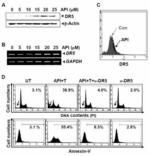
We next investigated whether DR5 expression by treatment with apigenin is involved in its synergetic effect on TRAIL-induced apoptosis. We treated with the indicated concentrations of apigenin in Hep3B cells for 24 h and DR5 expression was detected by western blot analysis. Apigenin induced DR5 protein (Fig. 3A) and mRNA (Fig. 3B) in Hep3B cells in a dose-dependent manner. Whether apigenin induces expression of DR5 on the cell surface was also investigated by flow cytometry. Fig. 3C showed that apigenin induced upregulation of DR5 on the cell surface. Furthermore, treatment with chimeric antibody against DR5 considerably decreased sub-G1 populations and annexin-V+ cells in apigenin/TRAIL-induced apoptosis (Fig. 3D). These results indicate that apigenin-induced upregulation of DR5 is crucial for the sensitization of TRAIL in Hep3B cells.
14
Fig. 3. Apigenin induced up-regulation of DR5 in Hep3B cells. (A) Hep3B cells were treated indicated concentration (0 - 25 μM) of apigenin for 24 h and western blot analysis of DR5 and β-actin as a loading control was conducted. (B) Hep3B cells were treated indicated concentration (0 - 25 μM) of apigenin for 24 h and RT-PCR analysis of DR5 and GAPDH as a loading control was conducted. (C) Hep3B cells were incubated with or without 25 μM of apigenin for 24 h, and flow cytometry was used to analyze the surface expression of DR5. (D) Hep3B cells were pre-treated with or without 15 μM of apigenin for 1 h followed by treatment with or without 25 ng/ml of TRAIL for 24 h in the presence of the indicated concentrations of DR5-specific blocking chimera antibody. Flow cytometry analyzed the DNA content (top) and annexin-V+ (bottom) of the cells. API, apigenin; T, TRAIL.
15
3.4. Apigenin-induced ROS protected TRAIL-induced apoptosis
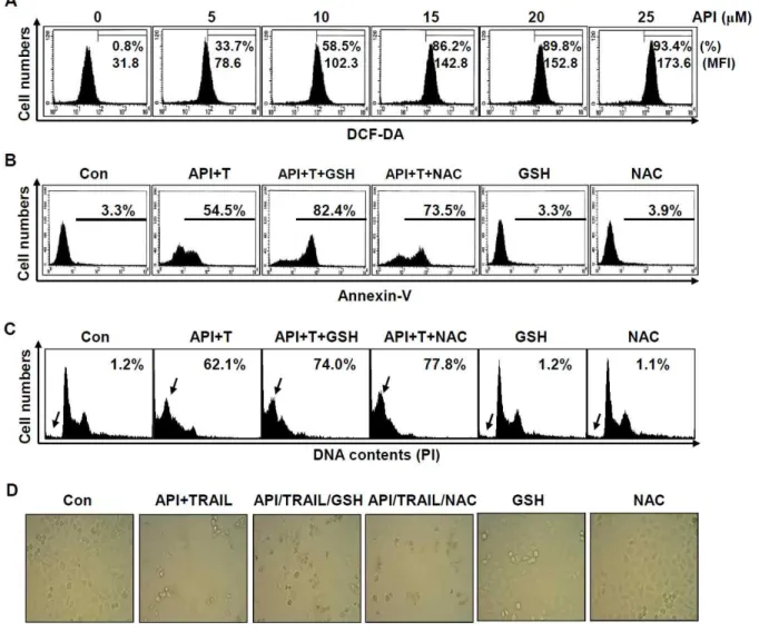
Recent several reports demonstrated that ROS increased DR5 expression in response to chemotherapy-induced stress, which consequently induced apoptosis [23, 24]. Therefore, whether apigenin regulates ROS generation, and which activates TRAIL-mediated apoptosis, was investigated. Fig. 4A showed that various concentrations of apigenin induced a significant increase of ROS generation from 1 h. To further confirm the role of ROS in apigenin/TRAIL-induced apoptosis, Hep3B cells were pretreated for 1 h with antioxidants such as GSH and NAC, and then treated with apigenin and TRAIL for an additional 24 h. As unexpected, DNA contents data showed that pretreatment with GSH and NAC considerably increased apigenin/TRAIL-induced apoptosis (Fig. 4B). Similar to the data of DNA contents, apigenin/TRAIL-induced annexin-V staining cells were increased in the presence of GSH and NAC compared to that in combined treatment with apigenin and TRAIL (Fig. 4C). Additionally, the combined treatment with apigenin and TRAIL enhanced total cell number and shrinkage of the cells in the presence of GSH and NAC under microscopy (Fig. 4D). These results indicate that apigenin stimulates ROS generation, which functions cellular protective effects in TRAIL-mediated apoptosis.
16
Fig. 4. Apigenin-induced ROS protected TRAIL-induced apoptosis. (A) Hep3B cells were stimulated with indicated concentration (0 - 25 μM) of apigenin for 1 h. Then, ROS generation was analyzed by DCFDA staining through detection of Flow cytometry. Hep3B cells were stimulated with 20 μM of apigenin and 25 ng/ml TRAIL for 24 h after pre-treatment with 2.5 mM of NAC and 2.5 mM of GSH 1 h. Then, Flow cytometry analyzed the DNA content (B) and annexin-V+ (C) of the cells. (D) Morphology of cells was monitored under light microscopy. API, apigenin; T, TRAIL
17
3.5. Apigenin transmits autophagy-mediated cell survival signal by generating ROS
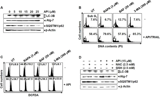
We next addressed whether apigenin-induced ROS activated autophagy in Hep3B cells. The conversion of LC-3B-1 to LC-3B-2, a hallmark of autophagy, was monitored in the Hep3B cells 24 h after apigenin treatment accompanied with a significantly increase of Atg-7 (Fig. 5A). In addition, autophagic protein, SQSTM1/p62, was gradually diminished in apigenin-treated condition. Interestingly, an autophagy inducer, RAPA, strongly potentiated apigenin/TRAIL-induced apoptosis (Fig. 5B), which suggests that RAPA boosters apigenin/TRAIL-induced apoptosis by increasing autophagy. Surprisingly, an autophagy inhitor, 3-MA, also significantly upregulated apigenin/TRAIL-induced apoptosis; however, no apoptotic change was observed in the presence of BAF. These results suggested that suitable autophagy attenuates apigenin/TRAIL-induced apoptosis.
We then examined whether ROS activates cell survival or death through autophagy formation. Both BAF and 3-MA alone have no effect of ROS generation; however, RAPA alone a little increased ROS generation and more multiplied with apigenin (Fig. 5C). In addition, both NAC and GSH inhibited apigenin-induced autophagy activation, which was shown via LC-3B conversion, regulation of SQSTM1/p62 and Atg-7 expression (Fig. 5D). These data indicate that apigenin-induced ROS generation stimulates the expression of autophagy proteins such as LC-3B, Atg-7 and SQSTM1/p62, which induces autophagy formation.
18
Fig. 5. Apigenin-induced autophagy protected TRAIL-induced apoptosis. (A) Hep3B cells were treated indicated concentration (0 - 25 μM) of apigenin for 24 h and western blot analysis of hallmarks of autophagy and β-actin as a loading control was conducted. (B and C) Hep3B cells were stimulated with 20 μM of apigenin and/or 25 ng/ml TRAIL for 24 h after pre-treatment with 3 μM of RAPA, 20 nM of BAF, and 300 μM of 3-MA for 1 h. Then, Flow cytometry analyzed the DNA content (B) and DCFDA intensity (C) of the cells. (D) Hep3B cells were stimulated with indicated concentration (15 μM) of apigenin and 25 ng/ml TRAIL for 24 h after pre-treatment with 2.5 mM of NAC and 2.5 mM of GSH 1 h. Then western blot analysis of hallmarks of autophagy and β-actin as a loading control was conducted. API, apigenin; T, TRAIL; RAPA, rapamycine; BAF, bafiolomycine A 1; MA, 3-Methyladenine
19
4. Discussion
Since TRAIL promotionally induced apoptosis to cancer cells but not normal cells, it is believed to possess brilliant potential as an anticancer chemotherapy. However, it reported that many cancer cells and the majority tumors originated from cancer patients has acquired resistance to TRAIL via abnormal protein expression [25]. Therefore, we, in the present study, investigated whether apigenin enhances TRAIL-mediated apoptosis. Especially, it is well-known that DR5 is necessary for the increase of death signal induced by TRAIL, resulting in the activation of caspase-8 [26]. Thus, it has been considered that upregulation of DR5 leads to effective apoptosis of cancer cells by direct binding with TRAIL. We found that apigenin significantly induced DR5 expression, which stimulates TRAIL-mediated apoptotic signal. DR5-specific blocking chimeric antibody inhibited apigenin/TRAIL-mediated apoptosis.
Autophagy is cellular process of recycling and clearance of cytoplasmic ingredients, such as protein aggregates and obsolete organelles by lysosomal degradation [27]. Under stress condition such as starvation and hypoxia, autophagy performs a cellular pro-survival mechanism via the production of alternative energy resources from fuel cellular metabolism, however, inordinate autophagy could contribute to cell death by autophagic cell death [28]. Our results found that apigenin induced autophagy activation, which consequently enhanced TRAIL-induced apoptosis by producing ROS; however, an autophagy inhibitor, 3-MA, enhanced apigenin/TRAIL-mediated apoptosis, but no changed by BAF, suggesting that apigenin-induced autophagy interfered TRAIL-induced apoptosis. On the other hand, an autophagy inducer, RAPA, enhanced apigenin/TRAIL-induced apoptosis, which means that considerable autophagy induction could converse cell fate to death. According to the present data, apigenin triggers TRAIL-mediated apoptosis and simultaneously activates suitable autophagy-mediated cell survival signal by producing ROS. However, another autophagy inhibitor, BAF, was not effective in apigenin/TRAIL-mediated apoptosis. Interestingly, 3-MA and BAF dissimilarly inactivates autophagy activation. 3-MA inhibits autophagosome formation in the early stage by phosphatidylinositol 3-kinases [29], whereas BAF blocks the fusion of autophagosome and lysosome (autolysosome) by inhibiting vacuolar ATPase (V-ATPase) located in the lysosomal membrane at the
20
late stage [30]. In the current study, we hypothesized that 3-MA restrains autophagosome formation, which sustains induced apoptotic proteins in the cytosol. Therefore, apigenin/TRAIL-mediated apoptosis amplified in the presence of 3-MA. However, BAF could not interrupt the formation of autophagosome induced by apigenin and TRAIL in the early stage, which could hold apoptotic proteins in the vacuoles without degradation of the proteins. We need further study whether apoptosis-inducing factors are trapped in the autophagosome, not autolysome, in the presence of apigenin.
We demonstrated that the activation of autophagy was upregulated by the generation of ROS, which protects apigenin/TRAIL-mediated apoptosis. Previous study suggested that ROS are mutagenic, which promotes cancer growth and survival [31]; however, our data showed that apigenin-induced ROS generation may inhibit TRAIL-mediated apoptosis. A number of studies also reported ROS suppress mTOR by activating AMP-activated protein kinase (AMPK) and suppressing AKT, inducing to autophagy-dependent cytotoxicity [32, 33]. On the other hand, contemplating that some cancer cells gain advantage of protective autophagy to survive, suppression of protective autophagy may be potential strategy for cancer chemotherapy [34].
Taken together, apigenin stimulates TRAIL-mediated apoptosis by inducing DR5 expression and coincides cell survival signal via ROS-mediated autophagy. The result is a good case of an antagonism between autophagy and apoptosis in cancer cells: protection of autophagy switches to apoptosis under apigenin treatment.
21
Reference
1. Abdulghani J, El-Deiry WS. TRAIL receptor signaling and therapeutics. Expert Opin Ther Targets 2010;14(10):1091-108.
2. Stegehuis JH, de Wilt LH, de Vries EG, Groen HJ, de Jong S, Kruyt FA. TRAIL receptor targeting therapies for non-small cell lung cancer: current status and perspectives. Drug Resist Updat 2010;13(1-2):2-15.
3. Johnstone RW, Frew AJ, Smyth MJ. The TRAIL apoptotic pathway in cancer onset, progression and therapy. Nat Rev Cancer 2008;8(10):782-98.
4. Bellail AC, Qi L, Mulligan P, Chhabra V, Hao C. TRAIL agonists on clinical trials for cancer therapy: the promises and the challenges. Rev Recent Clin Trials 2009;4(1):34-41.
5. Song JJ, An JY, Kwon YT, Lee YJ. Evidence for two modes of development of acquired tumor necrosis factor-related apoptosis-inducing ligand resistance. Involvement of Bcl-xL. J Biol Chem 2007;282(1):319-28.
6. Wenger T, Mattern J, Penzel R, Gassler N, Haas TL, Sprick MR, Walczak H, Krammer PH, Debatin KM, Herr I. Specific resistance upon lentiviral TRAIL transfer by intracellular retention of TRAIL receptors. Cell Death Differ 2006;13(10):1740-51.
7. van Geelen CM, Pennarun B, Le PT, de Vries EG, de Jong S. Modulation of TRAIL resistance in colon carcinoma cells: different contributions of DR4 and DR5. BMC Cancer 2011;11:39.
8. Klionsky DJ, Abdelmohsen K, Abeliovich H, Acevedo-Arozena A et al.Guidelines for the use and interpretation of assays for monitoring autophagy. Autophagy 2012;8(4):445-544. 9. Crighton D, Wilkinson S, O'Prey J, Syed N, Smith P, Harrison PR, Gasco M, Garrone O,
Crook T, Ryan KM. DRAM, a p53-induced modulator of autophagy, is critical for apoptosis. Cell 2006;126(1):121-34.
22
10. Rao S, Tortola L, Perlot T, Wirnsberger G, Novatchkova M, Nitsch R, Sykacek P, Frank L, Schramek D, Komnenovic V, Sigl V, Aumayr K, Schmauss G, Fellner N, Handschuh S, Glösmann M, Pasierbek P, Schlederer M, Resch GP, Ma Y, Yang H, Popper H, Kenner L, Kroemer G, Penninger JM . A dual role for autophagy in a murine model of lung cancer. Nat Commun 2014;5:3056.
11. Wu WK, Coffelt SB, Cho CH, Wang XJ, Lee CW, Chan FK, Yu J, Sung JJ. The autophagic paradox in cancer therapy. Oncogene 2012;31(8):939-53.
12. Mathew R, Karp CM, Beaudoin B, Vuong N, Chen G, Chen HY, Bray K, Reddy A, Bhanot G, Gelinas C, Dipaola RS, Karantza-Wadsworth V, White E. Autophagy suppresses tumorigenesis through elimination of p62. Cell 2009;137(6):1062-75
13. Degenhardt K, Mathew R, Beaudoin B, Bray K, Anderson D, Chen G, Mukherjee C, Shi Y, Gélinas C, Fan Y, Nelson DA, Jin S, White E. Autophagy promotes tumor cell survival and restricts necrosis, inflammation, and tumorigenesis. Cancer Cell 2006;10(1):51-64. 14. Yang ZJ, Chee CE, Huang S, Sinicrope FA. The role of autophagy in cancer: therapeutic
implications. Mol Cancer Ther 2011;10(9):1533-41.
15. Lamoureux F, Thomas C, Crafter C, Kumano M, Zhang F, Davies BR, Gleave ME, Zoubeidi A. Blocked autophagy using lysosomotropic agents sensitizes resistant prostate tumor cells to the novel Akt inhibitor AZD5363. Clin Cancer Res 2013;19(4):833-44. 16. Gupta SC, Hevia D, Patchva S, Park B, Koh W, Aggarwal BB. Upsides and downsides of
reactive oxygen species for cancer: the roles of reactive oxygen species in tumorigenesis, prevention, and therapy. Antioxid Redox Signal 2012;16(11):1295-322.
17. Moon DO, Park SY, Choi YH, Ahn JS, Kim GY. Guggulsterone sensitizes hepatoma cells to TRAIL-induced apoptosis through the induction of CHOP-dependent DR5: involvement of ROS-dependent ER-stress. Biochem Pharmacol 2011;82(11):1641-1650.
23
18. Scherz-Shouval R, Shvets E, Elazar Z. Oxidation as a post-translational modification that regulates autophagy. Autophagy 2007;3(4):371-3.
19. Duthie G, Crozier A. Plant-derived phenolic antioxidants. Curr Opin Clin Nutr Metab Care 2000;3(6):447-51.
20. Zheng PW, Chiang LC, Lin CC. Apigenin induced apoptosis through p53-dependent pathway in human cervical carcinoma cells. Life Sci 2005;76(12):1367-79.
21. Czyz J, Madeja Z, Irmer U, Korohoda W, Hülser DF. Flavonoid apigenin inhibits motility and invasiveness of carcinoma cells in vitro. Int J Cancer 2005;114(1):12-8.
22. Fang J, Zhou Q, Liu LZ, Xia C, Hu X, Shi X, Jiang BH. Apigenin inhibits tumor angiogenesis through decreasing HIF-1alpha and VEGF expression. Carcinogenesis 2007;28(4):858-64.
23. Jayasooriya RG, Choi YH, Hyun JW, Kim GY. Camptothecin sensitizes human hepatoma Hep3B cells to TRAIL-mediated apoptosis via ROS-dependent death receptor 5 upregulation with the involvement of MAPKs. Environ Toxicol Pharmacol 2014;38(3):959-67.
24. Yi L, Zongyuan Y, Cheng G, Lingyun Z, Guilian Y, Wei G. Quercetin enhances apoptotic effect of tumor necrosis factor-related apoptosis-inducing ligand (TRAIL) in ovarian cancer cells through reactive oxygen species (ROS) mediated CCAAT enhancer-binding protein homologous protein (CHOP)-death receptor 5 pathway. Cancer Sci 2014;105(5):520-7. 25. Mahalingam D, Szegezdi E, Keane M, de Jong S, Samali A. TRAIL receptor signalling and
modulation: Are we on the right TRAIL? TRAIL receptor signalling and modulation: Are we on the right TRAIL? Cancer Treat Rev 2009;35(3):280-8.
26. Nagata S. Apoptosis by death factor. Cell 1997;88:355-65.
27. White E. Deconvoluting the context-dependent role for autophagy in cancer. Nat Rev Cancer 2012;12(6):401-10
24
28. Baehrecke EH. Autophagy: dual roles in life and death? Nat Rev Mol Cell Biol 2005;6(6):505-10.
29. Nakahira K, Choi AM. Autophagy: a potential therapeutic target in lung diseases. Am J Physiol Lung Cell Mol Physiol 2013;305(2):L93-107.
30. Yamamoto A, Tagawa Y, Yoshimori T, Moriyama Y, Masaki R, Tashiro Y. Bafilomycin A1 prevents maturation of autophagic vacuoles by inhibiting fusion between autophagosomes and lysosomes in rat hepatoma cell line, H-4-II-E cells. Cell Struct Funct 1998;23(1):33-42.
31. Shibutani S, Takeshita M, Grollman AP. Insertion of specific bases during DNA synthesis past the oxidation-damaged base 8-oxodG. Nature 1991;349(6308):431-4.
32. Rahman M, Mofarrahi M, Kristof AS, Nkengfac B, Harel S, Hussain SN. Reactive oxygen species regulation of autophagy in skeletal muscles. Antioxid Redox Signal 2014;20(3):443-59.
33. Eom JM, Seo MJ, Baek JY, Chu H, Han SH, Min TS, Cho CS, Yun CH. Alpha-eleostearic acid induces autophagy-dependent cell death through targeting AKT/mTOR and ERK1/2 signal together with the generation of reactive oxygen species. Biochem Biophys Res Commun 2010;391(1):903-8.
34. Das G, Shravage BV, Baehrecke EH. Regulation and function of autophagy during cell survival and cell death. Cold Spring Harb. Perspect Biol. 2012;4(6):a008813.
25
Chapter Ⅱ.
Apigenin decreases cell viability and telomerase activity in human
leukemia cell lines
26
Abstract
Recent studies have shown that apigenin (40,5,7-trihydroxyflavone inhibits human malignant cancer cell growth through cell cycle arrest and apoptosis. However, the underlying relationship between apoptosis and telomerase activity in response to apigenin exposure is not well understood. In this study, we found that apigenin significantly induces direct cytotoxicity in human leukemia cells (U937, THP-1 and HL60) through activation of the caspase pathway. As we presumed, treatment with apigenin was found to increase the level of intracellular reactive oxygen species (ROS), whereas pretreatment with antioxidants, N-acetyl-cysteine (NAC) or glutathione (GSH), completely attenuated ROS generation. Surprisingly, these antioxidants did not promote recuperation from apigenin-induced cell death. We further showed that apigenin downregulates telomerase activity in caspase-dependent apoptosis and observed that apigenin dosing results in downregulation of telomerase activity by suppression of c-Myc-mediated telomerase reverse transcriptase (hTERT) expression. In addition, treatment of apigenin-dosed cells with the two antioxidants did not restore telomerase activity. Taken together, this data suggests that ROS is not essential for suppression of apigenin-mediated apoptosis associated with the activation of caspases and regulation of telomerase activity via suppression of hTERT. We conclude that apigenin has a direct cytotoxic effect and the loss of telomerase activity in leukemia cells.
27
1. Introduction
Telomeres are localized in the physical ends of eukaryotic chromosomes in a nucleic acid–protein complex. Disruption of the telomere structure, by telomeric DNA cleavage or loss of telomere binding protein functions, is associated with senescence and cell death, whereas telomere dysfunction also induces a state of constant cell proliferation [1]. Telomerase is a ribonucleoprotein complex with specialized reverse transcriptase activity which plays a crucial role in sustainment of telomere length and inducing cell immortalization, as found in many cancers [2]. Therefore, telomerase activity regulation has been considered as a strategy for control of senescence and cell death. Telomerase is structurally composed of an RNA subunit known as human telomerase RNA (hTERT) and a protein subunit known as human telomerase reverse transcriptase (hTERT), which plays an important enzymatic role in telomerase activity [3, 4]. Previous studies revealed that knockdown of hTERT completely suppressed cancer cell growth by telomerase inactivation, while overexpression of hTERT results in a significant decrease of flavonoid-mediated sensitivity in cancer cells [5, 6]. There have been numerous studies focused on hTERT regulation in inducing cancer cell apoptosis [7, 8], because cancer cells specifically expresses hTERT, but not normal cells. Most cancer cells exhibit pronounced activation of telomerase which prevents telomere shortening and subsequently leads to immortal cell characteristics and tumorigenesis [2, 9]. This finding suggests that telomerase activity inhibition can act as a cancer treatment strategy. Due to their biological activity, it has been demonstrated that flavonoids possess chemotherapeutic functions including cell cycle arrest and induction of apoptosis [10, 11]. In addition, published studies indicated that many flavonoids target downregulation of telomerase in the apoptosis mechanism of cancer cells, without concurrent cytotoxicity in normal cells [12, 13]. These results demonstrate that flavonoids are good candidates for diminishing the occurrence of tumorigenesis by telomerase activity suppression. Apigenin (40,5,7-trihydroxyflavone), present in common fruits and vegetables, has been identified as a chemopreventive agent [14, 15]. Recent studies have also demonstrated that apigenin inhibits cancer cell growth through cell cycle arrest and apoptosis in malignant human cancer cell lines [16, 17]. Reactive oxygen species (ROS) have been shown to be the
28
main regulators in apigenin-induced apoptosis [18, 19]. Interestingly, it has been reported that hTERT overexpression alleviates intracellular ROS production, and inhibits ROS-mediated apoptosis via regulation of mitochondrial function, suggesting that ROS generation enhances apoptosis via suppression of telomerase activity [20]. Nevertheless, the mechanisms underlying effects of apigenin on telomerase activity are not fully understood as related to ROS generation.
In this study, we reported for the first time that apigenin induces apoptosis and inhibits telomerase activity through downregulation of hTERT.
29
Materials & methods
2.1. Reagents and antibodies
Apigenin was purchased from Tocris (Ellisville, MO) and dissolved in DMSO (vehicle). Antibodies against caspase-3, caspase-9, hTERT, poly (ADP-ribose) polymerase (PARP), b-actin and c-Myc were purchased from Santa Cruz Biotechnology (Santa Cruz, CA). N-acetyl-cysteine (NAC), glutathione (GSH) and 3-(4,5-dimethyl-2-thiazolyl)-2,5-diphnyl-2H-tetrazolium bromide (MTT) were obtained from Sigma (St. Louis, MO).
2.2. Cell lines and culture
Human leukemia U937, THP-1 and HL60, human colorectal carcinoma HCT116, human hepatoma Hep3B cell and human prostate cancer PC3 were obtained from the American Type Culture Collection (Manassas, VA). Mouse splenocytes were obtained from ICR mouse (provided by Prof. Y. Jee at Jeju National University, Republic of Korea). Cells were maintained in RPMI-1640 medium supplemented with 10% heat-inactivated FBS and 1% penicillin–streptomycin (Sigma) in 5% CO2 at 37℃.
2.3. Cell culture and viability
Cells were seeded at 1×10⁵ cells/ml and then treated with the indicated concentrations of apigenin. In a parallel experiment, we pretreated the cells with 10 mM NAC or 5 mM GSH for 1 h at 37℃ and then administrated with 50 mM apigenin. After 24-h incubation, the cell number and viability were determined by trypan blue exclusion assay and MTT assay.
30
2.4. Flow cytometric analysis
Collected samples were absolutely homogenized with RNAiso Plus (Takara, Kyoto, Japan) reagent and for total RNA extracted following the manufacturer’s protocol. For prevent genomic DNA contamination, extracted total RNA was treated using RQ1 RNase-Free DNase (Promega, Madison, WI, USA). Total RNA concentration were measured using the Nano Vue (GE Healthcare, Ver.1.0.1, UK) and cDNA was synthesized using the 500 ng of total RNA an A260/280 ratio of 1.8-2.0 with Transcriptor First strand cDNA Synthesis kit (Roche Diagnostics, Mannheim, Germany). Synthesized cDNA was used experiment of real time-qPCR analysis.
2.5. Measurement ROS
Intracellular ROS generation was measured by flow cytometry following staining with 10 uM of 2`,7`-dichlorofluorescein diacetate (DCFDA, Sigma), which has been shown to be somewhat specific for detection of H2O2. After cells were collected, the fluorescence was analyzed using flow cytometer.
2.6. DNA fragmentation assay
Cells were treated with the indicated chemicals and then lysed on ice in a buffer containing 10 mM Tris–HCl (pH 7.4), 150 mM NaCl, 5 mM EDTA and 0.5% Triton X-100 for 30 min. Lysates were vortexed and cleared by centrifugation at 10,000g for 20 min. Fragmented DNA in the supernatant was extracted with an equal volume of neutral phenol:chloroform:isoamylalcohol (25:24:1, v/v/v) and analyzed electrophoretically on a 1.5% agarose gel containing ethidium bromide.
31
2.7. Western blot analysis
Total cell extracts were prepared using PRO-PREP protein extraction solution (iNtRON Biotechnology). Proteins were separated by SDS–PAGE and electrotransferred to nitrocellulose membranes (Amersham, Arlington Heights, IL). The detection of specific proteins was carried out with an ECL Western blotting kit (Amersham) according to the recommended procedure.
2.8. Telomerase activity assay
Telomerase activity was measured using a PCR-based telomeric repeat amplification protocol (TRAP) enzyme-linked immunosorbent assay (ELISA) kit (Boehringer Mannheim, Mannheim, Germany) according to the manufacturer’s description. In brief, cells were treated with apigenin, harvested, and approximately 1×10⁶ cells were lysed in 200 ul of lysis reagent and incubated on ice for 30 min. For the TRAP reaction, 2 ul of cell extract (containing 2 ug protein) was added to 25 ul of reaction mixture with the appropriate amount of sterile water to make a final volume of 50 ml. PCR was performed as follows: primer elongation (25℃ for 30 min), telomerase inactivation (94℃ for 5 min), product amplification by the repeat of 30 cycles (94℃ for 30 s, 50℃ for 30 s, and 72℃ for 90 s). Hybridization and the ELISA reaction were carried out following the manufacturer’s instructions.
2.9. RNA extraction and RT-PCR
Total RNA was isolated using Trizol reagent (GIBCO-BRL, Gaithersburg, MD) according to the manufacturer’s recommendations. Genes of interest were amplified from cDNA that was reverse-transcribed from 1 ug of total RNA using the One-Step RT-PCR Premix (iNtRON Biotechnology, Sungnam, Republic of Korea). Primers for hTERT sense (5`-CCG AAG AGT GTC TGG AGC
AA-32
3`); hTERT antisense (5`-GGA TGA AGC CGA GTC TGG A-3`); and glyceraldehydes-3-phosphate dehydrogenase (GAPDH) sense (5`-CCA CCC ATG GCA AAT TCC ATG GCA-3`); GAPDH antisense (5`-TCT AGA CGG CAG GTC AGG TCC ACC-3`) have been described previously [21]. PCR reaction was initiated at 94℃ for 2 min followed by 28 cycles of 94℃ for 1 min, 1min annealing temperature, 72℃ for 1 min followed by final extension at 72℃ for 5 min. Annealing temperatures for hTERT and GAPDH were 58 and 60℃, respectively. After amplification, PCR products were separated on 1.5% agarose gels and visualized by ethidium bromide fluorescence.
2.10. Electrophoretic mobility shift assay (EMSA)
The preparation of cytoplasmic and nuclear extracts was conducted using the NE-PER nuclear and cytoplasmic extraction reagents (Pierce, Rockford, IL). DNA–protein binding assays were carried out with nuclear extract. Synthetic complementary c-Myc (5`-GGA AGC AGA CCA CGT GGT CTG CTT CC-3`) binding oligonucleotides (Santa Cruz Biotechnology) were biotinylated using the biotin 30-end DNA labeling kit (Pierce) according to the manufacturer’s instructions and annealed for 1 h at room temperature. Binding reactions were carried out for 20 min at room temperature in the presence of 50 ng/ml poly(dI–dC), 0.05% Nonidet P-40, 5 mM MgCl2, 10 mMEDTA and 2.5% glycerol in 1_ binding buffer (LightShift™ chemiluminescent EMSA kit) with 20 fmol of biotin-end-labeled target DNA and 10 lg of nuclear extract. Assays were loaded onto native 4% polyacrylamide gels pre-electrophoresed for 60 min in 0.5×Tris borate/EDTA and before being transferred onto a positively charged nylon membrane (Hybond™-N+) in 0.5× Tris borate/EDTA at 100 V for 30 min. Transferred DNAs were cross-linked to the membrane at 120 mJ/cm2 and detected using horseradish peroxidase-conjugated streptavidin according to the manufacturer’s instructions.
33
2.11. Statistical analysis
All data from MTT assay, cell count, FACS analysis, RT-PCR, western blot analysis and telomerase activity were derived from at least three independent experiments. The images were visualized with Chemi-Smart 2000 (Vilber Lourmat, Cedex, France). Images were captured using Chemi-Capt (Vilber Lourmat) and transported into Adobe Photoshop (version 8.0). All data are presented as mean ± SE. Significant differences between the groups were determined using two-way ANOVA test. A value of ⁄p < 0.05 was accepted as an indication of statistical significance.
34
3. Results
3.1. Apigenin decrease leukemia cell viability
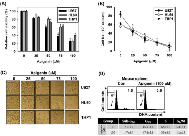
In order to investigate whether apigenin has anti-proliferative or cytotoxic effects, human leukemia cells (U937, THP-1 and HL60) were treated with indicated concentrations of apigenin for 24 h and the resulting number of viable cells remaining were assessed. Apigenin treatment significantly reduced leukemia cell viability in a dose-dependent manner (Fig. 1). According to MTT assay results, treatment with 100 uM apigenin significantly reduced viable cell percentages in all types of leukemia cells (Fig. 1A). Treatment with apigenin for 24 h also decreased human leukemia cell numbers in a dose-dependent manner, as illustrated by trypan blue assay (Fig. 1B). Under the light microscope, apigenin- treated cells exhibited a rounded and granulated morphology, and eventually degraded after treatment of up to 50 uM apigenin (Fig. 1C). To investigate whether apigenin has an influence on normal cells, we examined the cell cycle distribution in apigenin-treated mouse spleen cells. Cell cycle distribution revealed that treatment with 100 uM apigenin resulted in no sub-G1 DNA content in mouse splenocytes (Fig. 1D). This data indicated that apigenin inhibits cell proliferation and lead to cell death in leukemia cells, but did not have detrimental effect on normal cells.
35
Fig. 1. Apigenin decreases cell viability and proliferation in leukemia cell lines. Cells were seeded at 1×10⁵ cells/ml and treated with the indicated concentrations of apigenin for 24 h. Cell viability (A) and cell number (B) were determined by MTT assay and hemocytometer counts of trypan blue-excluding cells, respectively. (C) The morphologic cell changes over 24 h, observed by light microscopy (400×). (D) Splenocytes isolated from ICR mice were treated with or without apigenin (100 uM) for 24 h and their DNA content was analyzed by flow cytometry. Percentages of sub-G1 DNA contents were represented in each pannel. Data in the table represent percentage of each phase in cell cycle distribution. Data from three independent experiments are expressed as overall mean ± SE. Statistical significance was determined by the Student t-test (*, P < 0.05 vs. vehicle control).
36
3.2. Apigenin induces apoptosis via a caspase-dependent pathway
Further experiments were carried out to determine whether the apigenin-mediated anti-proliferative and cytotoxic effect on cells was closely associated with apoptotic cell death. Treatment with 50 uM apigenin resulted in approximate 25% increase in annexin-V intensity in U937 and HL60 leukemia cells (Fig. 2A, left panel). In a parallel experiment, we also analyzed the amount of sub-G1 DNA, which contain less DNA than cells in the G1 phase, to quantify the degree of cell death. Flow cytometric analysis indicated that treatment with 50 uM apigenin resulted in a 20% increase in accumulation of sub-G1 phase (Fig. 2A, right panel). Although THP-1 cells showed an increase in annexin-V intensity, these cells demonstrated some resistance to apigenin-mediated apoptosis compared to the other two types of leukemia cells. We next analyzed whether or not DNA fragmentation is induced by apigenin treatment. Following agarose gel electrophoresis of U937 cells treated with apigenin for 24 h, a typical ladder pattern of internucleosomal fragmentation was observed, following a dose-dependent manner (Fig. 2B). Moreover, concentrations above 50 uM apigenin resulted in a significant increase in caspase-3, caspase-9 and PARP cleavage, which are further indicative of apoptotic cell death. Apigenin-mediated apoptosis was also completely blocked by the pre-treatment of cells with pan caspase inhibitor z-VAD-fmk (Fig. 2D). Treatment with 50 uM apigenin reduced cell viability by approximate 60%, however presence of z-VAD-fmk increased cell viability to 87% (Fig. 2D). Taken together, these results suggest that apigenin induces apoptosis in human leukemia cells through caspase activation.
37
Fig. 2. Apigenin sensitizes apoptosis in leukemia cell line, but not in mouse spleen cells. Cells were seeded at 1×10⁵ cells/ml and treated with the indicated concentrations of apigenin for 24 h. (A) Apoptotic annexin-V+ (left panel) and DNA content population (right panel) were analyzed by flow cytometry. Percentages of annexin-V+ cells and sub-G1 DNA contents were represented. (B) Fragmented DNA was extracted and analyzed on 1.5% agarose gel stained by EtBr. (C) Cells treated with the indicated concentrations of apigenin for 24 h were then lysed for protein extraction. Equal amounts of cell lysates were separated by SDS–PAGE and specific protein detection was performed using Western blotting as indicated. β-actin was used as a loading control. (D) Splenocytes were isolated from mice and then treated with or without apigenin for 24 h. The DNA content of the splenocytes was then analyzed by flow cytometry. Data from three independent experiments are expressed as overall mean ± SE. Statistical significance was determined by the Student t-test (*, P < 0.05 vs. vehicle control).
38
3.3. ROS is not a main regulator in apigenin-induced apoptosis
To determine whether treatment with apigenin induces ROS-dependent apoptosis, the role of apigenin in increasing ROS generation in U937 cells was investigated by measuring the intracellular levels of H2O2 using DCFDA staining. In a parallel experiment, we also used ROS inhibitor NAC and GSH to evaluate their functional effects on ROS generation and apigenin-induced apoptosis. As expected, apigenin treatment significantly increased ROS generation, whereas this production was completely neutralized by pre-treatment with the two ROS inhibitors, NAC and GSH (Fig. 3A, left panel). Surprisingly, we observed that the two ROS inhibitors did not alleviate the appearance of an increased population of annexin-V positive cells (Fig. 3A, right panel). We also carried out trypan blue exclusion assay and PI staining in apigenin-mediated apoptosis to confirm the detailed function of ROS generation. Treatment with apigenin reduced cell viability by 40%. However, the presence of ROS inhibitors, NAC and GSH, had no influence in apigenin-induced cell death (Fig. 3B), a finding which was consistent with the annexin-V staining data. The result that these two ROS inhibitors induced hardly any reduction in sub-G1 population compared to the apigenin-treated group was confirmed with PI staining data (Fig. 3C). This data suggests that apigenin induces apoptosis is in leukemia cells.
39
Fig. 3. ROS is not essential for apigenin-induced apoptosis. U937 cells were treated with the indicated concentration of apigenin, NAC, or GSH, or treated with combined apigenin with NAC or GSH. (A) Intracellular ROS generation was determined by flow cytometry using the peroxide-sensitive dye DCFDA. The number means total mean fluorescent intensity. Apoptotic annexin-V+ population was analyzed by flow cytometry. (B) Cell count was determined by hemocytometer. (C) Cells were seeded at 1×10⁵ cells/ml and treated with the indicated concentrations of NAC and GSH for 24 h. DNA content of cells was analyzed by flow cytometry. Data from three independent experiments are expressed as overall mean ± SE. Statistical significance was determined by the Student t-test (*, P < 0.05 vs. vehicle control).
40
3.4. Apigenin suppresses telomerase activity and hTERT transcriptional level
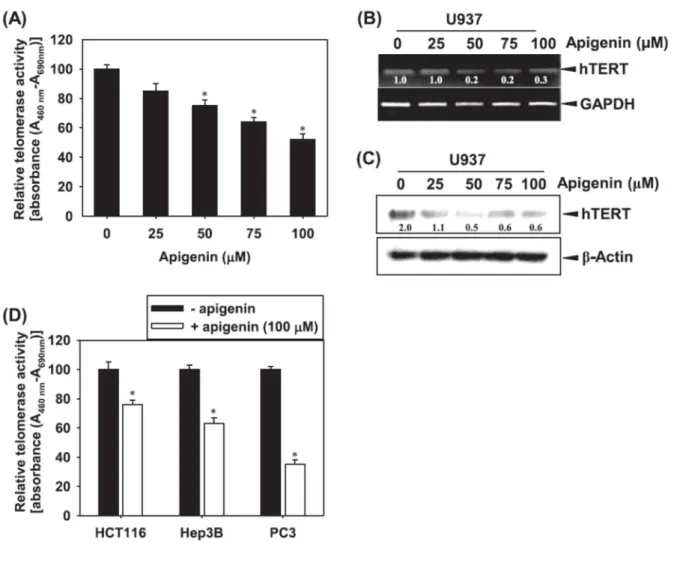
Cells were cultured in the presence of apigenin for 24 h and telomerase activity was measured using a TRAP-ELISA kit. Apigenin treatment resulted in a dose-dependent reduction of telomerase activity in U937 leukemia cells (Fig. 4A). We next attempted to reveal whether or not apigenin treatment is associated with hTERT expression. Dose-dependent treatment with apigenin reduced hTERT mRNA levels (Fig. 4B), as well as the resulting expressed protein level (Fig. 4C) in U937 cells. In addition, treatment with 100 uM apigenin-induced a decrease of telomerase activity in other cell lines including colon carcinoma HCT116, hepatoma Hep3B and prostate cancer PC3 (Fig. 4D). This data indicate that apigenin-induced apoptosis may be due to inhibition of telomerase activity resulting from downregulation of hTERT.
41
Fig. 4. Apigenin inhibits telomerase activity and hTERT expression. (A) Cells were treated with the indicated concentration of apigenin for 24 h. Telomerase activity of leukemia U937 cells was determined by TRAP-ELISA. (B) Total RNA was isolated using TRizol. Equal amounts of mRNA were subjected to RT-PCR using hTERT primer and visualized by EtBr staining. GAPDH was used as internal control. (C) Cells treated with the indicated concentrations of apigenin for 24 h were then lysed for protein extraction. Equal amounts of cell lysates were separated by SDS–PAGE and specific protein detection was performed using Western blotting as indicated. β-actin was used as a loading control. (D) Cells were treated with the indicated concentration of apigenin for 24 h. Telomerase activity of HCT116, Hep3B and PC3 cells were determined by TRAP-ELISA. Data from three independent experiments are expressed as overall mean ± SE. Statistical significance was determined by the Student t-test (*, P < 0.05 vs. vehicle control).
42
3.5. Apigenin suppresses c-Myc expression in leukemia cancer cells
The hTERT promoter site (-181 bp) has two c-Myc binding regions and c-Myc has previously been shown to directly regulate telomerase activity [22]. Therefore, we assessed the role of apigenin in regulation of the activity of c-Myc in U937 leukemia cells. We found that applications of apigenin in a timedependent (Fig. 5B) and a dose-dependent (Fig. 5A) manners, significantly affected c-Myc downregulation and DNA-binding activity. In addition, apigenin decreased both, c-Myc mRNA (Fig. 5C) and protein levels (Fig. 5D). Next, we assessed whether ROS generation regulates c-Myc-mediated telomerase activity. We found that apigenin alone significantly downregulated DNA-binding activity of c-Myc, but addition of NAC or GSH did not restore apigenininduced c-Myc activity (Fig. 5E). In addition, the downregulated level of apigenin-induced telomerase activity was sustained in the presence of NAC or GSH (Fig. 5F). These results indicated that apigenin-induced ROS generation is not related to suppression of c-Myc-dependent telomerase activity.
43
Fig. 5. Apigenin inhibits c-Myc DNA binding activity. (A) Cells were treated with the indicated concentration of apigenin for 24 h, (B) Treatment by 50 uM apigenin was performed at the indicated time point. c-Myc DNA binding activity was analyzed by LightShift™ chemiluminescent EMSA kit as a described in the Materials and Methods. (C) Total RNA was isolated using TRIzol. Equal amounts of mRNA were subjected to RT-PCR using hTERT primer, and then visualized by EtBr staining. GAPDH was used as an internal control. (D) Cells treated with the indicated concentrations of apigenin for 24 h were then lysed for protein extraction. Equal amounts of cell lysates were separated by SDS–PAGE and specific protein detection was performed using Western blotting as indicated. β-actin was used as a loading control. (E) U937 cells were treated with the indicated concentration of apigenin, NAC, or GSH, or combined apigenin with NAC or GSH, for 24 h. c-Myc DNA binding activity was analyzed by LightShift™ chemiluminescent EMSA kit. (F) Cells were treated with the indicated concentration of apigenin, NAC, or GSH, or combined apigenin with NAC or GSH, for 24 h. Telomerase activity of cells was determined by TRAP-ELISA. Data from three
44
independent experiments are expressed as overall mean ± SE. Statistical significance was determined by the Student t-test (*, P < 0.05 vs. vehicle control).
45
4. Discussion
Apoptosis is a process of programmed cell death essential for the elimination of unwanted cells in various biological systems and is also one of the key mechanisms employed by chemotherapeutic agents. Cells undergoing apoptosis exhibit a characteristic pattern of morphologic changes including cell shrinkage, condensation, nuclear fragmentation and blebbing [23]. Therefore, numerous researchers have attempted to discover novel chemicals which induce apoptosis in cancer cells, but without side effects such as apoptosis of healthy cells. Previous studies reported that apigenin induces apoptosis in various tumor cell lines through activation of a mitochondria-dependent pathway via intracellular generation of ROS as an activator [14, 15, 18, 24]. Although Lu et al. demonstrated the importance of ROS generation in apigenin-induced apoptosis, they did not identify the exact mechanism of the ROS generation process. Supporting the mechanism of functional generation of ROS, it was reported that exogenous catalase offers protection from apigenin-induced cell death, but other antioxidant compounds, such as butylated hydroxyanisole (BHA), butylated hydroxytoluene (BHT) and ascorbic acid, have no influence on cell death [25]. In contrast to this previous data, our study demonstrated that apigenin significantly inhibits proliferation and cell viability, as well as promotes the mechanisms of apoptosis via the mitochondria-dependent pathway, but not as an ROS-dependent mechanism. Because BHA, BHT and ascorbic acid are indirectly capable of promoting the scavenging of H2O2, catalase may have another function to induce cell survival in apigenin- induced apoptosis. In contrast, Lin et al. suggested that apigenin attenuates dopamine-induced apoptosis by intracellular generation of ROS [26]. Although we demonstrated an ROS-independent mechanism for apigenin-induced apoptosis, we need to exact further experiments to elucidate the impact of ROS because many conflicts to our findings have been published. Many studies reported that hTERT expression, in combination with oncogenes, are sufficient to induce normal human epithelial and fibroblast cells to become tumor cells, with the majority of cancer cells exhibiting high telomerase activity which enable their uncontrolled growth [27, 28]. Therefore, telomerase has been identified as a promising target for human cancer gene therapy, and its inhibition allows telomere shortening to occur in cancer cells which in turn is thought to trigger