Vol. 47, No. 4, pp. 551 - 557, 2006
The brain is particularly vulnerable to oxygen free radicals, and these radicals have been implicated in the pathology of several neurological disorders. In this study, the modulation of TNF-related apoptosis-inducing ligand (TRAIL) expression by oxidative stress was shown in LN215 cells, an astroglioma cell line. Hydrogen peroxide (H2O2) treatment increased TRAIL expression in LN215 cells and H2O2-induced TRAIL aug- mented apoptosis in Peer cells, a cell line sensitive to TRAIL- mediated cell death. Our findings suggest that the upregulation of TRAIL in astroglial cells may abrogate immune cell effector functions.
Key Words: Astroglial cells, hydrogen peroxide, TRAIL, cyclosporin A, apoptosis, oxidative stress, neuroimmunology
INTRODUCTION
TNF-related apoptosis-inducing ligand (TRAIL), a member of death ligand/receptor pairs, acts independently of Fas-mediated apoptosis. Like FasL, TRAIL induces rapid apoptosis in trans- formed cell lines of diverse origins.1,2 Death receptors (DR) 4 and DR5, which are receptors for TRAIL, mediate apoptosis by recruiting Fas-asso- ciated death domain (FADD) indirectly via the
TNF receptor-associated death domain (TRADD) and by the subsequent activation of capsase 8.3,4 TRAIL and DR4 are expressed in many human tissues, including the spleen, lung, and prostate.5 The human TRAIL promoter was cloned, and a number of transcription factor binding sites were identified, namely, the NFAT, AP-1, and Sp1 sequences, which are important for the expression of other TNF family members.6 DR4 and DR5 were expressed in normal brain tissue and glioma.7 Although TRAIL expression is detected in many human tissues, TRAIL is not always cyto- toxic due to the presence of decoy receptors. The decoy receptors, DcR1 and DcR2, have also been found in many types of normal cells. whereas many tumor cell lines do not express DcR1,2,5 which abrogates apoptotic signals by binding TRAIL, because DcR1 does not have death do- mains. DcR2 has a truncated form of cytoplasmic tail, which cannot transduce cell death signals.8
The relationship between reactive oxygen inter- mediate (ROIs) products and central nervous system (CNS) tumors is a current and important topic in light of their involvement in pathological conditions and possible therapeutic interventions.
Hydrogen peroxide (H2O2) has been implicated in cellular and tissue injury during pathologic con- ditions, such as ischemia-reperfusion injury, hyperoxia, and inflammation.9 In addition, the stimulation of various cells with either cytokines or phorbol ester increases the secretion of H2O2
into the extracellular space in vitro.10,11 High con- centrations of this diffusible ROI exert toxic effects on susceptible cells. However, low concentrations
Hydrogen Peroxide Upregulates TNF-Related Apoptosis- Inducing Ligand (TRAIL) Expression in Human Astroglial Cells, and Augments Apoptosis of T Cells
Daeho Kwon1 and In-Hong Choi2
1Department of Microbiology, Ajou University School of Medicine, Seoul, Korea;
2Department of Microbiology, Institute for Immunology and Immunological Diseases, Yonsei University College of Medicine, Seoul, Korea.
Received January 17, 2006 Accepted February 20, 2006
This work was supported by a research grant from KOSEF (RO1-2002-000-00210-0) and also by KOSEF, through the Natio- nal Core Research Center for Nanomedical Technology (R15-2004- 024-00000-0).
Reprint address: requests to Dr. In-Hong Choi, Department of Microbiology, Yonsei University College of Medicine, 134 Shinchon- dong, Seodaemum-gu, Seoul 120-752, Korea. Tel: 82-2-2228-1821, Fax: 82-2-392-7088, E-mail: [email protected]
of H2O2 alter cellular functions by modulating signal transduction in certain cells, including endothelial cells.12Oxidative injury occurs in vari- ous ways and has been studied in several diverse models, using different eliciting agents and biolo- gical preparations. The common feature of these models is that the primary activation involves radical formation; those radicals and related pro- ducts then generate a potent oxidant, which sub- sequently inactivates macromolecules. ROI-medi- ated damage has been generally associated with necrosis; however, some observations suggest that it has a role in apoptosis.13 Recent observations show that ROIs modulate the expression of Fas and FasL. The expression of Fas was upregulated in human endothelial cells under oxidative stress,14 and the upregulation of FasL has been reported in hepatoma cells,15 microglial cells,16 and glioma cells17,18 under conditions of oxidative stress.
In order to address the effects of oxidative stress in the brain, this study investigated the expression levels of TRAIL in human astroglial cells after H2O2 treatment.
MATERIALS AND METHODS
Culture of human astroglial cells and their treat- ment with H2O2 or cyclosporin A
LN215, an astroglioma cell line, was cultured in 10% FCS (Gibco BRL, Grand Island, NY, USA)- DMEM (Gibco BRL) containing 1% nonessential amino acids (Sigma Chemicals Co., St. Louis, MO, USA). The LN215 cell line was kindly provided by Dr. E. G. Van Meir (Department of Neurosurgery, Laboratory of Tumor Biology and Genetics, Lau- sanne, Switzerland). The cells were incubated with 200, 400, 600, and 800 M of Hμ 2O2(Sigma) for various times (1-24 hrs). Cyclosporin A (CsA, Sandoz, stock solution 50 mg/mL in DMSO) was added at the conc. of 10, 100 ng, 1, 5, and 10 gμ for 30 min before adding H2O2.
Isolation of total RNA and the synthesis of cDNA
1 × 10⁶ cells were cultured in a 75 cm2flask and
washed twice with PBS. H2O2was added in serum free RPMI medium. Total RNA was isolated with an RNeasy kit (Qiagen, Santa Claris, CA, USA) and 5 g of this RNA was used to synthesize cDNAμ with 0.1 O.D. of random hexamer (Pharmacia, Uppsala, Sweden) and 200 U of M-MLV reverse transcriptase (Gibco BRL). The cDNA produced was kept at -20 until used.
RT-PCR and RNase protection assay (RPA) RT-PCR was performed using the following primer sets: TRAIL primers (forward: 5'-CCC AAT gAC gAA gAg AgT ATg A -3'; reverse:
5'-ggA ATA gAT gTA gTA AAA CCC T-3. PCR conditions were as follows: denaturation at 94 for 30s, annealing at 55 for 30s, and extension at 72 for 30s for TRAIL. The buffer used for PCR was 10 mM Tris-HCl (pH 10), 2.0 mM MgCl2, 50 mM KCl with 1.25 U of Taq polymerase (Takara, Tokyo, Japan). After 27 cycles, an additional ex- tension at 72 for 10 min was carried out.
To detect TRAIL expressions, an RNase protec- tion assay was done using the RiboQuant multi- probe RNase protection assay system (Pharmingen, San Diego, CA, USA). Briefly, 15 g of total RNAμ was hybridized overnight with the in vitro-trans- lated 32P-labeled RNA probe; hAPO-3C kit and samples were treated with RNase A and pro- teinase.
Western blot
A total of 1 × 10⁶ LN215 cells were used for protein purification. Protein was extracted by ultrasonication and centrifuged to remove cell debris. The resulting supernatant was quantified using the Bradford method (Pierce, Rockford, IL, USA). Protein (50 g) was then loaded onto 12%μ Tris-HCL SDS-polyacrylamide gel (BioRad, Her- cules, CA, USA), and transferred to a nitrocellu- lose transfer membrane (Amersham Pharmacia Biotech, Piscataway, NJ, USA). TRAIL expression was determined using anti-TRAIL monoclonal Ab (Pharmingen) diluted 1:500 and anti-mouse rabbit Ab (Jackson Immuno Research, Baltimore, MD, USA) was used as a secondary Ab. Proteins were visualized using the ECL technique (Amersham Pharmacia Biotech.).
Flow cytometry analysis
Astroglial cells (2 × 10⁵cells/well) were plated in six-well (35-mm2) plates (Costar, Cambridge, MA, USA) and grown to 90% confluency. For analysis of TRAIL protein expression, cells were trypsinized, suspended in PBS containing 5% fetal bovine serum and 0.02% azide, incubated with PE-conjugated anti-human TRAIL antibody (Pharmingen), washed twice, fixed in 1% para- formaldehyde, and then analyzed with FACStar/
(Becton Dickinson, Mountain View, CA, USA).
Negative controls were incubated with goat anti- mouse IgG antibody conjugated to PE. Ten thou- sand cells were analyzed for each sample.
Detection of cell death by staining of annexin-V and PI
To study the expression of functional TRAIL in astroglial cells, 2 × 10⁵ LN215 cells were pre- treated with 800 M of Hμ 2O2for 18 h in serum free medium. Astroglial cells were washed to remove H2O2 and reacted with Peer cells, which were sensitive to TRAIL,19 for 24 hrs. Effector cells (LN 215 cells) and target cells (Peer cells) were then co-cultured at a ratio of 4 : 1. Cell death was mea- sured by staining with annexin V and PI. Control LN215 cells were cultured for 18 hrs in serum-free media without H2O2. Annexin V-FITC and PI (Trevigen, Gaithersburg, MD, USA) were used to detection of cell death.
RESULTS
The expression of TRAIL in human astroglial cells treated with H2O2
We hypothesized treatment with H2O2 would modulate the apoptosis-related gene expression of astroglial cells in central nerve system. To address this, human LN215 astroglioma cells were treated with H2O2 and the expression of TRAIL was detected by RT-PCR, RPA, western blot and flow cytometry.
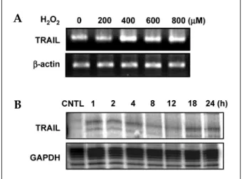
We experimented to determine whether treat- ment with H2O2 induced TRAIL expression in human astroglial cells. LN215 cells were treated
with varying concentrations of H2O2 (0-800 M)μ for 4 hrs (for mRNA) or 18 hrs (for protein). H2O2- mediated TRAIL induction was observed by RT- PCR (Fig. 1A) and western blot (Fig 2A). TRAIL expression was induced treatment of H2O2 and reached maximum expression at 800 M of Hμ 2O2
(Fig. 1A, 2A). LN215 cells were treated with 800 M of H
μ 2O2 for various times (0 - 24 hrs). The time course of the TRAIL expression was then deter- mined by RPA (Fig. 1B) or western blot (Fig 2C).
The gene expression of TRAIL was induced at 1 h and continued until 24 hrs (Fig. 1B). TRAIL pro- tein expression reached its maximum at 8 hrs and continued until 24 hrs (Fig. 2C). Also H2O2-media- ted TRAIL induction was observed by flow cytometry (Fig. 2B).
Inhibition of TRAIL expression by cyclosporin A in human astroglial cells treated with H2O2
Cyclosporin A (CsA), an immunosuppressive drug that targets calcineurin, blocked both RNA and protein expression of TRAIL in LN215 cells after treatment with H2O2(Fig. 3A, 3B). The most effective concentration of CsA started at 100 ng/
mL.
Fig. 1.Gene expression of TRAIL in human astroglial cells after treatment with hydrogen peroxide (H2O2) in a dose or time-dependent manner. (A) LN215 cells were treated with varying doses of hydrogen peroxide (0 to 800 M)μ for 4 hrs. (B) LN215 cells were incubated with hydrogen peroxide 800 M for various times (0 to 24μ hrs). Total RNA was measured for TRAIL mRNA by RT-PCR (A) or RNase protection assay (B). Cells not treated with H2O2
were used as control (CNTL). The data are representative of three experiments.
A
B
Fig. 3.Inhibition of TRAIL expression by cyclosporin A (CsA) in human astroglial cells after treatment with H2O2. (A) LN215 cells were pre-incubated with CsA (0.01 to 10 M) for 30 min, were treated with hydrogen peroxide (800 μ
M) for 4
μ hrs and then gene expression of TRAIL were measured by RT-PCR. Cells not treated with H2O2 were used as a control. -actin was used as the internal control.β The fold induction was calculated as follows: (TRAIL/ -β actin intensity in the study group)/(TRAIL/ -actin intenβ - sity in the control group). The fold induction value repre- sents mean ± SD of triplicate experiments. (B) LN215 cells were pre-incubated with CsA (1 M) for 30 min, wereμ treated with hydrogen peroxide (800 M) for 18 hrs andμ then expression of TRAIL were measured by western blot.
The data are representative of three independent experi- ments.
Fig. 2.Expression of TRAIL in human astroglial cells after treatment with H2O2in a dose or time-dependent manner.
(A) LN215 cells were treated with varying doses of hydrogen peroxide (0 to 800 M) for 18μ hrs. (C) LN215 cells were incubated with hydrogen peroxide 800 M forμ various times (0 to 24 hrs). For western blot, 20 g of totalμ protein was loaded in 12% gel and anti-TRAIL mono- clonal Ab was used as a primary Ab. (B) FACS analysis of TRAIL expression on LN215 were treated with 800 Mμ of hydrogen peroxide for 18 hrs. The data are represen- tative of three independent experiments.
Fig. 4.Induction of apoptosis in Peer cells by human astroglial cells treated with H2O2. LN215 cells were incubated with 800 M of Hμ 2O2 for 18 hrs in serum free medium. LN215 cells were washed to remove H2O2and then co-cultured with Peer cells for 24 hrs. Effector cells (LN215 cells) and target cells (Peer cells) were co-cultured at a ratio of 4 : 1. Cell death was measured by staining with annexin V. Control LN215 cells were not treated with H2O2, but maintained in serum free condition for 18 hrs. LN215 were pre-incubated for 30 min with CsA (1 g/mL), and incubated with Hμ 2O2. The fold induction value represents a mean ± SD of triplicate experiments.
A
B A
C B
The induction of the apoptosis of Peer cells by human astroglial cells treated with H2O2
To address the biological function of TRAIL in LN215 cells after H2O2 treatment, LN215 cells were co-cultured with Peer cells, a cell line sensi- tive to TRAIL mediated apoptosis. Annexin V staining of Peer cells showed an increase in apoptotic cells after H2O2 treatment, 64.2% versus 47.5% in the control group (Fig. 4). Peer cells treated with both CsA and H2O2 inhibited TRAIL induced apoptosis down to 49.8%, a level similar to control (Fig. 4).
DISCUSSION
Our study demonstrates that oxidative stress modulates the expression of TRAIL, which is in- volved in apoptosis. The up-regulation of FasL and Fas by IFN- or TNF- has been reported inγ α primary astrocytes,20 and H2O2increased Fas and FasL expression in astroglioma cell lines.18 Al- though Furuke et al. reported that H2O2 inhibits CD16-induced FasL expression21and that reactive oxygen species (ROS) suppress events upstream of FasL expression, another report suggested that oxidative stress induces FasL expression in hepa- toma and microglial cells.15,16 In other cell types, H2O2 may induce FasL expression by activating NF- B; Hκ 2O2 is known to be a potent activator of NF- B. Moreover, the CD95L promoter has beenκ described to contain NF- B binding sites, whichκ suggests that NF- B may regulate FasL expresκ sion.
In contrast to the limited expression of FasL, TRAIL mRNA has been detected in nearly all tissues. However, malignant astoroglioma cells express both TRAIL mRNA and protein.22,23In our study, TRAIL was detected constitutively in LN 215 cells, an astroglioma cell line, and its expres- sion was increased by H2O2.
In this study, CsA, a pharmacological inhibitor of the phosphatase calcineurin, blocked TRAIL expression at concentrations between 100 and 10 g/mL. These concentrations are reported to be μ
nontoxic to primary rat astrocytes,24 suggesting that NFAT might be involved in TRAIL ex- pression in human astrocytes. NFAT is known to be expressed in T cells and other immune cells.
However, several data show that cells of the ner- vous system, such as a neuronal cell line and rat astrocytes, express NFAT.24,25Moreover, the CsA- sensitive signaling pathway has been reported to regulate the survival of reactive astrocytes.24 Involvement of ROS in NFAT has been demon- strated in T cells.27,28 Also, 1.6 kb of the human TRAIL promoter was identified to have binding sites for such factors as NFAT, AP-1, and Sp1.6 Therefore, NFAT, under conditions of oxidative stress, may act as a transcription factor, signaling induction of TRAIL. Although H2O2 has been known to activate NF- B,κ 29,30 it is not certain whether the expression of TRAIL and its receptors is regulated by H2O2-activated NF- B. Althoughκ basal ROI in tumors can be lower than in normal brain tissue,31 it is important to keep in mind the effects of drug-induced apoptosis on tumor cells by ROI-producing chemotherapies.32
In our study, the upregulation of TRAIL sug- gests that some astroglial cells up-regulate TRAIL and may abrogate lymphocytes or other immune cells, thereby escaping immune surveillance. Fur- ther investigations into the regulation of TRAIL and TRAIL receptors are necessary to answer critical questions related to whether a specific gain of decoy receptors and death receptors in as- troglioma cells, as compared with non-neoplastic cells allows the selective therapeutic elimination of tumor cells in vivo.
Collectively our findings suggest that TRAIL may be inducible by hypoxia and other forms of oxidative stress, such as tumor-associated hypoxia or chemotherapy-induced hypoxia. Therefore, oxidative stress leading to the upregulation of TRAIL presumably plays an apoptotic role in pathological conditions of the brain.
REFERENCES
1. Wiley SR, Schooley K, Smolak PJ, Din WS, Huang CP, Nicholl JK, et al. Identification and characterization of a new member of the TNF family that induces apoptosis. Immunity 1995;3:673-82.
2. Pan G, Ni J, Wei YF, Yu G, Gentz R, Dixit VM. An antagonist decoy receptor and a death domain-con- taining receptor for TRAIL. Science 1997;277:815-8.
3. Mariani SM, Matiba B, Armandola EA, Krammer PH.
Interleukin 1 beta-converting enzyme related pro-
teases/caspases are involved in TRAIL-induced apop- tosis of myeloma and leukemia cells. J Cell Biol 1997;
137:221-9.
4. Wajant H, Johannes FJ, Haas E, Siemienski K, Schwenzer R, Schubert G, et al. Dominant-negative FADD inhibits TNFR60-, Fas/Apo1- and TRAIL-R/
Apo2-mediated cell death but not gene induction. Curr Biol 1998;8:113-6.
5. Pan G, O'Rourke K, Chinnaiyan AM, Gentz R, Ebner R, Ni J, et al. The receptor for the cytotoxic ligand TRAIL. Science 1997;276:111-3.
6. Wang Q, Ji Y, Wang X, Evers BM. Isolation and molec- ular characterization of the 5'-upstream region of the human TRAIL gene. Biochem Biophys Res Commun 2000;276:466-71.
7. Frank S, Kohler U, Schackert G, Schackert HK. Expres- sion of TRAIL and its receptors in human brain tumors.
Biochem Biophys Res Commun 1999;257:454-9.
8. Sheridan JP, Marsters SA, Pitti RM, Gurney A, Skubatch M, Baldwin D, et al. Control of TRAIL- induced apoptosis by a family of signaling and decoy receptors. Science 1997;277:818-21.
9. Halliwell B. Reactive oxygen species and the central nervous system. J Neurochem 1992;59:1609-23.
10. Lee YW, Kuhn H, Hennig B, Neish AS, Toborek M.
IL-4-induced oxidative stress upregulates VCAM-1 gene expression in human endothelial cells. J Mol Cell Cardiol 2001;33:83-94.
11. Zhang Z, Oliver P, Lancaster JR Jr, Schwarzenberger PO, Joshi MS, Cork J, et al. Reactive oxygen species mediate tumor necrosis factor alpha-converting, en- zyme-dependent ectodomain shedding induced by phorbol myristate acetate. FASEB J 2001;15:303-5.
12. Suhara T, Fukuo K, Sugimoto T, Morimoto S, Nakahashi T, Hata S, et al. Hydrogen peroxide induces up-regulation of Fas in human endothelial cells. J Immunol 1998;160:4042-7.
13. Jacobson MD. Reactive oxygen species and program- med cell death. Trends Biochem Sci 1996;21:83-6.
14. Bauer MK, Vogt M, Los M, Siegel J, Wesselborg S, Schulze-Osthoff K. Role of reactive oxygen intermedi- ates in activation-induced CD95 (APO-1/Fas) ligand expression. J Biol Chem 1998;273:8048-55.
15. Hug H, Strand S, Grambihler A, Galle J, Hack V, Stremmel W, et al. Reactive oxygen intermediates are involved in the induction of CD95 ligand mRNA expression by cytostatic drugs in hepatoma cells. J Biol Chem 1997;272:28191-3.
16. Vogt M, Bauer MK, Ferrari D, Schulze-Osthoff K. Oxi- dative stress and hypoxia/reoxygenation trigger CD95 (APO-1/Fas) ligand expression in microglial cells. FEBS Lett 1998;429:67-72.
17. Husain N, Chiocca EA, Rainov N, Louis DN, Zervas NT. Co-expression of Fas and Fas ligand in malignant glial tumors and cell lines. Acta Neuropathol (Berl) 1998;95:287-90.
18. Kwon D, Choi C, Lee J, Kim KO, Kim JD, Kim SJ, et al. Hydrogen peroxide triggers the expression of Fas/
FasL in astrocytoma cell lines and augments apoptosis.
J Neuroimmunol 2001;113:1-9.
19. Kayagaki N, Yamaguchi N, Nakayama M, Kawasaki A, Akiba H, Okumura K, et al. Involvement of TNF- related apoptosis-inducing ligand in human CD4+ T cell- mediated cytotoxicity. J Immunol 1999;162:2639-47.
20. Choi C, Park JY, Lee J, Lim JH, Shin EC, Ahn YS, et al. Fas ligand and Fas are expressed constitutively in human astrocytes and the expression increases with IL-1, IL-6, TNF- , or IFN- . J Immunol 1999;162:1889-95.α γ 21. Furuke K, Shiraishi M, Mostowski HS, Bloom ET. Fas ligand induction in human NK cells is regulated by redox through a calcineurin-nuclear factors of activated T cell-dependent pathway. J Immunol 1999;162:1988-93.
22. Weller M, Frei K, Groscurth P, Krammer PH, Yonekawa Y, Fontana A. Anti-Fas/APO-1 antibody- mediated apoptosis of cultured human glioma cells.
Induction and modulation of sensitivity by cytokines.
J Clin Invest 1994;94:954-64.
23. Rieger J, Ohgaki H, Kleihues P, Weller M. Human astrocytic brain tumors express APO2L/TRAIL. Acta Neuropathol (Berl) 1999;97:1-4.
24. Pyrzynska B, Lis A, Mosieniak G, Kaminska B.
Cyclosporin A-sensitive signaling pathway involving calcineurin regulates survival of reactive astrocytes.
Neurochem Int 2001;38:409-15.
25. Ho AM, Jain J, Rao A, Hogan PG. Expression of the transcription factor NFATp in a neuronal cell line and in the murine nervous system. J Biol Chem 1994;269:
28181-6.
26. Plyte S, Boncristiano M, Fattori E, Galvagni F, Paccani SR, Majolini MB, et al. Identification and characteri- zation of a novel nuclear factor of activated T-cells-1 isoform expressed in mouse brain. J Biol Chem 2001;
276:14350-8.
27. Martinez-Martinez S, Gomez del Arco P, Armesilla AL, Aramburu J, Luo C, Rao A, et al. Blockade of T-cell activation by dithiocarbamates involves novel mech- anisms of inhibition of nuclear factor of activated T cells. Mol Cell Biol 1997;17:6437-47.
28. Saccani S, Saccani A, Varesio L, Ghosh P, Young HA, Sica A. Divergent effects of dithiocarbamates on AP-1- containing and AP-1-less NFAT sites. Eur J Immunol 1999;29:1194-201.
29. Schieven GL, Kirihara JM, Myers DE, Ledbetter JA, Ucken FM. Reactive oxygen intermediates activate NF- B in a tyrosine kinase-dependent mechanism and in κ
combination with vanadate activate the p56lck and p59fyn tyrosine kinases in human lymphocytes. Blood 1993;82:1212-20.
30. Barchowsky A, Munro SR, Morana SJ, Vincenti MP, Treadwell M. Oxidant-sensitive and phosphorylation- dependent activation of NF-kB and AP-1 in endothelial cells. Am J Physiol 1995;13:L829-36.
31. Leaver HA, Williams JR, Ironside JW, Miller EP, Gregor A, Su BH, et al. Dynamics of reactive oxygen intermediate production in human glioma: n-6 essential fatty acid effects. Eur J Clin Invest 1999;29:220-31.
32. Lee YS, Wurster RD. Mechanism of potentiation of
LY83583-induced growth inhibition by sodium nitro- prusside in human brain tumor cells. Cancer Chemo- ther Pharmacol 1995;36:341-4.