Optimal Intravascular Ultrasound Criteria and Their Accuracy for Defining the Functional Significance of Intermediate Coronary Stenoses of Different Locations
Bon-Kwon Koo, MD, P
HD,* Hyoung-Mo Yang, MD,† Jun-Hyung Doh, MD, P
HD,‡
Hyunmin Choe, MD,‡ Sung-Yun Lee, MD, P
HD,‡ Chang-Hwan Yoon, MD, P
HD,§
Yun-Kyeong Cho, MD, P
HD,储 Chang-Wook Nam, MD, P
HD,储 Seung-Ho Hur, MD, P
HD,储 Hong-Seok Lim, MD, P
HD,† Myeong-Ho Yoon, P
HD,† Kyung-Woo Park, MD, P
HD,*
Sang-Hoon Na, MD, P
HD,* Tae-Jin Youn, MD, P
HD,§ Woo-Young Chung, MD, P
HD,¶
Seunghyun Ma, MD,# Sue-Kyung Park, MD, P
HD,# Hyo-Soo Kim, MD, P
HD,*
Seung-Jea Tahk, MD, P
HD†
Seoul, Gyeonggi-do, and Daegu, Korea
Objectives We performed this study to determine the optimal intravascular ultrasound (IVUS) crite- ria and to evaluate their accuracy for defining the functional significance of intermediate coronary stenoses in different locations of the coronary tree.
Background Presence of myocardial ischemia is the most important prognostic factor in patients with coronary artery disease and is determined by both the lesion severity and the amount of myo- cardium supplied.
Methods IVUS and fractional flow reserve (FFR) measurements were performed in 267 intermediate lesions located at the proximal or mid part of major epicardial coronary arteries. Optimal IVUS crite- ria and their diagnostic accuracy for functionally significant stenoses (FFR ⬍0.8) were assessed.
Results FFR was ⬍0.8 in 88 lesions (33%). The determinants of FFR were minimum lumen area (MLA) and lesion location. The diagnostic accuracy of MLA was highly variable according to the lo- cation of lesions. The best cutoff value of MLA to define the functional significance was 3.0 mm
2(area under the curve [AUC]: 0.81, 95% confidence interval [CI]: 0.68 to 0.91) for proximal left ante- rior descending artery (LAD) lesions and 2.75 mm
2for mid-LAD lesions located before the second diagonal branch (AUC: 0.76, 95% CI: 0.66 to 0.84). However, the appropriate MLA to predict the functional significance of lesions could not be found in other segments.
Conclusions When IVUS parameters are used to determine the functional significance of lesions in patients with intermediate coronary artery stenoses, different criteria should be used according to lesion location. In segments or vessels with anatomic variations, IVUS cannot be used for functional assessment of a stenosis. (Comparison of Fractional Flow Reserve and Intravascular Ultrasound;
NCT01133015) (J Am Coll Cardiol Intv 2011;4:803–11) © 2011 by the American College of Cardiology Foundation
From the *Department of Internal Medicine, Seoul National University Hospital, Seoul, Korea; †Department of Internal Medicine, Ajou University Medical Center, Gyeonggi-do, Korea; ‡Department of Internal Medicine, Inje University, Ilsan Paik Hospital, Gyeonggi-do, Korea; §Department of Internal Medicine, Seoul National University, Bundang Hospital, Gyeonggi-do, Korea;储Department of Internal Medicine, Keimyung University, Dongsan Medical Center, Daegu, Korea; ¶Department of Internal Medicine, Seoul National University, Borame Hospital, Seoul, Korea; and the #Department of Preventive Medicine, Seoul National University College of Medicine, Seoul, Korea. This study was supported by a grant from the Seoul National University Hospital (A062260), Innovative Research Institute for Cell Therapy, the Clinical Research Center for Ischemic Heart Disease (0412-CR02-0704-0001) sponsored by the Ministry of Health & Welfare, Republic of Korea, and the SNUH Research Fund (03-2010-0270). Dr. Koo has received lecture fees from St. Jude Medical. All other authors have reported that they have no relationships to disclose. Drs. Koo and Yang contributed equally to this work.
Manuscript received February 8, 2011; accepted March 31, 2011.
The presence of myocardial ischemia is the most important prognostic factor in patients with coronary artery disease (1).
The degree of stenosis seen on coronary angiography is considered the gold standard by which to assess the severity of coronary artery disease. However, in patients with intermediate stenosis, angiographic assessment does not accurately predict the functional significance of a lesion and is reported to have high inter- and intraindividual variability (2–5). Noninvasive functional tests can be helpful, but it is still difficult to find the culprit lesion in patients with multiple lesions or multivessel disease (6).
See page 812
Moreover, about half of patients that undergo coronary angiography do so without having a noninvasive functional test (7). Therefore, invasive assessment for these lesions is needed in a cardiac catheterization laboratory.
The 2 most frequently used in- vasive methods for coronary artery stenosis assessment are intravascular ultrasound (IVUS) and fractional flow reserve (FFR). IVUS can pro- vide accurate anatomical informa- tion, whereas FFR assesses the functional or physiological signifi- cance of a lesion. Previous studies have shown that there was a good correlation between FFR and min- imum lumen area by IVUS. In proximal lesions, IVUS criteria such as lumen area of 3 to 4 mm
2and/or percent area stenosis of 70% to 80%
are reported to accurately predict the functional significance of inter- mediate coronary stenoses (8–10).
Functional significance of a coronary stenosis is determined by both the severity of a stenosis and the amount of myocardium supplied. Therefore, when the functional sig- nificance of a lesion is assessed by lumen area measured by IVUS, different criteria should be applied according to lesion location and anatomical variations of the coronary artery. However, previous studies included only patients with proximal lesions or small vessel disease, and the sample sizes were too small to assess these differences. Therefore, we performed this study to determine the optimal IVUS criteria and to evaluate their accuracy for defining the functional significance of intermediate coronary stenoses in different locations of the coronary tree.
Methods
Patient population. This study was a multicenter, prospective registry conducted in 4 Korean centers. Patients who under-
went elective coronary angiography and had 30% to 70% de novo stenosis by visual estimation at the proximal or mid part of 3 major epicardial coronary arteries with available IVUS and FFR were consecutively enrolled. Patients were excluded if any of the following were present: acute ST-segment elevation myocardial infarction, regional wall motion abnormalities of a target vessel segment, additional stenosis (⬎30% by visual estimation) in the same vessel, left ventricular ejection fraction
⬍40%, primary myocardial or valvular disease, contraindica- tion to adenosine, left main coronary artery stenosis, presence of collateral vessels, or angiographically visible thrombus at a target lesion. In patients with acute coronary syndrome and previous myocardial infarction, only the nonculprit artery was tested. The study protocol was approved by the institutional review board at each participating center, and all patients provided written informed consent.
Study procedure. The target vessel was engaged using a guiding catheter of 5- to 7-F. Angiographic images were acquired after intracoronary administration of 100 to 200
g of nitroglycerin. FFR was measured using a 0.014-inch pressure guide wire (PressureWire, Radi Medical Systems, Uppsala, Sweden) as previously described (11). Hyperemia was induced with an intracoronary bolus administration (80
g in the left coronary artery, 40 g in the right coronary artery), intracoronary (240 g/min) or intravenous contin- uous infusion (140 g/kg/min) of adenosine (12–14). Le- sions with FFR ⬍0.8 were considered functionally signifi- cant. IVUS was performed in a standard fashion using an automated motorized pullback system (0.5 mm/s) with commercially available imaging catheters (Boston Scientific/
SCIMED, Minneapolis, Minnesota, or Volcano Corpora- tion, Rancho Cordova, California). Intracoronary nitroglyc- erin (100 to 200 g) was administered before the IVUS run or FFR measurement. The decision to treat the target lesion was left to the operator’s discretion after IVUS and FFR measurement.
Quantitative coronary angiography and IVUS analysis. Both quantitative coronary angiography (QCA) and IVUS anal- ysis were performed by an independent core laboratory at Seoul National University Cardiovascular Center. QCA was performed by a single experienced observer, who was blinded to the FFR value and IVUS findings. Using the guiding catheter for calibration and an edge detection system (CAAS 5.7 QCA system, Pie Medical, Maastricht, the Neth- erlands), the reference diameters, minimum lumen diameter and lesion length were measured, and the percentage diameter stenosis was calculated. Lesion location was determined ac- cording to the American Heart Association classification (15).
Minor modification was made to reflect the amount of myo- cardium supplied by the target coronary segment. Proximal left anterior descending coronary artery (LAD) was defined as the segment between the branching point of the left main stem and the first major branch (septal or diagonal branch). Mid-
Abbreviations and Acronyms
AUCⴝ area under the curve BCVⴝ best cutoff value CIⴝ confidence interval FFRⴝ fractional flow reserve
IVUSⴝ intravascular ultrasound LADⴝ left anterior descending coronary artery LCXⴝ left circumflex coronary artery
MLAⴝ minimum lumen area QCAⴝ quantitative coronary angiography
RCAⴝ right coronary artery
LAD was defined as one-half the distance from the first septal/
diagonal branch to the apex of the heart and was divided into mid-1 and mid-2 LAD according to the location of a second diagonal branch. When mid-LAD lesions were located distal to a second diagonal branch, it was classified as mid-2 LAD lesions. Mid-left circumflex coronary artery (LCX) was defined as the segment between the first and second obtuse marginal branches.
IVUS analysis was performed by 2 independent observers blinded to the FFR and QCA information. Quantitative analyses were performed using computerized planimetry soft- ware (echoPlaque, Indec Systems Inc., Santa Clara, California) as previously described (16). Minimum lumen area was mea- sured at the narrowest luminal cross section and the reference area at the most normal looking cross section within 10 mm of the lesion without an intervening side branch. Percent plaque burden was calculated as: 100 ⫻ (external elastic membrane area ⫺ lumen area)/external elastic membrane area. The vessel remodeling index was defined as the ratio of the vessel area at the site of minimum lumen area and that of reference site. Remod- eling was categorized as positive when the remodeling index was
⬎1.05 and negative when it was ⬍0.95.
Statistical analysis. Data are presented as mean ⫾ SD for continuous variables and frequency for categorical variables.
Comparison of continuous variables was performed using the Student t test. Analysis of discrete variables was per- formed using the chi-square test. Binary logistic regression analysis was performed to find the determinants of func- tionally significant coronary artery stenosis (FFR ⬍0.8).
Receiver-operator characteristic curve analysis was used to examine the angiographic and IVUS parameters as a pre- dictor of the functional significance of a lesion (FFR
⬍0.8). The resulting sensitivity and specificity were calculated. The best cutoff value (BCV) was determined by the maximum sum of sensitivity and specificity. The difference in % diameter stenosis and lesion length among the
lesions of different location was assessed by ANOVA test.
Differences between the groups were analyzed using the Student t test with the Bonferroni correction. All statis- tical analyses were performed using SAS version 9.2 (SAS Institute, Cary, North Carolina), and a p value of
⬍0.05 was considered statistically significant.
Results
Between March 2008 and May 2010, 300 patients with 315 lesions were enrolled. Forty-eight patients were excluded due to multiple lesions in the target vessel (n ⫽ 14), distal lesions (n ⫽ 16), collateral feeders (n ⫽ 5), or poor IVUS images (n ⫽ 13). Therefore, 267 lesions in 252 patients were finally included in this study.
Baseline clinical, angiographic, and IVUS characteris- tics are shown in Tables 1 and 2. FFR was ⬍0.8 in 88 lesions (33%). There was no difference in baseline clinical characteristics between patients with FFR ⬍0.8 or not.
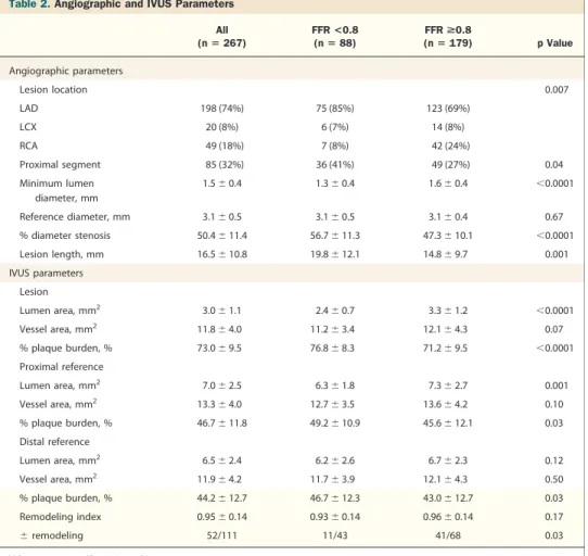
Lesions with FFR ⬍0.8 had less lumen diameter/area and more plaque, and were located more often in proxi- mal segments and the LAD than those lesions without functional significance.
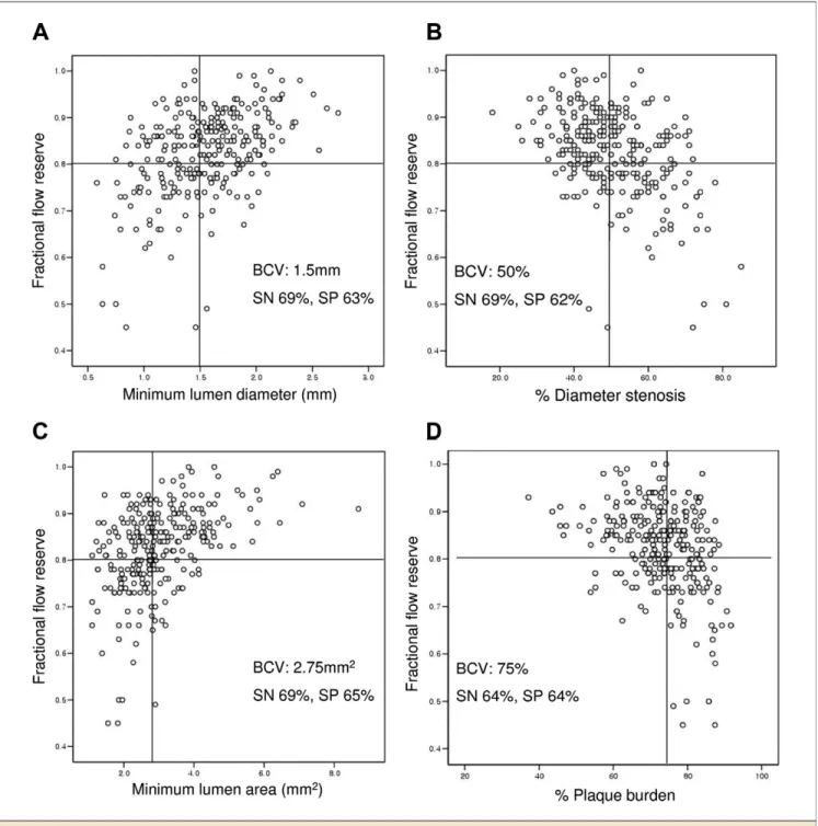
FFR versus angiographic and IVUS parameters. There was a positive correlation between FFR and minimum lumen diameter/lumen area, and a negative correlation between FFR and percent diameter stenosis/percent plaque burden (Fig. 1). BCV to predict the functional significance was 1.5 mm for angiographic minimum lumen diameter and 50%
for percent diameter stenosis. The BCV of IVUS parame- ters were minimum lumen area of 2.75 mm
2(sensitivity:
69%, specificity: 65%) and percent plaque burden of 75%
(sensitivity: 64%, specificity: 64%). The determinants of functionally significant stenosis were minimum lumen area and lesion location (Table 3). The size of a vessel was not a determinant of FFR.
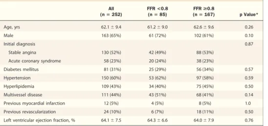
Table 1.Baseline Clinical Characteristics of the Patients All (nⴝ 252)
FFR <0.8
(nⴝ 85) FFR >0.8
(nⴝ 167) p Value*
Age, yrs 62.1⫾ 9.4 61.2⫾ 9.0 62.6⫾ 9.6 0.26
Male 163 (65%) 61 (72%) 102 (61%) 0.10
Initial diagnosis 0.87
Stable angina 130 (52%) 42 (49%) 88 (53%)
Acute coronary syndrome 58 (23%) 20 (24%) 38 (23%)
Diabetes mellitus 81 (31%) 25 (29%) 56 (34%) 0.57
Hypertension 150 (60%) 53 (62%) 97 (58%) 0.59
Hyperlipidemia 109 (43%) 34 (40%) 75 (45%) 0.50
Multivessel disease 111 (44%) 43 (51%) 68 (41%) 0.14
Previous myocardial infarction 12 (5%) 4 (5%) 8 (5%) 1.0
Previous revascularization 24 (10%) 6 (7%) 18 (11%) 0.50
Left ventricular ejection fraction, % 64.1⫾ 7.5 64.3⫾ 6.6 64.0⫾ 7.9 0.76 Values are mean⫾ SD or n (%). *Comparison between fractional flow reserve (FFR) ⬍0.8 versus FFR ⱖ0.8.
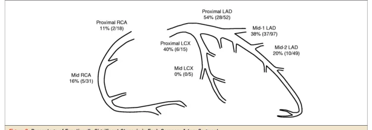
Influence of lesion location on the relation between FFR and minimum lumen area. The percentage of functionally sig- nificant stenosis was different in a different coronary artery segment (Fig. 2). Even though the angiographic percent diameter and lesion length were not different compared with other segments, the incidence of func- tionally significant stenosis was lowest in right coronary artery (RCA) lesions.
Receiver-operator characteristic curve analysis was per- formed to assess the diagnostic accuracy of minimum lumen area for the prediction of functional significance in lesions of different locations (Table 4). Sensitivity, specificity, and positive and negative predictive values of minimum lumen areas at a different location were shown in Figure 3. The area under the curve (AUC) was the largest in proximal LAD lesions (AUC: 0.81, 95% confidence interval [CI]: 0.68 to 0.91). The BCV for proximal LAD lesions was 3.0 mm
2, and its AUC was significantly larger than that of 4.0 mm
2(0.81 vs.
0.67, p ⫽ 0.03). In proximal LAD lesions, 7 out of 28 lesions with a minimum lumen area ⱖ3 mm
2had FFR ⬍0.8. Among them, 4 lesions had lesion length ⱖ20 mm. BCV and AUC for
mid-1 LAD lesions were 2.75 mm
2and 0.76 (95% CI: 0.66 to 0.84), respectively. In mid-1 LAD lesions, 10 out of 57 lesions with minimum lumen area ⱖ2.75 mm
2had FFR ⬍0.8, and 5 of them had lesion length ⱖ20 mm.
Appropriate minimum lumen area to predict the func- tional significance could not be found in LCX and mid-2 LAD lesions. In the mid-2 LAD segment, lesions located proximal to the last diagonal branch (n ⫽ 20) had a BCV of 2.3 mm
2. Its sensitivity and specificity were 93% and 87%, respectively. When the lesions were located distal to the last diagonal branch (n ⫽ 29), no appropriate anatomic criteria were found, and only 1 out of 14 lesions with minimum lumen area ⬍2 mm
2had FFR ⬍0.8.
Influence of a vessel size on the relation between FFR and minimum lumen area. When the lesions were divided accord- ing to a reference diameter of 3.0 mm, 31.8% (50 of 157) of large vessel lesions and 34.5% (38 of 110) of small vessel lesions had functional significance. When the relationship between IVUS and FFR was analyzed according to vessel size, BCV in large vessels was 3.0 mm
2(AUC: 0.70, 95% CI: 0.61 to 0.76) and its sensitivity was 76% and specificity, 62% (Fig. 4). BCV in
Table 2.Angiographic and IVUS Parameters All
(nⴝ 267) FFR <0.8
(nⴝ 88) FFR >0.8
(nⴝ 179) p Value
Angiographic parameters
Lesion location 0.007
LAD 198 (74%) 75 (85%) 123 (69%)
LCX 20 (8%) 6 (7%) 14 (8%)
RCA 49 (18%) 7 (8%) 42 (24%)
Proximal segment 85 (32%) 36 (41%) 49 (27%) 0.04
Minimum lumen diameter, mm
1.5⫾ 0.4 1.3⫾ 0.4 1.6⫾ 0.4 ⬍0.0001
Reference diameter, mm 3.1⫾ 0.5 3.1⫾ 0.5 3.1⫾ 0.4 0.67
% diameter stenosis 50.4⫾ 11.4 56.7⫾ 11.3 47.3⫾ 10.1 ⬍0.0001
Lesion length, mm 16.5⫾ 10.8 19.8⫾ 12.1 14.8⫾ 9.7 0.001
IVUS parameters Lesion
Lumen area, mm2 3.0⫾ 1.1 2.4⫾ 0.7 3.3⫾ 1.2 ⬍0.0001
Vessel area, mm2 11.8⫾ 4.0 11.2⫾ 3.4 12.1⫾ 4.3 0.07
% plaque burden, % 73.0⫾ 9.5 76.8⫾ 8.3 71.2⫾ 9.5 ⬍0.0001
Proximal reference
Lumen area, mm2 7.0⫾ 2.5 6.3⫾ 1.8 7.3⫾ 2.7 0.001
Vessel area, mm2 13.3⫾ 4.0 12.7⫾ 3.5 13.6⫾ 4.2 0.10
% plaque burden, % 46.7⫾ 11.8 49.2⫾ 10.9 45.6⫾ 12.1 0.03
Distal reference
Lumen area, mm2 6.5⫾ 2.4 6.2⫾ 2.6 6.7⫾ 2.3 0.12
Vessel area, mm2 11.9⫾ 4.2 11.7⫾ 3.9 12.1⫾ 4.3 0.50
% plaque burden, % 44.2⫾ 12.7 46.7⫾ 12.3 43.0⫾ 12.7 0.03
Remodeling index 0.95⫾ 0.14 0.93⫾ 0.14 0.96⫾ 0.14 0.17
⫾ remodeling 52/111 11/43 41/68 0.03
Values are mean⫾ SD, n (%), or n/N.
⫾ remodeling ⫽ positive/negative remodeling; FFR ⫽ fractional flow reserve; IVUS ⫽ intravascular ultrasound; LAD ⫽ left anterior descending coronary artery; LCX⫽ left circumflex coronary artery; RCA ⫽ right coronary artery.
small vessel was 2.5 mm
2(AUC: 0.61, 95% CI: 0.52 to 0.71) and its sensitivity was 61% and specificity, 63%. Positive predictive values of BCV in large and small vessels were 48%
and 46%, respectively. The sensitivity, specificity, and positive and negative predictive values of the criteria of lumen area ⬍3 mm
2and percent plaque burden ⬎75% were 58%, 75%, 52%, and 79%, respectively.
Discussion
The present study revealed that: 1) different IVUS criteria should be applied to discriminate the functional significance of lesions in different locations; and 2) the diagnostic accuracy of IVUS criteria could be variable according to lesion location and anatomic variation of the coronary artery.
Figure 1.Plots of Relationship Between FFR and Angiographic and IVUS Parameters
There was a positive correlation between fractional flow reserve (FFR) and minimum lumen diameter (A)/minimum lumen area (B), and a negative correlation between FFR and percent diameter stenosis (C)/percent plaque burden (D). The best cutoff value (BCV) to predict the functional significance was 50% for angiographic percent diameter stenosis and 2.75 mm2for minimum lumen area by intravascular ultrasound. SN⫽ sensitivity; SP ⫽ specificity.
The most commonly used invasive tools to evaluate intermediate coronary stenoses are IVUS and FFR. FFR is a physiological parameter that can discriminate the presence of myocardial ischemia. Outcomes of FFR-guided treat- ment in intermediate stenoses are reported to be favorable (17–19). Even though IVUS provides anatomic informa- tion, previous studies revealed that a lumen area cutoff value of 3 to 4 mm
2can accurately assess the physiological significance of lesions located at the proximal part of major coronary arteries (8 –10). However, as the presence of myocardial ischemia is determined by both the lesion severity and the amount of myocardium supplied, physio- logical significance of a minimum lumen area by IVUS should be different according to lesion location or the amount of myocardium supplied. For example, in a study by Jasti et al. (20), the optimal IVUS lumen area in left main coronary artery stenosis was 5.9 mm
2. Moreover, as the bifurcating structure of the coronary artery follows the law of flow conservation (21), there cannot be a single IVUS cutoff point for the functional significance of stenoses in different locations. In our study, the determinants of func- tional significance of a lesion were lesion severity (minimum lumen area) and lesion location.
Previous studies could not provide adequate IVUS criteria for the functional significance of lesions in different loca- tions because early studies included only proximal lesions (8 –10), and 2 recent studies (PHANTOM [Physiologic and Anatomical Evaluation Prior to and After Stent Im- plantation in Small Coronary Vessels] and IDEAS [Intra- vascular Ultrasound Diagnostic Evaluation of Atheroscle- rosis in Singapore] trials) (22,23) included only small vessels. In our study, when all lesions were analyzed to- gether, the BCV of minimum lumen area and percent plaque burden were 2.75 mm
2and 75%, respectively. When lesions were divided according to their reference diameter, large vessels (ⱖ3 mm) had a BCV of 3.0 mm
2, and small vessels, 2.5 mm
2. However, the diagnostic accuracy was relatively low, and the positive predictive value was ⬍50%
for all these values. In the IDEAS study (23), investigators compared IVUS parameters and FFR in small vessel lesions
Table 3.Determinants of Functionally Significant Coronary Artery Stenosis (FFR <0.8)
OR 95% CI p Value
Minimum lumen area 0.35 0.19–0.66 0.001
Proximal segment (vs. mid) 2.97 1.20–7.32 0.02
LAD lesion (vs. non-LAD) 3.40 1.24–9.30 0.02
Other included variables: reference vessel diameter (ⱖ3.0 mm), diagnosis, multivessel disease, angio- graphic lesion length (ⱖ20 mm), percent plaque burden, left ventricular ejection fraction, history of previous myocardial infarction, method of adenosine administration, remodeling index.
CI⫽ confidence interval; OR ⫽ odds ratio; other abbreviations as inTable 2.
Figure 2.Percentage of Functionally Significant Stenosis in Each Coronary Artery Segment
The percentage of functionally significant stenosis was different in a different coronary artery segment. Even though the angiographic percent diameter and lesion length were not different compared with other segments, the incidence of functionally significant stenosis was lowest in right coronary artery lesions.
LAD⫽ left anterior descending coronary artery; LCX ⫽ left circumflex coronary artery; RCA ⫽ right coronary artery.
Table 4.BCV and AUC in ROC Curves of MLA in Prediction of
Functionally Significant Stenosis in Different Locations and Vessel Sizes
BCV, mm2 AUC 95% CI
Lesion location
Proximal LAD (n⫽ 52) 3.0 0.81 0.68–0.91
Mid-LAD (n⫽ 146) 2.5 0.64 0.56–0.72
Mid-1 LAD (n⫽ 97) 2.75 0.76 0.66–0.84
Mid-2 LAD (n⫽ 49) NA
Right coronary artery (n⫽ 49) 3.0 0.68 0.53–0.81 Left circumflex artery (n⫽ 20) NA
Vessel size
ⱖ3.0 mm (n ⫽ 157) 3.0 0.70 0.61–0.76
⬍3.0 mm (n ⫽ 110) 2.5 0.61 0.52–0.71
BCA⫽ best cutoff value; MLA ⫽ minimum lumen area; NA ⫽ not available; other abbreviations as inTables 2and3.
and proposed IVUS criteria for functionally significant stenosis. However, 59% of the lesions were located at proximal segments in this study despite the fact that all
lesions had a reference diameter ⬍3.0 mm. In our study, only 6% of lesions with a reference diameter ⬍3.0 mm were located at proximal segments (proximal LAD: 10 cases,
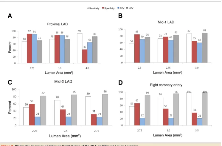
Figure 3.Diagnostic Accuracy of Different Cutoff Points of the MLA at Different Lesion Locations
The diagnostic accuracy of the minimum lumen area (MLA) was highly variable according to the location of lesions. The best cutoff value (BCV) of MLA to define the functional significance was 3.0 mm2for the proximal left anterior descending artery (LAD) lesions (A) and 2.75 mm2for mid-LAD lesions located before the second diagonal branch (mid-1 LAD) (B). Shown also are plots of the cutoff values of the mid-2 LAD (C) and right coronary artery (D). NPV⫽ negative predictive value; PPV ⫽ positive predictive value.
Figure 4.Diagnostic Accuracy of Different Cutoff Points of the MLA According to the Size of Vessel
The BCV in large (A) and small (B) vessels were 3.0 mm2and 2.5 mm2, respectively. PPV of BCV were⬍50% in both large and small vessels.
Abbreviations as inFigure 3.
proximal LCX: 6 cases). In another study, which compared the anatomical parameters and FFR in lesions located at small vessels (⬍2.8 mm), most lesions (92%) were located at nonproximal segments, and there was no correlation be- tween any IVUS parameters and FFR (22). Therefore, it seems to be more appropriate to propose different IVUS criteria according to the lesion location rather than the size of a vessel.
In our study, the diagnostic accuracy of the lumen area was variable according to the location of lesions. This variability seems to reflect the anatomic variation of coro- nary arteries. Diagnostic accuracy was higher in lesions at the proximal LAD, which usually has fewer anatomic variations. However, diagnostic accuracy was lower in seg- ments with relatively higher anatomic variations. When all mid-LAD lesions were analyzed together, diagnostic accu- racy was fair, but the positive predictive value of a BCV (2.5 mm
2) was only 48%. The diagnostic accuracy of the mini- mum lumen area became better after the mid-LAD lesions were divided according to the location of diagonal branches, which reflects the difference in the amount of myocardium supplied. Diagnostic accuracy was fair in mid-LAD lesions located before the second diagonal branch. In mid-2 LAD lesions, appropriate IVUS criteria could be found only in lesions proximal to the last diagonal branch. In LCX and RCA lesions, appropriate anatomic values to predict the functional significance were difficult to find due to variabil- ity in the bifurcating structure of coronary trees and in dominance between the LCX and RCA. In our study, even though the angiographic percent diameter was not different compared with other segments, the incidence of function- ally significant stenosis was lowest in RCA lesions.
Because this study was not designed to evaluate the outcomes of FFR-guided versus IVUS-guided revascular- ization strategies, the discrepancy between anatomic and physiological parameters cannot be regarded as evidence of superiority of FFR over IVUS in patients with intermediate stenosis. However, this study demonstrated that when IVUS is used to assess the functional significance of inter- mediate lesions, the operators should consider the lesion location and the amount of myocardium supplied by the target segment as well as the minimum lumen area. This consideration may translate into a more accurate physiolog- ical assessment of these lesions, with a concomitant reduc- tion in unnecessary revascularizations.
Study limitations. First, although the number of patients was larger than previous studies, the number of LCX lesions in our study was relatively small. Second, even though intracoronary nitrate was given before each IVUS run, the possibility of catheter-induced spasm in distal lesions or small vessel lesions could not be completely excluded. Third, when the LAD lesions were classified, septal branches were not considered except for the proximal LAD. Because the size and location of septal branches were so variable, it was
hard to set the valid segmentation according to the location of a septal branch. Fourth, the cutoff points of minimum lumen diameter suggested in our study cannot be applied to lesions with multiple stenoses. FFR or the functional significance of a lesion is determined by total atherosclerotic burden in a target vessel. Fifth, various methods of adeno- sine administration were allowed in our study. Because some reports suggested that the hyperemic efficacy of bolus administration of adenosine was less than that of other methods (14,24), there is a possibility of underestimating lesion severity by FFR. Sixth, because this study was done in Korean patients, the numbers proposed in this study may be different in a Western population. Finally, clinical outcomes could not be evaluated in this study. The outcomes of FFR-guided versus IVUS-guided revascularization strategy need to be evaluated in future randomized trials.
Conclusions
When anatomic parameters are used to determine the functional significance of lesions in patients with interme- diate stenosis, different cutoff values should be used accord- ing to lesion location and the amount of myocardium supplied by the target segment. In segments or vessels with anatomic variations, IVUS cannot be used for functional assessment of a stenosis.
Reprint requests and correspondence: Dr. Hyo-Soo Kim, De- partment of Internal Medicine, Seoul National University Hospi- tal, Yongon-dong 28, Jongno-gu, 110-744 Seoul, Republic of Korea. E-mail: [email protected]; or Dr. Seung-Jea Tahk, De- partment of Cardiology, Ajou University Medical Center, San 5, Wonchon-dong, Yeongtong-gu, Suwon, Gyeonggi-do 443-721, Republic of Korea. E-mail: [email protected].
REFERENCES