|
J BD
Journal of Breast DiseaseSynchronous Bilateral Breast Carcinoma in a Patient with Cowden Syndrome with PTEN Mutation: A Case Report
Sun Young Kwon, Soo Hyun Yeo1, Jung Sook Ha2, Sun Hee Kang3
Departments of Pathology, 1Radiology, 2Laboratory Medicine, and 3General Surgery, Keimyung University School of Medicine, Daegu, Korea
Cowden syndrome (CS), also known as multiple hamartomas syndrome, is a rare hereditary autosomal dominant disorder caused by a ger- mline mutation in the phosphatase and tensin homolog (PTEN) gene mapped on chromosome 10. The clinical features of CS are variable, primarily presenting as mucocutaneous lesions (99%). A mucocutaneous lesion, such as trichilemmoma of the face or keratosis of the ex- tremities, is an important diagnostic marker for CS. CS has been reported to increase the incidence of benign and malignant neoplasms in the breast, thyroid, and gastrointestinal tract. The risk of developing malignancy in individuals with CS is up to 10 times higher than general population throughout an entire life time.
Key Words: Breast neoplasms, Cowden syndrome, Mutation, PTEN gene
INTRODUCTION
Cowden syndrome (CS), or multiple hamartoma syndrome, is a rare hereditary autosomal dominant disorder caused by a mutation in the phosphatase and tensin homolog (PTEN) gene on chromosome 10 with characteristic hamartoma of multiple organs [1-3]. PTEN is a tu- mor suppressor gene that controls phosphate, which mediates prolif- eration, progression, and apoptosis in the cell proliferation [4]. The in- cidence of CS is 1/200,000 individuals, and this syndrome is very diffi- cult to diagnose clinically. The clinical features of CS are variable, pre- senting primarily as mucocutaneous lesions (99%). A mucocutaneous lesion, such as trichilemmoma of the face or keratosis of the extremi- ties, is an important diagnostic marker for CS [5,6]. CS has been re- ported to increase the incidence of benign and malignant neoplasms in the breast, thyroid, and gastrointestinal tract [1-3,7]. The risk of de- veloping breast cancer is up to 10 times higher in individuals with CS than in the general population throughout an entire life time [8]. The risk of developing breast cancer is up to 25%–50% times higher in in- dividuals with CS than in the general population. Herein, we present a case of CS with synchronous bilateral breast cancer.
CASE REPORT
A 27-year-old woman presented with ill-defined firm masses in both breasts without palpable axillary nodes. She had no family histo- ry of breast carcinoma. However, she was previously diagnosed with CS at the age of 16 years. She had macrocephaly and multiple polyps in the oral cavity, pharynx, and gastrointestinal tract, as well as benign breast tumors and a nodular goiter of the thyroid gland when diag- nosed. She underwent a right lobectomy of the thyroid gland and both breast masses were excised at the age of 18 years. Histologically, the right thyroid mass revealed follicular adenoma with atypia. The pathologic analysis of the bilateral breast masses in both breasts indi- cated a phyllodes tumor and tubular adenoma. She was followed up with breast ultrasonography every 1–2 years; an imaging study showed multiple Breast Imaging-Reporting and Data System (BI-RADS) cate- gory 3 masses in both breasts that had not changed. Nevertheless, at the age of 27 years, she again presented with masses in both breasts.
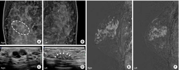
Physical examination revealed multiple 2.5 cm-sized ill-defined and firm masses in the upper outer quadrant of both breasts without pal- pable axillary lymph nodes. Mammography and breast ultrasonogra- phy were performed, and they showed that segmentally distributed pleomorphic microcalcifications in the left upper outer quadrants (Figure 1A and 1B) and multiple hypoechoic mass lesions in both breasts (Figure 1C and 1D). The radiologist concluded suspicious le- sions of BI-RADS category 4a in both breasts. The patient then under- CASE REPORT
J Breast Dis 2018 December; 6(2): 79-83 https://doi.org/10.14449/jbd.2018.6.2.79
Correspondence: Sun Hee Kang
Department of General Surgery, Keimyung University School of Medicine, 1095 Dalgubeol-daero, Dalseo-gu, Daegu 42601, Korea
Tel: +82-53-250-7922, Fax: +82-53-250-7211, E-mail: [email protected] Received: Feb 12, 2018 Revised: May 31, 2018 Accepted: Aug 1, 2018
Figure 2. Low power microscopic-findings of the right (A, H&E stain, ×40) and left (C, ×100) breast masses showed atypical ductal proliferation with cribriform, solid, papillary, and micropapillary patterns. Myoepithelial cells were intact and central comedonecrosis was found in the right breast (A, arrows). High power microscopic-findings of the right breast mass (B, ×400) demonstrated high grade ductal carcinoma in situ (DCIS) with enlarged nuclei with prominent nucleoli and increased mitotic figures (B, arrows). However, in the left breast mass (D, ×400) showed low grade DCIS.
C A
D B Figure 1. Mammography showed 17-mm sized, lobulated nodule with tiny calcification at upper central portion of right breast (A, dashed line) and segmental distributed, pleomorphic microcalcifications at left upper outer quadrant (B). Ultrasonography showed that multiple hypoechoic mass lesions in right breast (C) and microcalcification in left breast (D, arrows). Magnetic resonance imaging showed diffusely distributed, heterogenous, nodular non-mass enhancement, about 9.0-cm size area at both breast upper outer quadrant (E, F).
A
E F
C
B
D
Right
Right Right
Left
Left Left
went ultrasonography-guided core biopsies of the suspicious lesions in both breasts, the pathologic report revealed ductal carcinoma in situ (DCIS) of both breasts. To decide on surgery, we performed breast magnetic resonance imaging, which revealed diffusely distributed, heterogenous, and nodular non-mass enhancement of approximately a 9.0-cm sized area in both breast upper outer quadrants (Figure 1E and 1F). According to this finding, we performed the bilateral skin-sparing mastectomy with immediate breast reconstruction.
Breast reconstruction was performed using implants by plastic sur- geons.
The final pathologic report showed that macroscopically, bilateral breast tissues bilaterally showed ill-demarcated, pale tan, and diffuse firm and nodular lesions without definite masses. Microscopically, the tumor from the right breast consisted of DCIS of intermediate grade that was predominantly cribriform type (Figure 2A and 2B).
Different from the tumor in the right breast, the tumor in the left breast revealed DCIS of high-grade with various types, such as papil-
lary, cribriform, solid, and comedo-type (Figure 2C and 2D).
Immunohistochemistry (IHC) of estrogen receptor showed posi- tive in both breast tumors (Figure 3A and 3B). IHC of human epider- mal growth factor receptor 2 (HER2) revealed negativity in the right breast tumor (Figure 3C). However, in the left breast, IHC of HER2 showed positive membranous staining (Figure 3D). IHC of PTEN re- vealed positive nuclear staining in both breast masses (Figure 3E and 3F).
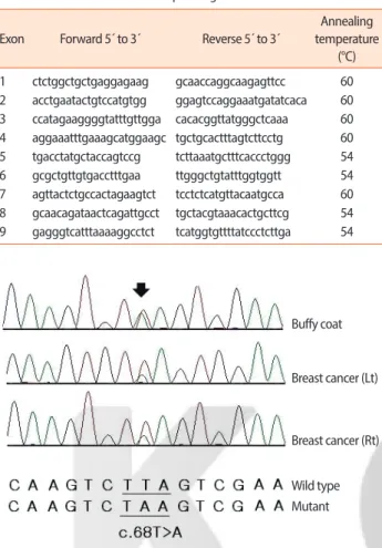
The mutational analysis of PTEN was performed by using poly- merase chain reaction (PCR) amplification and direct sequencing of all nine exons of the gene, including the splicing regions. Blood and cancer tissue samples were obtained from Keimyung University Dongsan Hospital Biobank, Korea. Informed consent was obtained, and the protocol was approved by the Institutional Review Board of Dongsan Medical Center (DSMC 2014-10-051). Genomic DNA was extracted, and PCR was performed using Smart Tag Pre-mix (Solgent Co., Daegu, Korea). PCR primers for PTEN were designed using
Figure 3. Immunohistochemistry (IHC) of estrogen receptor showed positive in both breast tumors (A, right; B, left; × 200). IHC of human epidermal growth fac- tor receptor 2 (HER2) in the right breast tumor showed negative expression (C, ×400). However, HER2 expression in the left breast tumor showed positive mem- branous staining (D, ×400). IHC of PTEN in both breast tumors showed strong nuclear expression (E, right; F, left; × 200).
A
D
B
E
C
F
Primer3 (http://frodo.wi.mit.edu/primer3) (Table 1). Products were purified and bidirectionally sequenced using a BigDye Terminator v3.1 Cycle Sequencing kit (Applied Biosystems, Foster City, USA) on an ABI 3730XL DNA analyzer (Applied Biosystems). For all sequenc- ing results, traces were reviewed by the Sequencherv5.2.4 software (http://genecodes.com/). DNA sequencing analysis detected the het- erozygous transversion c.68T>A in exon 1 from both breast cancer tissues and peripheral blood (Figure 4). This was a pathogenic non- sense variant resulting in the substitution of a residue of leucine on co- don 23 with a stop codon (leu23Ter).
The patient did not require additional treatment for bilateral DCIS and no further routine breast examinations were performed because of the bilateral mastectomy. However, further imaging studies are necessary for early detection of secondary malignancies, e.g., uterine or renal malignancy during the follow up period.
DISCUSSION
CS, also known as multiple hamartoma syndrome, is an autosomal dominant disorder caused by a germline inactivating mutation of the PTEN gene located on chromosome 10q23.31-3 [1-3]. The clinical di- agnostic criteria for multiple hamartoma syndrome have been estab- lished and revised; major and minor criteria are listed in Table 2 [9].
CS is suspected if a person meets three major criteria which must in- clude either macrocephaly, Lhermitte-Duclos disease [10], or gastroin- Table 1. Primers used for direct sequencing
Exon Forward 5´ to 3´ Reverse 5´ to 3´
Annealing temperature
(°C) 1 ctctggctgctgaggagaag gcaaccaggcaagagttcc 60 2 acctgaatactgtccatgtgg ggagtccaggaaatgatatcaca 60 3 ccatagaaggggtatttgttgga cacacggttatgggctcaaa 60 4 aggaaatttgaaagcatggaagc tgctgcactttagtcttcctg 60 5 tgacctatgctaccagtccg tcttaaatgctttcaccctggg 54 6 gcgctgttgtgacctttgaa ttgggctgtatttggtggtt 54 7 agttactctgccactagaagtct tcctctcatgttacaatgcca 60 8 gcaacagataactcagattgcct tgctacgtaaacactgcttcg 54 9 gagggtcatttaaaaggcctct tcatggtgttttatccctcttga 54
Table 2. Revised PTEN hamartoma tumor syndrome clinical diagnostic crite- ria
Major criteria Breast cancer
Endometrial cancer (epithelial) Thyroid cancer (follicular)
Gastrointestinal hamartomas (including ganglioneuromas, but excluding hyperplastic polyps; ≥ 3)
Lhermitte-Duclos disease (adult)
Macrocephaly (≥ 97 percentile: 58 cm for females, 60 cm for males) Macular pigmentation of the glans penis
Multiple mucocutaneous lesions (any of the following):
Multiple trichilemmomas (≥ 3, at least one biopsy proven)
Acral keratoses (≥ 3 palmoplantar keratotic pits and/or acralhyperkeratotic papules)
Mucocutaneous neuromas (≥ 3)
Oral papillomas (particularly on tongue and gingiva), multiple (≥ 3) or biopsy proven or dermatologist diagnosed
Minor criteria
Autism spectrum disorder Colon cancer
Esophageal glycogenic acanthosis (≥ 3) Lipomas (≥ 3)
Mental retardation (i.e., IQ≤ 75) Renal cell carcinoma Testicular lipomatosis
Thyroid cancer (papillary or follicular variant of papillary) Thyroid structural lesions (e.g., adenoma, multinodular goiter)
Vascular anomalies (including multiple intracranial developmental venous anomalies)
Operational diagnosis in an individual (either of the following) 1. Three or more major criteria, but one must include macrocephaly,
Lhermitte-Duclos disease, or gastrointestinal hamartomas; or 2. Two major and three minor criteria
Operational diagnosis in a family where one individual meets revised PTEN hamartoma tumor syndrome clinical diagnostic criteria or has a PTEN mutation:
1. Any two major criteria with or without minor criteria; or 2. One major and two minor criteria; or
3. Three minor criteria
Adapted from Pilarski R, et al. J Natl Cancer Inst 2013;105:1607-16, with per- mission of Oxford University Press [9].
Figure 4. Sequence analysis of tumor tissue revealed a PTEN mutation (c.68 T>A transversion).
Buffy coat
Breast cancer (Lt)
Breast cancer (Rt)
Wild type Mutant
testinal hamartomas, or two major and three minor criteria. In fami- lies where one individual meets the clinical diagnostic criteria of CS or has a PTEN mutation, CS is suspected if a person has either any two major criteria with or without minor criteria, one major and two mi- nor criteria, or three minor criteria. With the identification of PTEN involvement in CS, the incidence of the syndrome has increased to 1/200,000 individuals.
Herein, we reported a woman with CS presenting with thyroid fol- licular adenoma, multiple intestinal hyperplastic polyps, and syn- chronous bilateral breast DCIS over 10 years. The syndrome was di- agnosed when the patient was 16 years old and she underwent bilater- al mastectomy when she was 27 years old. It is critical to note that ma- lignant transformation can occur in young patients with CS, in the form of bilateral and synchronous lesions of the breast. As was the case for this patient, adequate follow-up examinations for the organs known to undergo malignant transformation in CS should be per- formed in patients with early-onset. When benign lesions are ob- served, the possibility of transformation into malignant tumors should be acknowledged, and follow-up examinations should be per- formed consistently over the patient’s life time [11,12]. We advise that clinical management of all patients with CS should consist of an early multidisciplinary surveillance program initiating from the time of di- agnosis.
CONFLICT OF INTEREST
The authors declare that they have no competing interests.
ACKNOWLEDGMENTS
The biospecimen and data used for this study were provided by the Biobank of Dongsan Hospital, a member of the Korea Biobank Net- work.
REFERENCES
1. Rhei E, Kang L, Bogomolniy F, Federici MG, Borgen PI, Boyd J. Mu- tation analysis of the putative tumor suppressor gene PTEN/
MMAC1 in primary breast carcinomas. Cancer Res 1997;57:3657-9.
2. Lynch ED, Ostermeyer EA, Lee MK, Arena JF, Ji H, Dann J, et al. In- herited mutations in PTEN that are associated with breast cancer, Cowden disease, and juvenile polyposis. Am J Hum Genet 1997;61:
1254-60.
3. Hanssen AM, Fryns JP. Cowden syndrome. J Med Genet 1995;32:
117-9.
4. Porto AC, Roider E, Ruzicka T. Cowden syndrome: report of a case and brief review of literature. An Bras Dermatol 2013;88(6 Suppl 1):52-5.
5. Tellechea O, Cardoso JC, Reis JP, Ramos L, Gameiro AR, Coutinho I, et al. Benign follicular tumors. An Bras Dermatol 2015;90:780-96.
6. Ng E, Terushkin V, Meehan SA, Ho R, Pomeranz MK. Cowden syndrome presenting with trichilemmomas. Dermatol Online J 2016;22:33-5.
7. Cameselle-Teijeiro J, Fachal C, Cabezas-Agrícola JM, Alfonsín- Barreiro N, Abdulkader I, Vega-Gliemmo A, et al. Thyroid pathology findings in Cowden syndrome: a clue for the diagnosis of the PTEN hamartoma tumor syndrome. Am J Clin Pathol 2015;144:322-8.
8. Peiró G, Adrover E, Guijarro J, Ballester I, Jimenez MJ, Planelles M, et al. Synchronous bilateral breast carcinoma in a patient with Cowden syndrome: a case report with morphologic, immunohisto- chemical and genetic analysis. Breast J 2010;16:77-81.
9. Pilarski R, Burt R, Kohlman W, Pho L, Shannon KM, Swisher E.
Cowden syndrome and the PTEN hamartoma tumor syndrome:
systematic review and revised diagnostic criteria. J Natl Cancer Inst 2013;105:1607-16.
10. Riegert-Johnson DL, Gleeson FC, Roberts M, Tholen K, Youngborg L, Bullock M, et al. Cancer and Lhermitte-Duclos disease are common in Cowden syndrome patients. Hered Cancer Clin Pract 2010;8:6.
11. Schultz KA, Rednam SP, Kamihara J, Doros L, Achatz MI, Wasserman JD, et al. PTEN, DICER1, FH, and their associated tumor susceptibility syndromes: clinical features, genetics, and surveillance recommendations in childhood. Clin Cancer Res 2017;23:e76-82.
12. Ngeow J, Eng C. PTEN hamartoma tumor syndrome: clinical risk assessment and management protocol. Methods 2015;77-78:11-9.