• 검색 결과가 없습니다.

Effects of PNF and Respiratory Muscle Endurance Training on Lung Function and Physical Activity in Chronic Stroke Patients

N/A
N/A
Protected

Academic year: 2021

Share "Effects of PNF and Respiratory Muscle Endurance Training on Lung Function and Physical Activity in Chronic Stroke Patients"

Copied!
10
0
0

로드 중.... (전체 텍스트 보기)

전체 글

(1)

| Abstract |

Purpose: The purpose of the study was to assess the effects of proprioceptive neuromuscular facilitation (PNF) training and respiratory muscle endurance training on pulmonary function and activity in chronic stroke patients.

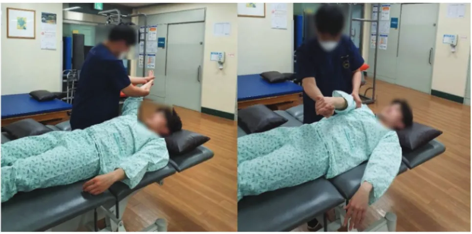
Methods: The participants were 25 chronic stroke patients. They were assigned to two groups: a PNF and respiratory muscle training group (experimental group; n = 12) and a conservative training group (control group; n = 13). The experimental group completed 50 minutes (30 minutes of conventional physical therapy, 10 minutes of PNF training, and 10 minutes of respiratory muscle endurance training). The control group also completed 50 minutes (30 minutes of conventional physical therapy and 20 minutes on a full-body workout machine). Pulmonary function and activity were measured before and after the intervention, using Cosmed to analyze pulmonary function and 6MWT as clinical evaluation indicators.

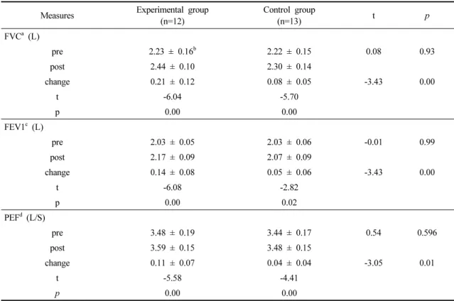
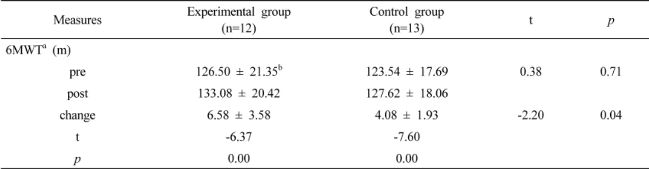
Results: Both groups showed significant within-group differences on all tests before and after the intervention; the experimental group showed greater improvement on all tests.

Conclusion: The findings confirm that PNF training and respiratory muscle endurance training have a positive effect on pulmonary function and activity index in chronic stroke patients.

Key Words: Stroke, Rehabilitation, Respiration, Pulmonary function

†Corresponding Author : Dong-Hoon Kim ([email protected])

Original Article Open Access

고유수용성 신경근 촉진법과 호흡근 지구력 훈련이 만성 뇌졸중 환자의 폐 기능과 신체활동량에 미치는 영향

김경훈⋅김동훈

1)

김천대학교 물리치료과

Effects of PNF and Respiratory Muscle Endurance Training on Lung Function and Physical Activity in Chronic Stroke Patients

Kyoung-Hun Kim, P.T, Ph.D⋅Dong-Hoon Kim, P.T, Ph.D Department of Physical Therapy,Gimcheon university, Proffessor Received: March 20, 2020 / Revised: April 13, 2020 / Accepted: April 14, 2020

ⓒ 2020 Journal of Korea Proprioceptive Neuromuscular Facilitation Association

This is an Open Access article distributed under the terms of the Creative Commons Attribution Non-Commercial License

(http://creativecommons.org/licenses/by-nc/3.0) which permits unrestricted non-commercial use, distribution, and reproduction

in any medium, provided the original work is properly cited.

(2)

Ⅰ. 서 론

뇌졸중은 뇌 조직의 허혈, 출혈에 의해 운동, 시야결 손, 감각장애, 언어상실 등과 함께 지적 장애 등의 동반 되어 환자와 가족들의 삶의 질을 감소시키는 만성 질 환으로(Darcy, 2006), 고혈압이나 당뇨, 스트레스, 심장 질환 등의 이유로 발생된다(Song, 2012).

뇌졸중은 손상 부위와 정도의 차이가 있지만 일반 적으로 편측 사지의 마비, 감각 손상, 운동 손상, 인지 와 언어 장애, 연하 장애, 시⋅지각적 등의 문제를 보인 다(Mercier et al., 2001). 특히 Carr와 Shephered (1998)는 비대칭적인 자세와 비정상적인 신체균형 및 체중 이 동능력의 감소 그리고 섬세한 기능을 수행하는 특정 운동능력의 소실 등으로 기립과 보행에 장애가 생긴 다고 하였다. 뇌졸중 환자는 균형 조절에 어려움을 갖게 되고, 서 있는 동안 자세동요가 증가하며, 비 마비 측으로 체중부하가 증가하여 양측으로 동일한 체중 분배가 어려워져 안정적인 신체이동 능력이 감소된다 (Srivastava et al., 2009).

고유수용성신경근촉진법(PNF)은 Herman Kabat과 Margaret Knott에 의해 시작된 신경생리학적 치료로써 근육과 관절 안의 고유수용성감각을 자극을 통해 신 경근의 움직임을 증진시키기 위해 고안된 운동방법으 로(Kofotolis et al., 2005), “쉽게 운동 한다(making easier)”는 의미를 가지며, 운동방향의 촉진과 기능 회 복은 환자의 안정성, 가동성, 협응된 움직임, 신경근 조절 등을 증진시키는데 도움이 된다(Kisner & Colby, 2013).

신체 활동은 앉아서 자세를 유지하는 쉬운 동작을 포함하여 보다 더 많은 에너지를 사용하여 근골격계 에 의해 만들어지는 모든 신체 동작으로 정의되고 있 으며(Horn et al., 2005), 뇌졸중 환자들은 발병 후, 적어 도 일주일에 150분 이상 최소 3일 중간강도로 활동하 는 것을 추천한다고 하였다(Gordon et al., 2004). 하지 만 이러한 신체 활동에 대하여 신경근골격계 손상으 로 인해 보행장애 및 근 약화, 심혈관계 저하가 나타난 다(Baert et al., 2012; Moore et al., 2013). 뇌졸중 급성기

에는 신체적 상태악화(decoditioning)로 불사용 위축 발생과 더불어 심폐능력이 저하되어 활동 시 높은 에 너지가 요구되며(Ryan et al., 2000), 마비측 근육 조직 특성을 해롭게 변화시키는 잠재성 뿐만 아니라 대사 성 증후군을 악화시키는 요인이 된다(DeDeyne et al.,2004; Landin et al., 1977). 뇌졸중 발병 이 후 6개월에 서 뇌졸중 환자의 기능적인 향상이 두드러지지만 (Kwakkel et al., 2004), 신체적인 활동의 범위는 여전히 제약되어진다 (West & Bernhardt, 2012). 뇌졸중 환자 들의 활동량은 저하되어있으며(Rand et al., 2009), 선행 연구에서는 뇌졸중 환자의 58%는 정상인의 활동량 수준에 미치지 못하는 것으로 나타났다(Eng et al., 2004). 또한 발병 이 후, 적극적으로 운동하는 비율이 전반적으로 감소된다고 하였다(Shaughnessy et al., 2006).

뇌졸중 환자의 심폐능력과 활동에 대한 연구들을

살펴보면 West와 Bernhardt (2011)는 병원에 입원중인

뇌졸중 환자들의 활동에서 활동시간이 48.1%이며, 홀

로 있는 시간은 53.7%, 침상에 있는 시간은 56.5%,

물리치료를 받는 시간은 63.2분, 작업치료시간은 57분

으로 뇌졸중 환자의 활동량이 현저하게 낮다고 제시

하였다. 또한 뇌졸중 환자를 대상으로 심폐증진을 위

한 연구에서 가로막-호흡 환자군, 흡기근 환자군과 통

제군을 비교했을 때 흡기근 환자군에서 1초간 노력성

호기량(forced expiratory volume at one second, FEV1),

노력성폐활량(forced vital capacity, FVC), 폐활량(vital

capacity, VC), 노력성호기중간유량(forced expiratory

rate, FEF 25-75%), 최대노력환기량(maximal voluntary

ventilation, MVV)이 다른 두 군보다 유의한 증진이

나타났다고 하였다(Sutbeyaz et al., 2010). Kim과 Choi

(2011)는 정상인, 아급성 뇌졸중 환자, 만성 뇌졸중

환자의 심폐기능 및 보행의 비교에서 세 군 모두 폐활

량, 노력 폐활량, 1초 노력성 호기량이 유의한 차이가

나타났다고 보고하였으며, 보행에서도 유의한 차이가

나타났다고 보고하였다. 또한 Kathleen 등(2009)은 지

속적 활동에서 뇌졸중 환자의 균형 및 보행, 심혈관계

능력의 증진에 대한 영향에서 심혈관계 및 보행의 유

(3)

의한 차이가 나타났다고 하였다.

그러나, 현재 우리나라의 뇌졸중 환자 재활은 일반 적으로 뇌졸중환자의 재활은 균형과 보행, 위팔 기능 등 특정 기능의 향상에 초점이 맞춰져 있어 뇌졸중 환자의 현재 심폐능력 측정과 관련되거나 심폐능력 향상을 위한 중재방법 연구는 적은 실정이며, 심폐계 통이나 활동성에는 적절한 치료가 이루어지지 않고 있다. 또한 뇌졸중 환자의 심폐 근육에 대하여 기능의 회복, 안정성, 가동성, 신경근 조절, 협응 움직임 등에 장점이 있는 고유수용성 신경근 촉진법을 이용한 치 료는 미미하며, 또한 호흡근 기기 훈련을 병행하여 진행한 연구, 신체 활동성의 변화에 대한 연구는 매우 적다. 호흡근 기기 훈련은 호흡 근력과 운동 능력이 향상에 및 일상생활 증진에 효과가 있다고 하였다 (Beckerman et al., 2005). 따라서 고유수용성 신경근 촉진법과 호흡근 지구력 훈련을 통해 만성 뇌졸중 환 자 심폐기능 및 신체활동성에 대한 연구를 통해 뇌졸 중 환자의 심폐능력 증진을 알아보았다.

Ⅱ. 연구 방법

1. 연구 대상

대한민국에 S시에 위치한 2곳의 병원에서 치료를 받는 환자 25명을 선정하였다. 선정 조건은 뇌졸중으 로 처음 진단받고 6개월 경과한 자, 기립과 균형에 대한 근 골격 손상이 없는 자, 보조도구 없이 보행이 가능한 자, 원뿔 주변을 8자 모양으로 지팡이와 보조도 구를 사용하여 보행이 가능한 자, 환자와 보호자가 연구에 목적을 이해하며 참여에 동의한 자이며, 제외 조건은 체성 감각이 결손된 환자, 내과적 질환(간헐성 파행증, 허혈성 심장질환)자, 늑골부위의 손상이 있는 자이다.

대상자들을 고유수용성 신경근 촉진법과 호흡근 훈련군과 보존적치료군으로 컴퓨터 프로그램을 통해 무작위로 나누어 배치하였다.

2. 측정방법 및 도구

1) 폐기능 검사

폐 기능 검사는 Cosmed (MicroQuark, COSMED Srl, RM, Italy)를 사용하여 측정하였다. 대상자는 바르게 앉은 자세에서 측정을 시작하였으며, 측정 변수로는 최대 노력성 호기 곡선을 측정 후, 노력성폐활량 (FVC), 1초 노력성호기량(FEV1), 최대 호기 속도(PEF) 이다. 측정에서 중 입이 닿는 부위는 마우스피스 사용 하였으며, 1명의 대상자가 측정이 끝나면 위생을 위해 즉시 분리하여 알코올 소독을 실시하였다.

2) 신체활동성 검사

신체활동성 검사는 6분 보행 검사(6-minute walk test, 6MWT)를 통해 실시하였으며, 6MWT는 6분동안 대상자가 이동한 총 거리를 측정하는 도구이다.

6MWT는 실내에서 1 m 간격으로 테이프를 통해 표시 를 한 트랙에서 측정자는 대상자가 보행을 시작하고 6분 후에 스톱워치를 통해 이동거리를 표시한다.

6MWT는 뇌졸중 환자의 근지구력 및 기능적 보행 정 도를 볼 수 있는 검사방법이며 높은 신뢰도 r=0.91를 보이는 측정방법이다(Mossberg, 2003).

3. 실험 절차

본 연구는 고유수용성 신경근 촉진법과 호흡근 훈

련군(12명)과 보존적치료군(13명)으로 나누어 하루 50

분 주 3회 8주 동안 훈련을 실시하였다. 참가자 및

보호자에게 연구 전 서면으로 연구의 목적, 연구방법,

비밀유지를 설명하였으며, 원한다면 언제든지 참여

철회가 가능하다고 설명 후 진행하였다.

(4)

1) 고유수용성 신경근 촉진법과 호흡근 지구력 훈 련군(experimental group)

실험군은 보존적치료를 30분간 시행하였으며, 고 유수용성 신경근 촉진법 10분과 호흡근 훈련10분을 시행하였다.

고유수용성 신경근 촉진 운동은 PNF 개념에서 사 용된 기본 절차를 따랐다. 본 연구에서 사용된 패턴은 위팔의 굽힘 - 벌림 - 팔꿈치가 확장된 상태에서의 가쪽돌림과 폄 - 모음 - 팔꿈치가 확장된 안쪽돌림이 며, 특정 기술로서 길항근의 반전을 사용하였다(Adler et al., 2000). 대상자는 매트에서 바로 눕고 중재자는 대상자의 옆에 위치한다. 굽힘 - 벌림 - 팔꿈치가 확장 된 상태에서의 가쪽돌림에서 중재자는 먼쪽 손으로 환자 손등에 잡기한다. 몸쪽 손은 대상자의 아래팔 먼쪽에 노뼈와 자뼈 측면을 잡기한다. 이후 대상자에 게 패턴 움직임을 충분히 알려준 뒤 “손가락을 펴고 손목을 올리고, 팔 올리세요”라고 지시하면서 근육의 활성화되면서 움직임이 나타나도록 한다. 대상자의 시선은 손끝을 따라 가도록 하고, 이때 패턴을 적용할 때에 날숨을 유도하도록 구두 명령을 하였다. 폄 - 모 음 - 팔꿈치가 확장된 안쪽돌림에서 중재자는 먼쪽 손으로 대상자 손바닥쪽 두 번째 손허리뼈와 다섯 번 째 손허리뼈에 잡기한 후 반대쪽으로 압력을 가한다.

몸쪽손은 아래팔에서 손목 가까운 부위에 엄지는 노

뼈면, 손가락은 자뼈면에 잡기한다. 이후 대상자에게 패턴 움직임을 충분히 알려준 뒤 “주먹 쥐고 당기세 요” 라고 지시하면서 근육 활성화와 움직임이 나타나 도록 유도한다. 이때 운동 기법으로 동적반전과 안정 적 반전을 사용하였다. 한 방향에서 반대방향으로 움 직임을 주는 기법으로 지구력 향상, 피로의 예방과 감소 등의 효과가 있다(Adler et al., 2000). 한쪽 패턴 10회 3세트 운동 후 반대 방향 패턴을 동일한 방법으로 실시하였으며, 중재 세트 사이 대상자의 피로도를 줄 이기 위해 휴식 시간을 제공하였다. 비 마비측의 중재 이후 마비측 중재를 시행하였으며, 대상자의 기능에 따라 촉진과 보조를 맞추어 진행하였다. 이 운동은 PNF 국제 코스를 이수하고, 5년 이상의 물리치료 경력 이 있는 중재자에 의해 실시되었다.

호흡근 훈련은 RESPIFITS (Eumedics Medizintechnik GmbH, Wien, Austria)를 사용하여 실시하였다. 이 장치 는 프로그램 카드가 삽입된 본체와 손잡이 마우스 피 스, 연결 장치인 투명 튜브로 구성되어 있다. 대상자는 바르게 앉은 뒤 본체에 화면을 보면서 호흡 훈련을 시행하였다. 중재 중 대상자가 피로감을 느끼거나 어 지러움을 호소하면 준비된 옆의 매트에서 휴식을 하 였다.

Fig. 1. Proprioceptive neuromuscular facilitation exercise.

(5)

2) 보존적치료군(control group)

대조군은 보존적치료(관절 움직임, 근력운동, 보행 훈련 등)를 30분간 시행하였으며, 이후 선행 연구를 바탕으로 전신운동훈련기(신광, 대한민국)를 20분간 실시하였다(Kim et al., 2014). 전신운동훈련기에 대상 자는 앉아서 개인의 상태에 맞추어 패달을 밟으며 훈 련을 실시하였다.

4. 자료 분석

본 연구의 변수에 통계적 분석은 SPSS 20.0(IBM Co., USA) 소프트웨어 프로그램을 사용하였으며, 참 여 대상자의 일반적 특성 중 성별, 뇌졸중 유형, 마비 측은 카이제곱 검정을 통해, 키. 체중, 나이, 훈련 이전 의 종속변수의 동질성은 독립표본 t-검정을 통해 검정 하였다. 중재에 따른 집단 내 전⋅후의 차이는 대응표 본 t-검정을 통해 시행하였으며, 집단 간 차이는 독립 표본 t-검정으로 분석하였다. 중재를 통한 효과를 알아 보기 위해 평균값의 차이인 변화량을 사용하du 분석 하였다. 모든 통계학적 유의수준은 α=0.05로 하였다.

Ⅲ. 연구 결과

1. 중재에 따른 폐 기능의 변화

실험군과 대조군의 중재 전 후 폐 기능의 결과는 Table 2와 같다. FVC의 측정 결과, 실험군은 2.23±

0.16L에서 2.44±0.10L로 유의하게 증가하였으며 (p<0.05), 대조군은 2.22±0.15L에서 2.30±0.14L로 유의 한 증진이 있었다(p<0.05), 군간 비교에서 실험군이 대조군에 비해 통계학적으로 유의한 차이가 있었다 (p<0.05).

FEV의 측정 결과, 실험군은 2.03±0.05L에서 2.17±

0.09L로 유의하게 증가하였으며(p<0.05), 대조군은 2.03±0.06L에서 2.07±0.09L로 유의한 증진이 있었다 (p<0.05), 군간 비교에서 실험군이 대조군에 비해 통계 학적으로 유의한 차이가 있었다(p<0.05).

PEF의 측정 결과, 실험군은 3.48±0.19L에서 3.59±

0.15L로 유의하게 증가하였으며(p<0.05), 대조군은 3.44±0.17L에서 3.48±0.15L로 유의한 증진이 있었다 (p<0.05), 군간 비교에서 실험군이 대조군에 비해 통계 학적으로 유의한 차이가 있었다(p<0.05).

Parameters Experimental group (n=12) Control group

(n=13) t/x 2 p

Age (years) 49.33 ± 10.12 a 50.54 ± 6.39 -0.36 0.73

Height (㎝) 168.20 ± 6.04 167.78 ± 7.86 0.15 0.88

Weight (kg) 66.14 ± 6.75 68.52 ± 7.15 -0.85 0.40

Gender

Male 7(58.3%) 6(46.2%) 0.37 0.54

Female 5(41.7%) 7(58.3%)

Hemiplefic side

Right 4(33.3%) 6(46.2%) 0.43 0.51

Left 8(66.7%) 7(58.3%)

Type of lesion

Infarction 7(58.3%) 7(58.3%) 0.05 0.82

Hemorrhagic 5(41.7%) 6(46.2%)

a Mean ± standard deviation

Table 1. General characteristics of subjects (N=25)

(6)

2. 중재방법에 따른 활동성 변화

실험군과 대조군의 중재 전 후 활동성의 결과는 Table 3와 같다. 6MWT의 측정 결과, 실험군은 126.50±

21.35m에서 133.08±20.42m로 유의하게 증가하였으며 (p<0.05), 대조군은 123.54±17.69m에서 127.62± 18.06m 로 유의한 증진이 있었다(p<0.05), 군간 비교에서 실험 군이 유의한 차이가 있었다(p<0.05).

Ⅳ. 고 찰

뇌졸중 발병 이후 뇌졸중 환자에게 발생하는 근력 약화는 매일 활동하는 수행 능력에 영향을 미치고 이

는 지역사회 참여를 저하시키며(Brogårdh & Lexell, 2012), 이러한 신경근의 장애만이 기능 회복에 저하시 키는 것이 아니며, 심혈관질환 또는 심폐능력 제한 역시 회복을 방해한다(Roth, 1993). 뇌졸중 환자에게 동반되는 심혈관계 문제는 임상적으로 매우 중요한 요인이며 이는 뇌졸중 환자의 재활과 지속되는 건강 에 많은 영향을 미친다(Macko et al., 1997). 심폐능력의 저하는 뇌졸중, 심혈관질환, 고혈압과 사망률에 위험 을 증가시키며(Pang et al., 2005), 이러한 모든 사망원 인과 연관이 있는 심폐능력은 최대산소섭취량 또는 최고산소섭취량으로 측정한다(Vincent et al., 2002). 뇌 졸중 환자의 심폐능력은 평가 및 치료계획에 중요한 부분이며, 심폐능력 증진의 효과를 알아보고자 중재 에 따른 차이를 알아보았다. 실험군과 대조군 모두 Measures Experimental group

(n=12) Control group

(n=13) t p

FVC a (L)

pre 2.23 ± 0.16 b 2.22 ± 0.15 0.08 0.93

post 2.44 ± 0.10 2.30 ± 0.14

change 0.21 ± 0.12 0.08 ± 0.05 -3.43 0.00

t -6.04 -5.70

p 0.00 0.00

FEV1 c (L)

pre 2.03 ± 0.05 2.03 ± 0.06 -0.01 0.99

post 2.17 ± 0.09 2.07 ± 0.09

change 0.14 ± 0.08 0.05 ± 0.06 -3.43 0.00

t -6.08 -2 .82

p 0.00 0.02

PEF d (L/S)

pre 3.48 ± 0.19 3.44 ± 0.17 0.54 0.596

post 3.59 ± 0.15 3.48 ± 0.15

change 0.11 ± 0.07 0.04 ± 0.04 -3.05 0.01

t -5.58 -4.41

p 0.00 0.00

a Forced vital capacity

b Mean ± standard deviation

c Forced exporatory volum at one second

d Peak expiratory flow

Table 2. Comparison of the pulmonary function between the two groups (N=25)

(7)

50분의 중재를 받았으며, 대조군의 중재 방법은 현재 뇌졸중 환자 재활에서 일반적으로 쓰이고 있는 중재 방법으로 심폐체력을 향상을 위해 일주일에 3-5회, 회 당 20-60분의 유산소운동을 American College of Sports Medicine (ACSM, 2006)에서 권장하는 것을 기반으로 실시하였다(ACSM, 2006).

본 연구는 만성 뇌졸중 환자에게 고유수용성 신경 근 촉진법과 호흡근훈련을 병행한 군(12명)과 보존적 치료와 전신운동훈련을 시행한 대조군(13명)을 통해 폐 기능과 활동량에 미치는 영향을 알아본 결과, 두 군 군내에서 증진이 있었으며, 군간 비교에서 PR군이 더 향상이 있었다.

Sutbeyaz 등(2010)의 연구에서는 뇌줄중 환자를 대 상으로 흡기근 훈련군, 호흡 재교육 훈련군, 대조군으 로 중재 적용 후 폐기능을 알아본 결과 흡기근 훈련군 에서 1초간 노력성 호기량, 노력성 폐활량이 유의하게 증가하였다고 하였으며, Kim (2006)의 연구에서는 체 간 스트레칭을 겸한 허리 안정화 운동이 노력성 폐활 량에서 유의하게 증진이 나타났다고 보고 하여 본 연 구의 결과와 일치하였다. Dean와 Frownfelter (2006)은 가슴벽 움직임의 손상과 비대칭, 호흡근육의 마비의 정도로 인해 호흡 효율성 및 호흡 메커니즘의 변화가 발생하고 이것을 해결하기 위해서는 가슴벽의 확장 및 환기, 폐 용량과 용적을 적절하게 유지시켜야 할 것이라고 제시하였다. 따라서 날숨 근육으로서 노력 성 날숨에 깊이 관여하는 배 근육의 최대 수축 능력을

향상시키는 것은 호흡기능 향상에 매우 중요한 요인 이라고 할 수 있다(Widdicombe & Fontana, 2006). 본 연구의 고요수용성 신경근 촉진법 위팔 움직임 패턴 은 체간의 가동성을 증진시키며, 팔의 근력뿐 아니라 배 근육(배속빗근, 배바깥빗근)의 강화를 야기한다 (Kendal et al., 1983). 또한 호흡근 지구력 기구를 이용 한 훈련 역시 배 근육의 수축능력에 증진을 일으키며, 이러한 중재 방법을 통해 호흡능력 증진에 영향을 미 쳤을 것으로 생각된다.

본 연구의 고유수용성 신경근 촉진법과 호흡근 지 구력훈련의 활동성 변화를 알아보기 위한 6MWT에서 두 군 모두 증진이 나타났으며, PR군에서 더욱 향상이 나타났다. Kathleen 등(2009)은 뇌졸중 환자에게 하루 종일 만보기를 착용하고 지속적인 신체활동을 통해 균형, 보행과 건강에 미치는 영향에서 6MWT 항목의 유의한 증진이 있었으며, 또한 Kim 등(2014)의 연구에 서 뇌졸중 환자를 대상으로 실험군의 호흡근 지구력 훈련을 통해 대조군의 전신운동훈련과 비교하여 6분 보행에서 유의한 증진이 나타났다는 연구와 일치하였 다. Li 등(2012)의 연구에서는 뇌졸중 환자의 심혈관계 질환의 위험과 신체활동에 대한 코호트 분석 결과 높 은 수준의 활동을 할수록 심혈관계 이점이 있었다고 하여 기능적 보행 능력의 향상이 나타난 본 연구의 결과와 일치하는 것을 확인할 수 있었다.. 뇌졸중 환자 는 호흡 근육의 저하와 더불어 강직, 가슴벽 가동성 감소가 활동성 저하에 영향을 미치게 되는데, 본 연구 Measures Experimental group

(n=12) Control group

(n=13) t p

6MWT a (m)

pre 126.50 ± 21.35 b 123.54 ± 17.69 0.38 0.71

post 133.08 ± 20.42 127.62 ± 18.06

change 6.58 ± 3.58 4.08 ± 1.93 -2.20 0.04

t -6.37 -7.60

p 0.00 0.00

a 6 Minute walking distance

b Mean ± standard deviation

Table 3. Com parison of the 6MWT between the two groups (N=25)

(8)

에서 고유수용성 신경근 촉진법의 패턴 적용을 통해 흉곽의 가동성을 증가시킨 상태에서 능동적으로 지구 력 훈련의 호흡운동을 실시함으로써 호흡근육이 보다 효율적으로 활성화되어 활동성의 증진이 나타난 것으 로 생각된다.

본 연구는 연구대상자를 선정기준을 통해 충족한 대상자에 대해 연구를 실시하여 본 연구에서 나타난 결과를 모든 뇌졸중 환자에게 일반화하는 것은 제한 이 있으며, 중재 이외에 대상자들의 일상생활에 대해 완전히 통제하지 못해 외부 요인이 대상자의 기능에 대한 영향을 배제할 수 없다. 또한 G-power를 사용하 여 대상자 표본을 선정하지 않았으며, 단일맹검법을 적용하여 평가를 실시하였다. 앞으로는 보다 다양한 고유수용성 신경근 촉진법 패턴, 호흡근 훈련 방식, 평가의 다양성 등을 고려하여 보다 다양한 연구가 이 루어져야 할 것이다.

Ⅴ. 결 론

본 연구에서 만성 뇌졸중 환자에게 고유수용성 신 경근 촉진법과 호흡근 지구력 훈련을 통해 폐 기능과 활동량에 미치는 효과에 대해 알아보았으며, 그 결과 만성 뇌졸중 환자의 폐 기능과 활동량에 긍정적인 영 향을 미침을 확인하였다. 만성 뇌졸중 환자에게 폐 기능과 활동량의 회복은 독립적인 일상생활에 중요한 요소이다. 하지만 임상의 재활과정에서 이를 회복하 는 것은 많은 어려움이 있는 실정이다. 뇌졸중 환자에 게 고유수용성 신경근 촉진법과 지구력 훈련을 통해 뇌졸중 환자가 보다 간단하며, 폐 기능에 자극이 되도 록 하였다. 본 연구는 만성 뇌졸중 환자의 폐 기능 회복을 위한 중재 방법을 제시하여, 장기간의 재활 치료가 요구되는 뇌졸중 환자에게 적용하는 것에 의 의가 있다.

References

Adler SS, Beckers D, Buck M. PNF in practice an illustrated guide, 4th ed. Heidelberg. Spinger. 2016.

Baert I, Feys H, Daly D, et al. Are patients 1 year post-stroke active enough to improve their physical health?

Disability and rehabilitation. 2012;34(7):574-580.

American College of Sports Medicine. ACSM’s guidelines for exercise testing and prescription, seventh edition.

Philadelphia. Lippincott Williams & Wilkins. 2006.

Beckerman M, Magadle R, Weiner M et al. The effects of 1 year of specific inspiratory muscle training in patients with COPD. Chest. 2005;128(5):3177–3182.

Brogårdh C, Lexell J. Effects of cardiorespiratory fitness and muscle-resistance training after stroke. Physical Medicine and Rehabilitation. 2012;4(11):901-907.

Carr JH, Shepherd RB. Neurological rehabilitation optimizing motor performance. Oxford. Butterworth-Heinemann.

1998.

Darcy AU. Neurological rehabilitation, 5th ed. St. Louis. Mosby Elsevier. 2006.

Dean E, Frownfelter D. Cardiovascularand pulmonary physical therapy-evidenceand practice, 4th ed. Philadelphia.

Mosby. 2006.

DeDeyne PG, Hafer-Macko CE, Ivey FM, et al. Muscle molecular phenotype after stroke is associated with gait deficit severity. Muscle Nerve. 2004;30(2):

209-215.

Eng JJ, Dawson AS, Chu KS. Submaximal exercise in persons with stroke: test-retest reliability and concurrent validity with maximal oxygen consumption. Archives of Physical Medicine and Rehabilitation. 2004;85(1):

113-118.

Gordon NF, Gulanick M, Costa F, et al. Physical activity

and exercise recommendations for stroke survivors

an american heart association scientific statement from

the council on clinical cardiology, subcommittee on

(9)

exercise, cardiac rehabilitation, and prevention; the council on cardiovascular nursing; the council on nutrition, physical activity, and metabolism; and the stroke council. Stroke. 2004;35(5):1230-1240.

Horn SD, DeJong G, Smout RJ, et al. Stroke rehabilitation patients, practice, and outcomes: is earlier and more aggressive therapy better? Archives of Physical Medicine and Rehabilitation. 2005;86(12):101-114.

Kathleen M, Goldberg AP, Treuth MS, et al. Progressive adaptive physical activity in stroke improves balance, gait, and fitness: preliminary results. Topics in Stroke Rehabilitation. 2009;16(2):133-139.

Kwakkel G, van Peppen R, Wagenaar RC, et al. Effects of augmented exercise therapy time after stroke: a meta-analysis. Stroke; a journal of Cerebral Circulation. 2004;35(11):2529-2539.

Kendall FP, McCreary EK, Kendall HO. Muscles testing and function. Baltimore. Williams & Wilkins. 1983.

Kim CB, Choi JD. Comparison of pulmonary and gait function in subacute or chronic stroke patients and healthy subjects. The Journal of Korean Society of Physical Therapy. 2011;23(5):23-28.

Kim JH, Park JH, Yim JE. Effects of respiratory muscle and endurance training using an individualized training device on pulmonary function and exercise capacity in stroke patients. Respiratory exercise in stroke patients. 2014;20:2543-2549.

Kim SH. The effects of exercises for lumbar stabilization and trunk muscle stretching on the reduction of lower back pain and increase of lung capacity in people working sitting on sitting on the chair. Pochon CHA University. Dissertation of Doctorate Degree. 2006.

Kisner C, Colby LA. Therapeutic exercise foundation and techniques, 6th ed. Philadelphia. F.A. Davis Company.

2013.

Kofotolis N, Verbas IS, Vamvakoudis E, et al. Proprioceptive neuromuscular facilitation training induced alterations

in muscle fiber type and cross sectional area. British Journal of Sports Medicine 2005;39(3): e11.

Landin S, Hagenfeldt L, Saltin B, et al. Muscle metabolism during exercise in hemiparetic patients. Clinical Science and Molecular Medicine. 1977;53(3):

257-269.

Li J, Siegrist J. Physical activity and risk of cardiovascular disease—a meta-analysis of prospective cohort studies. InIntional Journal of Environmental Research and Public Health. 2012;9(2):391-407.

Macko RF, Katzel LI, Yataco A, et al. Low-velocity graded treadmill stress testing in hemiparetic stroke patients.

Stroke. 1997;28(5):988-992.

Mercier L, Audet T, Hebert R, et al. Impact of motor, cognitive, and perceptual disorders on ability to perform activities of daily living after stroke. Stroke. 2001;32(11):

2602-2608.

Moore SA, Hallsworth K, Plotz T, et al. Physical activity, sedentary behaviour and metabolic control following stroke: a cross-sectional and longitudinal study. PLoS hub for clinical trials. 2013;8(1):e55263.

Mossberg KA. Reliability of a timed walk test in persons with acquired brain injury. American Journal of Physical Medicine and Rehabilitation. 2003;82(5):

385-390.

Pang MY, Eng JJ, Dawson AS. Relationship between ambulatory capacity and cardiorespiratory fitness in chronic stroke: influence of stroke-specific impairments. Chest. 2005;127(2):495-501.

Rand D, Eng JJ, Tang PF, et al. How active are people with stroke? Use of accelerometers to assess physical activity. Stroke. 2009;40(1):163-168.

Roth EJ. Heart disease in patients with stroke. Part 1:

classification and prevalence. Archives of Physical Medicine and Rehabilitation. 1993;74(7):752-760.

Ryan AS, Dobrovolny CL, Silver KH, et al. Cardiovascular

fitness after stroke: role of muscle mass and gait

(10)

deficit severity. Journal of Stroke and Cerebrovascular Diseases. 2000;9(4):185-91.

Shaughnessy M, Resnick, BM, Macko RF. Testing a model of post? Stroke exercise behavior. Rehabilitation Nursing. 2006;31(1):15-21.

Song BK. The effect of tactile stimulation on two point discrimination, hand function, and ADL in impaired characteristics of stroke patient. Journal of the Korean Society of Physical Medicine. 2012;7(4):481-491.

Srivastava A, Taly AB, Gupta A, et al. Post-stroke balance training: role of force platform with visual feedback technique. Journal of the Neurological Sciences.

2009;287(1-2):89-93.

Sutbeyaz ST, Koseoglu F, Inan L, et al. Respiratory muscle training improves cardiopulmonary function and

exercise tolerance in subjects with subacute stroke:

a randomized controlled trial. Clinical Rehabilitation.

2010;24(3):240-250.

Verheyden G, Vereeck L, Truijen S, et al. Trunk performance after stroke and the relationship with balance, gait and functional ability. Clinical rehabilitation.

2006;20(5):451-458.

Vincent KR, Braith RW, Feldman RA, et al. Improved cardiorespiratory endurance following 6 months of resistance exercise in elderly men and women.

Archives of Internal Medicine. 2002;162(6):673-678.

West T, Bernhardt J. Physical activity in hospitalised stroke patients. Stroke Research and Treatment. 2012;1-13.

Widdicombe J, Fontana G. Cough: what’s in a name? European

Respiratory Journal. 2006;28(1):10-15.

수치

Fig.  1.  Proprioceptive  neuromuscular  facilitation  exercise.
Table  1.  General  characteristics  of  subjects  (N=25)
Table  2.  Comparison  of  the  pulmonary  function  between  the  two  groups  (N=25)
Table  3.  Com parison  of  the  6MWT  between  the  two  groups                     (N=25)

참조

관련 문서

In other words, kinesio tape did not show maximizing the inspiratory muscle exercise effect to improve endurance and pulmonary function.. Key words: inspiratory

Purpose: The study aimed to determine the effect of proprioceptive neuromuscular facilitation (PNF) lower trapezius muscle strengthening exercise on pain, cervical range of

Purpose: This study aimed to compare the elector spine muscle tone using the irradiation of the proprioceptive neuromuscular facilitation (PNF) arm pattern according

Purpose: The purpose of this study was to compare the strength and walking ability of chronic stroke patients following either proprioceptive neuromuscular facilitation

Purpose: The purpose of this study was to investigate the effects of a modified constraint induced movement therapy (mCIMT) using proprioceptive neuromuscular facilitation (PNF)

Purpose: The purpose of this study is to examine the effects of proprioceptive neuromuscular facilitation (PNF) exercises on the neck disability index and deep neck flexor

Purpose: The purpose of this study was to determine the immediate effects of using the proprioceptive neuromuscular facilitation (PNF) contract-relax technique in the

Purpose: The purpose of this study was both to examine the effects of proprioceptive neuromuscular facilitation (PNF) lower leg taping and treadmill training on the gait