KOR J CEREBROVASCULAR SURGERY June 2OO9 Vol. 11 No 2, page 81-4
81 Arterial dissections have been well recognized at the
cervical portion of the internal carotid artery and extracranial vertebral artery as an important cause of stroke, especially in young and middle-aged patients and accounting for 10 to 25 percent of such cases.8) They are usually a consequence of trauma or spontaneous. Collagen vascular disease may play an important role in the pathogenesis of spontaneous arterial dissection, particularly when multiple vessels are involved.1) Multiple dissections should therefore prompt thorough investigation including potential risk factors, family history, and pathologic examination of skin connective tissue to assess for primary arterial disease. We describe a patient who presented with neurologic symptoms consistent with a stroke and found to have dissecting aneurysms of carotid and
basilar arteries following minor craniocervical trauma without known underlying collagen vascular disease. The mechanism and treatment of the dissections are discussed.
Case Report
A 21-year-old man driving a car was involved in a head-on collisihead-on with a truck. He was wearing a three-point retractable shoulder harness seat belt. He had a transient loss of consciousness just after the crash. On admission, neurological findings showed an oriented patient with severe left-sided hemiparesis and dysarthria. Babinski’s sign was present on the left side. There was ecchymosis over the course of the safety belt. The past medical history was unremarkable. On examination he was hemodynamically stable and laboratory findings were normal. Electrocardiogram and radiograms of chest, skull and cervical spine showed normal findings.
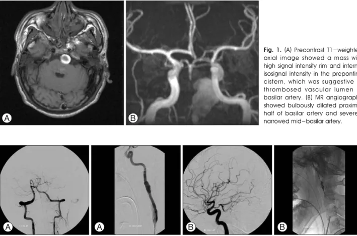
T1-weighted MRI showed mass with high signal intensity rim and internal isosignal intensity in the prepontine cistern, which was suggestive of thrombosed vascular lumen of
Coexisting Dissecting Aneurysms of the Internal Carotid and
Basilar Arteries Following Flexion Injury Case Report
-1Department of Neurosurgery, Gangnam Severance Hospital, Yonsei University, College of Medicine, 2Department of Radiology, Gangnam Severance Hospital, Yonsei University, College of MedicineBong Ju Moon,MD
1∙Chang Ki Hong,MD
1∙Sang Hyun Suh,MD
2Jung Yong Ahn,MD
1∙Jin Yang Joo,MD
1ABSTRACT
Traumatic or sponataneous arterial dissections have been well recognized at the cervical portion of the internal carotid artery and extracranial vertebral artery as an important cause of stroke, especially in young and middle-aged patients. Multiple arterial dissections following craniocervical injury are exceedingly rare. We describe a patient with brain stem infarction caused by basilar occlusion secondary to basilar artery dissection, associated with left ICA dissecting aneurysm after following minor craniocervical trauma without known underlying arteriopathy. (Kor J Cerebrovascular Surgery 11(2):81-4, 2009)
KEY WORDS : Arterial dissection∙Internal carotid artery∙Basilar artery.
논문접수일 : 2009년05월 20일 심사완료일 : 2009년06월 21일
교신저자 : Chang Ki Hong MD, Department of Neurosurgery, Gangnam Severance Hospital 146-92, Dogok-dong, Kangnam-gu Seoul, 135-720, Republic of Korea
전화 : (02) 2019-3390�전송 : (02) 3461-9229 E-mail : [email protected]
basilar artery (Fig. 1A). T2-weighted MRI revealed high-signal change in dorsal pons and upper medulla, and the anterior inferior cerebellar artery (AICA) territory of cerebellum, suggestive of recent infarction, and mass effect on the brain stem was prominent. T1-weighted gadolinium-enhanced MRI showed a distinct enhancement at the same region of the BA occlusion. There is no signal change on supratentorial brain parenchyma. MR angiography showed bulbously dilated proximal half of BA and severely narrowed mid-BA (Fig. 1B). Cerebral angiograms showed complete occlusion at just distal to the confluence site (Fig. 2A), and irregularly fusiform aneurysmal dilatation of cervical portion of the left ICA, consistent with dissection (Fig. 2B). A carotid angiogram showed retrograde contrast filling into the distal half of the BA with preserved patency of posterior cerebral arteries and superior cerebellar arteries
(Fig. 2C). Therefore, BA dissection was treated conservatively with antiplatelet medications and ICA dissection was planned for stent placement.
On day 7, stents deployment for left ICA dissection was successfully achieved. First stent (Precise, Cordis Co, 6mm × 40mm) was introduced into the dissecting aneurysm. After stent placement, angiography showed persistent filling of aneurysm, but the speed of contrast filling was delayed. Then second stent (Precise, Cordis Co, 6mm × 40mm) was deployed in overlapping fashion. The final carotid angiograms showed minimal contrast filling of aneurysm (Fig. 2D). The postoperative course was uneventful. On day 11, the patient was discharged with antiplatelet medications and transferred to military hospital for rehabilitation.
At a 17-month follow-up examination, left-sided hemiparesis was much improved but mild dysarthria still
Coexisting Dissecting Aneurysms of the Internal Carotid and Basilar Arteries Following Flexion Injury Case Report
-82 Kor J Cerebrovascular Surgery 11(2):81-4, 2009
Fig. 2. (A) Vertebral angiogram showed complete occlusion of the basilar artery at just distal to the confluence site. (B) Angiogram of
the left internal carotid artery showed irregularly fusiform aneurysmal dilatation of cervical portion of the left internal carotid artery, consistent with dissection. (C) Angiogram of the right internal carotid artery showed retrograde contrast filling into the distal half of the basilar artery with preserved patency of posterior cerebral arteries and superior cerebellar arteries. Distal half of the basilar artery was not opacified. (D) Angiogram after placement of double overlapping stents covering dissected segment showed persistent contrast filling in the dissecting pseudoaneurysm.
Fig. 1. (A) Precontrast T1-weighted
axial image showed a mass with high signal intensity rim and internal isosignal intensity in the prepontine cistern, which was suggestive of thrombosed vascular lumen of basilar artery. (B) MR angiography showed bulbously dilated proximal half of basilar artery and severely narrowed mid-basilar artery.
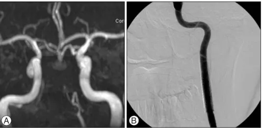
remained, but MRI showed further shrinkage of the BA dissecting aneurysm without evidence of recanalization of the occluded BA (Fig. 3A). Vertebral angiograms showed no recanalization of the occluded BA and left carotid angiograms revealed well-restored luminal configuration of left ICA without contrast filling of aneurysm (Fig. 3B).
Discussion
Arterial dissection typically occurs in young adults, with
an annual incidence of 2.6 per 100,000.3,9)It is usually a
consequence of trauma, either major trauma such as car accident with whiplash injury or more frequently, minor or even ‘trivial’. A concomitant traumatic factor in the pathogenesis of dissection is possible through internal mechanical forces exerted on the neck or head by unusual positions or rapid head turning. For example, a wide spectrum of sport and fitness activities, chiropractic manipulation, violent coughing or nose-blowing can precede arterial dissection. In our case, a sustained hyperflexion status might give rise to arterial dissection.
In multiple dissections, even in the setting of known minor trauma, an underlying collagen vascular disease should be suspected. ICA dissection has been associated with heritable connective tissue disorders such as Ehlers-Danlos syndrome type IV and Marfan’s syndrome, suggesting that a defect in a single extracellular matrix component may be responsible for a more fragile vessel wall which under certain conditions
could give rise to a dissection.7,9) Because of frequent mild
phenotype expression, the primary underlying heritable connective tissue disorder is often diagnosed only after the
occurrence of dissection.5)
Our patient’s sudden neurological deterioration was associated with brain stem infarction caused by BA and its branch occlusions secondary to BA dissection. Definitive diagnosis depends mainly on demonstration of intramural
hematoma.2) MRI can directly demonstrate an intramural
hematoma, the signal intensity of which varies with the age of the hematoma. MRI and angiography are modalities that complement each other, and thus we consider both to be necessary for accurate diagnosis.
Although the ICA dissecting aneurysm was asymptomatic, stents implantation was achieved to prevent extension of dissection and thromboembolic complications. BA occlusion due to BA dissection initially was characterized by a severe neurological impairment. A therapeutic strategy for unruptured BA dissection has not been established, however, anticoagulation therapy for intracranial dissecting aneurysm has not been widely advocated because it may aggravate the
risk of rupture.4) Sequential radiological studies revealing
neither subsequent ischemic insult nor aneurysmal dilatation justified conservative treatment for BA dissection as previous reports, which revealed spontaneous healing of the
BA dissections in case of unruptured BA dissection.6) The
indications of treatment for ischemic BA dissections would be limited to the following two situations : 1) A persistent pseudolumen indicating a risk of future ischemic insults and 2) aneurysmal growth occurring as a sequela of dissection indicating a risk of future hemorrhage. In our patient, neither subsequent ischemic insult nor aneurysmal dilatation was observed during the 17-month follow-up period. Therefore, non-surgical treatment appeared to be justified. Furthermore,
Bong Ju Moon∙Chang Ki Hong∙Sang Hyun Suh∙Jung Yong Ahn∙Jin Yang Joo
83 Kor J Cerebrovascular Surgery 11(2):81-4, 2009
Fig. 3. (A) Follow-up MR angiography
showed significantly shrunken thrombosed aneurysm at proximal basilar artery. (B) At a 17-month follow-up, carotid angiogram of the left side revealed well-restored luminal configuration of left internal carotid artery without contrast filling of aneurysm.
Coexisting Dissecting Aneurysms of the Internal Carotid and Basilar Arteries Following Flexion Injury Case Report
-84 Kor J Cerebrovascular Surgery 11(2):81-4, 2009
a rich collateral circulation developed, and spontaneous healing of the dissected pseudolumen was demonstrated. REFERENCES
11) Eachempati SR, Sebastian MW, Reed RL, 2nd: Posttraumatic
bilateral carotid artery and right vertebral artery dissections in a patient with fibromuscular dysplasia: case report and review of the literature. J Trauma 44:406-9, 1998
12) Kitanaka C, Tanaka J, Kuwahara M, Teraoka A: Magnetic
resonance imaging study of intracranial vertebrobasilar artery dissections. Stroke 25:571-5, 1994
13) Leys D, Lucas C, Gobert M, Deklunder G, Pruvo JP: Cervical
artery dissections. Eur Neurol 37:3-12, 1997
14) Maeda K, Usui M, Tsutsumi K, Iijima A: Spontaneous
occlusion of a giant basilar tip aneurysm and a basilar artery due to the dissection of both structures: case report. Surg
Neurol 48:606-9, 1997
15) Mayer SA, Rubin BS, Starman BJ, Byers PH: Spontaneous
multivessel cervical artery dissection in a patient with a substitution of alanine for glycine (G13A) in the alpha 1 (I) chain of type I collagen. Neurology 47:552-6, 1996
16) Nakatomi H, Nagata K, Kawamoto S, Furusho JI: Basilar
artery occlusion due to spontaneous basilar artery dissection in a child. Acta Neurochir (Wien) 141:99-104, 1999
17) North KN, Whiteman DA, Pepin MG, Byers PH:
Cerebrovascular complications in Ehlers-Danlos syndrome type IV. Ann Neurol 38:960-4, 1995
18) Schievink WI: Spontaneous dissection of the carotid and
vertebral arteries. N Engl J Med 344:898-906, 2001
19) Schievink WI, Bjornsson J, Piepgras DG: Coexistence of
fibromuscular dysplasia and cystic medial necrosis in a patient with Marfan’s syndrome and bilateral carotid artery dissections. Stroke 25:2492-6, 1994