31
Immune Network 서 론
대장암(colorectal cancer)은 서구사회에서는 두 번째로 흔한 암이며 국내에서도 현재 남녀 모두 4대 암에 포함 될 정도로 발생률이 매우 현저하게 증가하고 있다. 5%
전후의 대장암은 유전인자에 의해 발생하지만 식생활의
급격한 서구화, 특히 동물성지방이나 단백질의 과다섭 취 등의 환경인자가 대장암의 발생에 더 많은 영향을 미 치는 것으로 알려져 있다(1-3). 대장암 환자의 경우 절제 수술과 방사선 치료의 개발과 새로운 항암 요법에 의해 생존율이 증가하였지만 여전히 수술 환자의 50%에서 재 발이 일어난다. 따라서 다른 종류의 암과 마찬가지로 대 장암에 대한 새로운 치료수단으로 signal-transduction inhibitor, vaccine, antivascular drug, gene therapy 등을 이 용한 치료법이 개발되고 있다. 특히 gene therapy는 최근 에 human genome project에서 얻어진 많은 정보를 이용 하여 주목을 받고 있다. Gene therapy의 예로는 gene
HHD Mice를 이용한 대장암세포유래 펩타이드 특이적 CD8
+T 세포의 입양전이
1한동대학교 생명과학부, 2AIDS-retrovirus Department, Antiviral Cellular Unit, Pasteur Institute, Paris, France, 3Department of Immunology, The Weizmann Institute of Science, Rehovot 76100, Israe
정헌순
1․안인숙
1․도형기
1․Francois A. Lemonnier
2․Boaz Tirosh
3․Esther Tzehoval
3․Ezra Vadai
3․Lea Eisenbach
3․도명술
1Adoptive Transfer of Colon Cancer Derived Peptide-specific CD8
+T Cells in HHD Mice
Hun-Soon Jung
1, In-Sook Ahn
1, Hyung-ki Do
1, Francois A. Lemonnier
2, Boaz Tirosh
3, Esther Tzehoval
3, Ezra Vadai
3, Lea Eisenbach
3and Myoung-Sool Do
11
School of Life and Food Sciences, Handong Global University, Pohang, Korea,
2AIDS-retrovirus Department, Antiviral Cellular Unit, Pasteur Institute, Paris, France,
3Department of Immunology, The Weizmann Institute of Science, Rehovot 76100, Israel
ABSTRACT
Background: 1-8D gene is a member of human 1-8 interferon inducible gene family and is shown to be overexpressed in fresh colon cancer tissues. Three peptides 1-6, 3-5 and 3-7 derived from 1-8D gene were shown to have immunogenicity against colon cancer. Methods: To study tumor immunotherapy of these peptides we established an adoptive transfer model. Db-/-×β2 microglobulin (β2m) null mice transgenic for a chimeric HLA-A2.1/Db-β2m single chain (HHD mice) were immunized with irradiated peptide-loaded RMA-S/HHD/B7.1 transfectants. Spleens were removed after last immunization, and splenocytes were re-stimulated in vitro. Lymphocytes from vaccinated HHD mice were transferred together with IL-2 to the tumor bearing nude mice that were challenged S.C. with the HCT/HHD/B7 colon carcinoma cell line that was found to grow in these mice. Results: Peptide 3-5 was found to be highly effective in CTL activity. Adoptively transferred anti-peptide 3-5 cytolytic T lymphocytes caused signi- ficant retardation in tumor growth. Conclusion: This study shows that peptide 3-5 can be the most effective candidate for the vaccine of adoptive immunotherapy against colon cancer. (Immune Network 2004;4(1):31-37)
Key Words: 1-8Dgene, HHD mice, colon caner, cytotoxic T lymphocyte, adoptive transfer
책임저자:도명술, 한동대학교 생명과학과
ꂕ 791-708, 경북 포항시 북구 흥해읍 남송리 3번지 Tel: 054-260-1301, Fax: 054-261-1306
E-mail: msdo@handong.edu
본 연구는 과기부의 한이생명공학기반조성사업(M6-0223-01-0003) 에 의하여 지원을 받음.
replacement, virus-directed enzyme-prodrug therapy, im- mune manipulation, virotherapy가 있다(4). 대장암 치료에 대한 immunogenetic strategy는 lymphoproliferative cyto- kine과 종양항원(tumor-associated antigen)을 이용하여 암 에 대한 면역원성(immunogenicity)을 증가시키는 것이다.
하지만 지금까지 대장암의 종양항원으로는 Carcinoemb- ryonic antigen (CEA), Ep-CAM 등만이 알려진 상태이다.
Zhang L, et al (1997)은 같은 환자로부터 얻은 정상 대 장 조직과 대장암 조직을 비교하여 정상 대장보다 5배 더 많이 발현되는 26개의 유전자를 얻었다(5). 이 Data를 이용하여 최근에 HLA-A2.1과 결합하는 500개의 추정적 인 종양 항원 펩타이드를 합성하였다. 여기서 면역학적 으로 관련 있는 MHC Class I restricted 펩타이드를 구별 하기 위하여 HHD마우스를 이용하였다. Humanized mu- rine 모델인 HHD마우스는 murine MHC가 knockout되었 고 대신 HLA-A2.1가 들어가서 single chain의 human MHC를 가지고 있다. 따라서 이 쥐는 종양항원의 HLA- A2.1 restricted epitopes에 대하여 HLA-A2.1 restricted CTL response를 나타낼 수 있으며 각 펩타이드들의 anti- tumor efficacy를 보여 줄 수 있다(6,7). 이 쥐를 이용하여 실험한 결과 이 중 7개의 펩타이드가 면역원성(immu- nogenic)이 있었고 그 중에서도 세 개의 펩타이드(1-6, 3-5, 3-7)가 가장 면역원성이 높았다.1 이 세 개의 펩타이 드는 모두 휴먼 1-8D 유전자부터 나온 것이며 이는 바로 1-8 interferon inducible gene군 중의 하나이다.1 휴먼 1-8 유전자 군은 인터페론 type I (αβ)과 II(γ)에 의해 유도 되어 만들어진다. 이 군에는 1-8U, 1-8D, 9-27 유전자가 있으며, 이들은 하나의 사람 genomic DNA 단편에서 분 리되었다(8). 세 개의 유전자는 120 bp의 동일한 염기서 열을 가지고 있으며 coding region의 대부분에서도 매우 높은 유사성을 가지고 있으며 인트론의 위치도 모두 동 일하다. 또한 아미노산의 배열과 펩타이드의 구조에서 도 서로 매우 유사성이 높게 나타났다. 하지만, 인터페론 에 대한 각각의 유전자들의 활성은 다양한 interferon- inducible factor와 constitutive factor와 interferon- stim- ulable response elements (ISREs)의 상호 반응 정도에 따 라 차이점이 나타난다. 1-8 유전자의 기능적 역할에 대 하여서는 지금까지 잘 알려져 있지 않다.
특별히 본 연구에서는 대장암에 대한 면역학적 치료 의 가능성을 알아보기 위하여 입양 전이(adoptive tran- sfer) 방법을 사용하였다. 1-8D 유전자로 부터 유래된 세 가지 펩타이드(1-6, 3-5, 3-7)를 HHD 마우스에 주사한 후 비장을 제거하여 lymphocyte를 얻은 후 in vitro에서 각 펩타이드와 anti-CD3 Ab와 IL-2를 함께 넣어 재자극시켰 다. 그 후 CTL assay를 이용하여 각 펩타이드들에 특이
1. 논문투고 중
적인 CTL 생산을 확인하고 각 CTL을 HCT 대장암세포 주가 자라고 있는 누드마우스에 입양전이시켰다. 본 연 구의 결과로 3-5펩타이드가 입양전이 치료에서 대장암에 대한 가장 좋은 펩타이드 백신 후보물질로 나타났다.
재료 및 방법
마우스. 대장암 종양항원에 특이한 CTL생산을 위하여 각 그룹당 8∼10마리씩의 12주 된 암컷 HHD 마우스 (H-2Db-/-×β2m-/- double knockout, HLA-A2.1/Db-β 2m monochain 형질전환)를 준비하였다. 또한 입양전이 실험 을 위하여 각 그룹당 10마리씩의 10주 된 암컷
CD1nude/nude (CD1nu/nu)-누드 마우스가 이스라엘의 와이즈
만 연구소에서 specific pathogen free (SPF) 조건하에 사 육되었다.
암 세포주. HCT세포는 HLA class I molecules이 발현되 지 않는 휴먼 대장암 세포주이며 10% FCS와 1% sodium pyruvate와 40μg/ml gentamicin이 들어간 DMEM medium 에서 배양하였다. HCT/HHD클론은 HCT세포에 HHD유 전자를 전이시킨 것이고 HCT/HHD/B7.1클론은 HCT/
HHD에 murine B7.1유전자를 전이시켜 얻은 것이다.
RMA-S세포는 C57BL/6마우스에서 기원된 TAP-2 defic- ient lymphoma이며 10% FCS와 antibiotics가 들어간 RPMI 1640 medium에서 배양하였다. RMA-S/HHD/B7.1 클론은 RMA-S세포에 HHD와murine B7.1 유전자를 전이 시켜 얻은 것이다. EL4세포는 C57BL/6N 마우스에서 기 원된 lymphoma이며 EL4/HHD는 EL4세포에 HHD 유전 자를 전이한 것이다.
RMA-S/HHD/B7에 펩타이드 첨가. PBS로 TAP-2 defic- ient RMA-S/HHD/B7를 세 번 씻은 후에 세포 표면에 HHD monochain을 안정적으로 발현시키기 위하여 26oC 에서 하룻밤 동안 배양하였다. 합성시킨 각 펩타이드 1-6, 3-5, 3-7, PAP (30μg peptide/ml)을 5×10⁵ 세포에 첨 가시킨 후 Opti-MEM (Life Technologies Inc.)배지에서 26oC, 2시간 동안 배양시킨 후 37oC에서 2∼3시간 동안 배양하였다.
1-6 펩타이드의 아미노산 서열은 EMLKEEQEV이고, 3-5펩타이드의 경우는 LILGIFMTI이고, 3-7펩타이드는 KCLNIWALI이며 PAP펩타이드는 ILLWQPIPV이다.
HHD마우스 면역. 먼저 2×10⁶ HCT/HHD/B7.1과 각 펩 타이드가 로딩된 RMA-S/HHD/B7.1 (4×10⁶/ml)을 irrad- iation (5000 rad)시켰다. 양성 대조군인 HCT/HHD/B7.1과 각 펩타이드가 로딩된 RMA-S/HHD/B7.1을 일주일 간격 으로 세 번에 걸쳐 각각 다른 그룹의 HHD 마우스에 접 종(i.p.)하였다. 마지막 접종 후 10일이 지난 뒤 비장을 제거하여 비장세포를 얻었다. In vitro에서 irradiation (5000 rad)과 mitomycin C (80μg/ml)를 처리한 107 cells/
ml HCT/HHD/B7.1을 비장세포와 함께 4일 동안 37oC,
5% CO2 배양기에서 배양시킴으로써 재자극시키는 데 사용하였다. 마찬가지로 Opti-MEM배지에서 비장세포에 각 펩타이드(1-6, 3-5, 3-7, PAP) 50μg/ml를 넣어 4일 동 안 37oC, 5% CO2 배양기에서 재자극을 시켜서 활성세포 로 만들었다. 재자극된 비장세포들은 10% FCS, 2 mM glutamine, combined antibiotics, 1 mM sodium pyruvate, 25 mM HEPES, 5×10⁻⁵ M β-mercaptoethanol, 1% nonessen- tial amino acids가 들어있는 RPMI-HEPES배지에서 배양 되었다. 살아있는 활성세포들은 Lympholyte-M gradient 에 의해 분리되었으며, 35S-methionine으로 표지시킨 표 적세포와 함께 다른 비율로 섞어 부유시킨 뒤 cytolytic assays를 하였다. 표적세포는 HCT/HHD와 HCT/HHD/
B7.1과 EL4/HHD가 사용되었다. 표적세포 EL4/HHD의 경우엔 각 펩타이드(50μg /ml)를 첨가하여 37oC에서 4시 간 동안 배양하였다. % specific lysis는 다음 공식으로 구 하였다.
Count - Spontenous
%Specific lysis= ꠏꠏꠏꠏꠏꠏꠏꠏꠏꠏꠏꠏꠏꠏꠏꠏꠏꠏꠏꠏꠏꠏꠏꠏ ×100 Total - Spontenous
FACS analysis. Cytolytic assays 이후 활성세포에 anti- CD3 Ab와 IL-2 (100 unit)를 첨가하여 배양시킨 뒤 CTL (Cytotoxic T Lymphocyte)의 생산을 FACS (Fluorescence- activated cell sorter)를 이용하여 확인하였다. FITC- labeled anti CD4, FITC-labeled anti CD8a, FITC-labeled mouse IgG2b의 단일 클론 항체들을 사용하였다. 5-10×
106의 활성세포들을 0.5% BSA와 0.1% Na-Azide가 들어 있는 PBS용액(0.5% BSA-0.1% azide-PBS)에 현탁 후 단 일 클론 항체를 넣고 4oC에서 30분간 배양하였다. 0.5%
BSA-0.1% azide-PBS용액 3 ml을 넣은 후에 1,000 rpm, 4oC, 10분간 원심 분리한 뒤 상층액을 제거하였다. 0.1%
azide가 들어있는 PBS용액 0.5 ml을 넣은 후 FACS를 사 용하여 분석하였다.
입양전이 실험. 각 그룹당 10마리씩의 CD1nu/nu-누드마 우스를 irradiation (400 R)을 시킨 후 CD1nu/nu-누드마우스 에 5×10⁶ HCT/HHD/B7.1 세포를 접종(S.C.)하여 마우스 에서 암이 성장하도록 하였다. 일주일 후 암세포를 가지 고 있는 마우스에 11×106의 각 펩타이드에 특이적인 CTL과 대장암 세포주(HCT/HHD/B7.1)에 특이적인 CTL 을 암 주위에 주사하였다. 음성 대조군에는 PBS를 주사 하였다. 그 후 IL-2 (1,000 unit)를 하루에 두 번씩 일주일 동안 암 주위에 주사하였다. 일주일에 두 번씩 암의 지름 을 측정하면서 암의 성장을 관찰하였다.
결 과
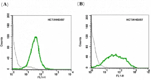
HCT는 HLA Class I을 발현하지 않는 사람의 대장암 세포주이며 1-8D 유전자를 발현하는 세포주이다. 이에 입양전이(adoptive transfer)연구를 위해 HHD와 murine B7.1을 발현하는 HCT/HHD/B7.1 transfectant를 만들어 안 정적인 발현을 조사하였다(Fig. 1). Fig. 1의 (A)는 HHD 유전자의 발현을, (B)에서는 murine B 7.1의 발현을 FACS를 사용하여 분석한 결과이다. HCT 대장암 세포주 에서 HHD 유전자와 murine B 7.1유전자가 안정적으로 발현 되는 것을 확인하였으며 이 세포를 앞으로의 실험 에서 계속적으로 사용하였다.
TAP-2 deficient RMA-S/HHD/B7.1세포에 1-8D유전자에 서 유래된 세 가지 펩타이드인 1-6, 3-5, 3-7 그리고 PAP 펩타이드를 첨가하여 배양하고 RMA-S/HHD/B7.1의 HLA와 각 epitope이 안정적으로 결합하도록 하였다.
PAP는 전립선 암의 특이적인 종양 항원 펩타이드인데 본 실험에서는 음성 대조군으로 사용되었다.
HCT/HHD/B7.1 세포와 peptide-HLA complex를 형성한 RMA-S/HHD/B7.1 세포를 가지고 각 그룹의 HHD 마우 스의 복강에 접종시켰다. 비장에서 얻은 lymphocyte를 in
vitro에서 HCT/HHD/B7.1 세포와 각 펩타이드로 재자극
시킨 후 cytolytic assay에 이용하였다. Cytolytic assay에서Figure 1. HCT/HHD/B7.1 cells highly express HHD and B7.1.
Cells were stained with FITC con- jugated anti-HLA Ab (A), purified anti-mouse CD80 (B7.1) and FITC conjugated goat anti-rat IgG (B) and analyzed by flow cytometry.
표적세포는 35S-methionine으로 표지시킨 HCT/HHD와 HCT/HHD/B7.1와 각 펩타이드로 자극시킨 EL4/HHD가 사용되었다.
세 개의 1-8D에서 유래된 펩타이드 중 3-5 펩타이드에 의해 만들어진 CTL이 다른 펩타이드에 비해 매우 강한 CTL 활성을 보여주었다(Fig. 2). 3-5 펩타이드로 자극시 킨 표적세포 EL4/HHD에 대하여 Effector/Target 100:1 에서 90%에 가까운 세포상해를 보여주었고 12.5:1의 낮은 비율에서도 60%의 세포상해를 보여주었다.
1-6 펩타이드에 특이적인 CTL의 경우는 약한 세포 상 해 활성을 보여주었다. 하지만 3-7 펩타이드에 특이적인 CTL은 세포 상해 활성을 보여주지 않았다.
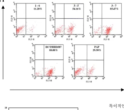
다음으로 1-8D 유전자에서 유래된 각 펩타이드에 의 해 만들어진 lymphocyte가 immunotherapeutic potential을 가지고 있는지 알아보기 위하여 입양 전이 모델을 사용 하였다. HHD 마우스로부터 얻은 각 펩타이드에 대한 비 장세포를 cytolytic assay 이후 in vitro에서 anti-CD3 Ab를 코팅시킨 plate에서 IL-2를 첨가하여 T 세포를 활성화시 킨 후 FACS analysis를 이용하여 CD8+ T cell의 percen- tage를 결정하였다(Fig. 3). 각 펩타이드에 대한 CD8+ T
cell의 분포를 조사하여 입양전이를 위한 CTL의 양을 11
×106 CD8+/mouse로 동일하게 계산하였다.
피하주사로 HCT/HHD/B7.1 세포를 접종하여 암이 유 발된 CD1nu/nu-누드마우스에 각 펩타이드에 특이적인 CTL (11×106 CD8+/mouse)과 HCT/HHD/B7.1에 특이적 인 CTL (11×106 CD8+/mouse)을 각 해당 그룹의 누드마 우스의 암세포 주변에 주사하여 CTL을 입양전이시켰고 대조군에게는 PBS를 사용하였다. 일주일 동안 하루에 두 차례씩 IL-2 (1,000 unit)를 암세포 주변에 주사하였으 며 40일 동안 일주일에 두 번씩 암세포의 지름을 측정함 으로써 암세포의 성장을 관찰하였다(Fig. 4).
실험의 결과로 1-8D 유전자에서 유래된 세 가지 펩타 이드 중 3-5 펩타이드에 의해 야기된 특이적인 CTL이
in vivo에서 확실히 암의 성장을 억제시켰다. 양성 대조
군으로 사용된 HCT/HHD/B7.1에 의해 야기된 특이적인 CTL의 경우 암의 크기가 가장 작았다. 그러나 다른 펩타 이드 1-6, 3-7에 의해 야기된 특이적인 CTL을 주사한 그 룹의 경우는 PBS만을 주사한 그룹과 동일하게 암의 성 장을 억제하지 못하였으며 암의 크기도 거의 차이가 나 지 않았다. 또한 PBS와 IL2를 주사한 그룹에서도 마찬가 Figure 2. 1-8D peptides can mount CTL responses. HHD mice were immunized with HCT/HHD/B7.1 and with peptide loaded on RMA-S/HHD/B7.1. In vitro cytolytic assay were performed using EL4/HHD, HCT/HHD and HCT/HHD/B7.1 as a target. CTL assay were performed at a different E:T ratio. (A) anti-1-6 peptide specific CTL effector and X, HCT/HHD/B7.1 target, and ▲, HCT/HHD target, and ◆, EL4/HHD target pulsed with 1-6 peptide, and ■, EL4/ HHD target pulsed with PAP peptide. (B) anti-3-5 peptide specific CTL effector and X, HCT/HHD/B7.1 target, and ▲, HCT/HHD target, and ◆, EL4/HHD target pulsed with 3-5 peptide, and ■, EL4/ HHD target pulsed with PAP peptide. (C) anti-3-7 peptide specific CTL effector and X, HCT/HHD/B7.1 target, and ▲, HCT/HHD target, and ◆, EL4/HHD target pulsed with 3-7 peptide, and ■, EL4/ HHD target pulsed with PAP peptide. (D) anti-HCT/HHD/B7.1 specific CTL effector and X, HCT/HHD/B7.1 target, and ▲, HCT/HHD target, and ■, EL4/HHD target pulsed with PAP peptide.0 20 40 60 80 100
100:1 50:1 25:1 12.5:1
(A) 1-6
0 20 40 60 80 100
100:1 50:1 25:1 12.5:1
(B) 3-5
0 2 0 4 0 6 0 8 0 1 0 0
1 0 0 :1 5 0 :1 2 5 :1 1 2 .5 :1
(C) 3-7
0 20 40 60 80 100
100:1 50:1 25:1 12.5:1
(D) HCT/HHD/B7 0
20 40 60 80 100
100:1 50:1 25:1 12.5:1
(B) 3-5
0 2 0 4 0 6 0 8 0 1 0 0
1 0 0 :1 5 0 :1 2 5 :1 1 2 .5 :1
(C) 3-7
0 20 40 60 80 100
100:1 50:1 25:1 12.5:1
(D) HCT/HHD/B7
Effector : Target Ratio
% S pecif ic L ysis
지로 암의 성장을 억제하지 못하였다.
고 찰
입양 전이를 이용한 면역학적 치료는 in vitro에서 생 산된 종양항원에 대한 allogenic CTL을 암세포를 가지고 있는 호스트에 주입하여 암을 퇴화시키고자 하는 것이 다. 대부분의 경우에 CTL의 활성을 증가시키기 위하여 adjuvants로서 IL-2(9)와 IFN-α(10)와 IFN-β(11)와 inte- rleukin-6(11,12) 등을 함께 사용하며 PBMC와 TIL이 용해 성 세포의 소스로서 사용된다. 하지만 melanoma와 달리 carcinoma의 경우 in vitro에서 PBMC로부터 CTL을 얻기 가 어렵다는 기술적인 문제들이 있다. 이러한 문제점에 대한 대체적인 전략으로 또한 대장암처럼 종양항원과
특이적인 CTL클론이 거의 알려지지 않은 경우에 면역 학적인 치료 전략으로 HHD 마우스를 이용한 입양 전이 모델이 이용될 수 있다.
HHD 마우스의 HLA-A2.1 single chain 구조는 HLA- A2.1의 α1, α2 도메인과 H-2Db의 α3 transmembrane과 cytoplasmic 도메인으로 구성되어 있으며 linker에 의해 human β2m cDNA가 연결되어 있다. 따라서 펩타이드를 이용한 백신개발을 위하여 새로운 HLA-A2.1 restricted CTL epitope을 밝혀내기에 유용한 humanized murine 모 델이다. 최근에 HHD 마우스를 이용한 이러한 전략이 바 이러스 항원과 종양 항원으로부터 CTL epitope를 밝혀내 기에 효과적이라는 것이 증명되었다(13-19).
본 실험에서도 HHD 마우스를 이용하여 대장암의 종 양 항원과 관련된 HLA-A2.1 restricted CTL response를 유 도하기 위해 HCT 대장암 세포주에 HHD와 costimulatory B7.1을 전이시킨 HCT/HHD/B7.1을 표적 암세포로 이용 하였다. B7.1 (CD80)에 의한 CTL표면의 CD28수용체 자 극은 항 세포사멸 Bcl유전자의 발현(20)과 Interleukin-2 의 생산(21,22)을 통하여 항원 특이적인 T세포의 생존과 증식을 활성화시킨다. 따라서 HCT/HHD/B7.1세포는 HHD 마우스에서 HCT/HHD 또는 HCT 세포보다 더 강 한 CTL response를 보여줄 수 있다. 그러나 누드 마우스 를 HCT/HHD/B7.1 세포로 접종하여 암 성장을 보았을 때 HCT/HHD를 접종한 누드 마우스와 비슷한 성장을 보 여 주어(data not shown) 입양전이 실험을 위하여 HCT/
HHD/B7.1 세포를 사용할 수 있었다. 그러나 B7.1의 발현 으로 인한 다른 관련성을 제하기 위하여 HCT/HHD 세포 를 누드마우스에 접종한 입양 전이 실험이 앞으로 더 요 구된다. 또한 대장암에 대한 잠재적인 종양 항원의 특징 Figure 3. CTLs were restimulated with anti-CD3 Ab and IL2 in vitro. Cells were stained with FITC conjugated anti-CD4 Ab, FITC conjugated anti-CD8 Ab and FITC conjugated mouse IgG2b. Cells were selected by gating on live cells using PI.
The percentages of CD8+ were deter- mined.
CD8 PI
CD8 PI
Figure 4. Nude mice were challenged S.C. with HCT/HHD/
B7.1 cells. Lymphocytes from vaccinated HHD mice were transferred together with IL-2 to the tumor bearing nude mice and the growth of the tumor was monitored twice a week.
을 가지고 있는 1-8D 유전자로부터 유래된 세 가지 펩타 이드(1-6, 3-5, 3-7)가 대장암에 대하여 CTL response를 일 으킬 수 있는지 알기 위하여 RMA-S/HHD/B7.1 세포에 첨가하여 HHD 마우스에 주사하였다. 3-5 펩타이드에 의 해 만들어진 CTL이 표적세포 EL4/HHD에서 매우 효율 이 높은 세포 상해 활성을 일으킴을 cytolytic assay를 통 해 알 수 있었다(Fig. 2). 더 나아가 입양 전이 모델을 세 워 3-5 펩타이드에 의해 만들어진 CTL이 누드 마우스에 서 대장암의 성장을 억제함을 보여 주었다(Fig. 4). 이는
in vitro에서 가장 강한 활성을 보여준 3-5 펩타이드에 의
해 만들어진 CTL이 cytolytic assay 이후 anti-CD3 Ab와 IL-2의 자극에 의해 CTL이 더욱 완전하게 활성화되어 invivo 실험에서도 역시 강한 활성을 나타냄을 보여준다.
하지만 나머지 펩타이드들의 경우는 HHD 마우스에서 CTL response를 충분히 유도하지 못하였고 in vitro의 재 자극에 의해서도 충분한 활성화가 일어나지 않음을 알 수 있다.
본 실험에서 항원전달시스템(antigen delivery system) 으로 TAP-2 deficient RMA-S/HHD/B7.1을 이용하였다.
하지만 최근에는 면역학적 치료에서 항원에 대한 운반 체로 professional APC인 dendritic cell (DC)에 더욱 관심 이 증가하고 있다. 외부의 항원과 펩타이드로 자극시킨 mature DC가 항종양 면역 반응을 효과적으로 향상시킨 다는 것이 증명되었다(23-25). 최근에는 사람과 동물연 구에서 종양항원으로 자극시킨 mature DC가 효과적으로 CTL response를 일으키고 암의 성장을 억제함을 보여주 었다(26-29). 따라서 본 실험에서도 HHD마우스의 골수 로부터 얻어진 autologous DC에 1-8D 유전자로부터 유래 된 각 펩타이드를 첨가한 후 HHD마우스에 주사함으로 더욱 더 효과적인 CTL활성과 암의 성장 억제를 기대해 볼 수 있다.
앞서 언급하였듯이 휴먼 1-8 유전자 군에는 1-8U, 1-8D, 9-27 유전자가 있으며 인터페론에 위해 유도되며 서로 상동성이 매우 높다. 9-27은 integral membrane protein으로 세포의 성장 조절과 관련되어 있다(30). 1-8U 는 대장암과 관련된 colitis와 ulcerative colitis의 점막에서 발현되었다(31). p53 negative 세포주에서 1-8D와 9-27 발 현이 radiation에 의해 interferon independent mechanism으 로 유도되었다(32). 또한 1-8 유전자 군의 발현이 ang- iotensin II 처리에 의해 human adrenocortical carcinoma cell line에서 유도되어 다른 신호전달 경로가 있음을 보 여주었다(33). 하지만 그 외 1-8D 유전자에 대하여 지금 까지 알려진 정보가 거의 없다. 이러한 때에 본 연구는 대장암에 대한 잠재적인 종양 항원으로 1-8D 펩타이드 (1-6, 3-5, 3-7)의 특성을 알고자 실험하였고 특히 3-5 펩 타이드에 의해 만들어진 CTL이 in vitro와 in vivo에서 대 장암 세포주와 대장암에 대하여 강한 면역 반응을 나타
냄을 보여주었다. 이러한 결과는 대장암에서 1-8D 유전 자, 특히 3-5 펩타이드가 immunodominant 종양 항원 역 할을 한다는 것을 보여주는 것이다. 따라서 대장암의 면 역특이 요법으로 대장암 특이 항원 즉, 1-8D 유전자의 3-5 펩타이드에 의해 유래된 CTL을 통한 항암치료 효과 의 가능성을 보여주고 있다.
참 고 문 헌
1. Fearon ER, Vogelstein B: A genetic model for colorectal tumor- igenesis. Cell 61;759-767, 1990
2. Bodmer WF, Bailey CJ, Bodmer J, Bussey HJ, Ellis A, Gorman P, Lucibello FC, Murday VA, Rider SH, Scambler P, et al:
Localization of the gene for familial adenomatous polyposis on chromosome 5. Nature 328;614-616, 1987
3. Cottrell S, Bicknell D, Kaklamanis L, Bodmer WF: Molecular analysis of APC mutations in familial adenomatous polyposis and sporadic colon carcinomas. Lancet 340;626-630, 1992
4. Kerr D: Clinical development of gene therapy for colorectal can- cer. Nat Rev Cancer 3;615-622, 2003
5. Zhang L, Zhou W, Velculescu VE, Kern SE, Hruban RH, Hamilton SR, Vogelstein B, Kinzler KW: Gene expression profiles in normal and cancer cells. Science 276;1268-1272, 1997 6. Pascolo S, Bervas N, Ure JM, Smith AG, Lemonnier FA,
Perarnau B: HLA-A2.1-restricted education and cytolytic activity of CD8(+) T lymphocytes from beta2 microglobulin (beta2m) HLA-A2.1 monochain transgenic H-2Db beta2m double knockout mice. J Exp Med 185;2043-2051, 1997
7. Firat H, Garcia-Pons F, Tourdot S, Pascolo S, Scardino A, Garcia Z, Michel ML, Jack RW, Jung G, Kosmatopoulos K, Mateo L, Suhrbier A, Lemonnier FA, Langlade-Demoyen P: H-2 class I knockout, HLA-A2.1-transgenic mice: a versatile animal model for preclinical evaluation of antitumor immunotherapeutic stra- tegies. Eur J Immunol 29;3112-3121, 1999
8. Lewin AR, Reid LE, McMahon M, Stark GR, Kerr IM: Molecular analysis of a human interferon-inducible gene family. Eur J Biochem 199;417-423, 1991
9. Schmidt-Wolf IG, Finke S, Trojaneck B, Denkena A, Lefterova P, Schwella N, Heuft HG, Prange G, Korte M, Takeya M, Dorbic T, Neubauer A, Wittig B, Huhn D: Phase I clinical study applying autologous immunological effector cells transfected with the interleukin-2 gene in patients with metastatic renal cancer, colorectal cancer and lymphoma. Br J Cancer 81;1009-1016, 1999
10. Mahvi DM, Madsen JA, Witt PL, Sondel PM: Interferon alpha enhances expression of TAG-72 and carcinoembryonic antigen in patients with primary colorectal cancer. Cancer Immunol Immunother 40;311-314, 1995
11. Dansky-Ullmann C, Salgaller M, Adams S, Schlom J, Greiner JW:
Synergistic effects of IL-6 and IFN-gamma on carcinoembryonic antigen (CEA) and HLA expression by human colorectal carc- inoma cells: role for endogenous IFN-beta. Cytokine 7;118-129, 1995
12. Tsang KY, Kashmiri SV, Qi CF, Nieroda C, Calvo B, De Filippi R, Greiner JW, Primus FJ, Schlom J: Transfer of the IL-6 gene into a human colorectal carcinoma cell line and consequent enhancement of tumor antigen expression. Immunol Lett 36;179-185, 1993
13. Firat H, Tourdot S, Ureta-Vidal A, Scardino A, Suhrbier A, Buseyne F, Riviere Y, Danos O, Michel ML, Kosmatopoulos K, Lemonnier FA: Design of a polyepitope construct for the induction of HLA-A0201-restricted HIV 1-specific CTL re- sponses using HLA-A*0201 transgenic, H-2 class I KO mice.
Eur. J. Immunol 10;3064-3074, 2001
14. Carmon L, Bobilev-Priel I, Brenner B, Bobilev D, Paz A, Bar-Haim E, Tirosh B, Klein T, Fridkin M, Lemonnier F, Tzehoval E, Eisenbach L: Characterization of novel breast carcinoma-associated BA46-derived peptides in HLA-A2.1/D (b)-beta2m transgenic mice. J.Clin. Invest 110;453-462, 2002 15. Graff-Dubois S, Faure O, Gross DA, Alves P, Scardino A,
Chouaib S, Lemonnier FA, Kosmatopoulos K: Generation of CTL recognizing an HLA-A*0201-restricted epitope shared by MAGEA1,-A2, -A3, -A4, -A6, -A10, and -A12 tumor antigens:
implication in a broad-spectrum tumor immunotherapy. J. Im- munol 169;575-580, 2002
16. Scardino A, Gross DA, Alves P, Schultze JL, Graff-Dubois S, Faure O, Tourdot S, Chouaib S, Nadler LM, Lemonnier FA, Vonderheide RH, Cardoso AA, Kosmatopoulos K: HER-2/neu and hTERT cryptic epitopes as novel targets for broad spectrum tumor immunotherapy. J. Immunol 168;5900-5906, 2002 17. Passoni L, Scardino A, Bertazzoli C, Gallo B, Coluccia AM,
Lemonnier FA, Kosmatopoulos K, Gambacorti-Passerini C:
ALK as a novel lymphoma-associated tumor antigen: identifica- tion of 2 HLA-A2.1-restricted CD8+ T-cell epitopes. Blood 99;2100-2106, 2002
18. Scardino A, Alves P, Gross DA, Tourdot S, Graff-Dubois S, Angevin E, Firat H, Chouaib S, Lemonnier F, Nadler LM, Cardoso AA, Kosmatopoulos K: Identification of HER-2/neu immunogenic epitopes presented by renal cell carcinoma and other human epithelial tumors. Eur. J. Immunol 11;3261-3270, 2001
19. Brinster C, Muguet S, Lone YC, Boucreux D, Renard N, Fournillier A, Lemonnier F, Inchauspe G: Different hepatitis C virus nonstructural protein 3 (Ns3)-DNA- expressing vaccines induce in HLA-A2.1 transgenic mice stable cytotoxic T lym- phocytes that target one major epitope. Hepatology 34;1206- 1217, 2001
20. Boise LH, Minn AJ, Noel PJ, June CH, Accavitti MA, Lindsten T, Thompson CB: CD28 costimulation can promote T cell survival by enhancing the expression of Bcl- XL. Immunity 3;87-98, 1995
21. Harding FA, Allison JP: CD28-B7 interactions allow the induc- tion of CD8+ cytotoxic T lymphocytes in the absence of exog- enous help. J Exp Med 177;1791-1796, 1993
22. Malek TR, Yu A, Scibelli P, Lichtenheld MG, Codias EK: Broad programming by IL-2 receptor signaling for extended growth to multiple cytokines and functional maturation of antigen-activated T cells. J Immunol 166;1675-1683, 2001
23. Celluzzi CM, Mayordomo JI, Storkus WJ, Lotze MT, Falo LD
Jr: Peptide-pulsed dendritic cells induce antigen-specific CTL- mediated protective tumor immunity. J Exp Med 183;283-287, 1996
24. Paglia P, Chiodoni C, Rodolfo M, Colombo MP: Murine dendritic cells loaded in vitro with soluble protein prime cytotoxic T lymphocytes against tumor antigen in vivo. J Exp Med 183;
317-322, 1996
25. Young JW, Inaba K: Dendritic cells as adjuvants for class I major histocompatibility complex-restricted antitumor immunity. J Exp Med 183;7-11, 1996
26. Nestle FO, Alijagic S, Gilliet M, Sun Y, Grabbe S, Dummer R, Burg G, Schadendorf D: Vaccination of melanoma patients with peptide- or tumor lysate-pulsed dendritic cells. Nat Med 4;328- 332, 1998
27. Thurner B, Haendle I, Roder C, Dieckmann D, Keikavoussi P, Jonuleit H, Bender A, Maczek C, Schreiner D, von den Driesch P, Brocker EB, Steinman RM, Enk A, Kampgen E, Schuler G:
Vaccination with mage-3A1 peptide-pulsed mature, monocyte- derived dendritic cells expands specific cytotoxic T cells and induces regression of some metastases in advanced stage IV melanoma. J Exp Med 190;1669-1678, 1999
28. Bellone M, Cantarella D, Castiglioni P, Crosti MC, Ronchetti A, Moro M, Garancini MP, Casorati G, Dellabona P: Relevance of the tumor antigen in the validation of three vaccination strategies for melanoma. J Immunol 165;2651-2656, 2000
29. Schreurs MW, Eggert AA, de Boer AJ, Vissers JL, van Hall T, Offringa R, Figdor CG, Adema GJ: Dendritic cells break toler- ance and induce protective immunity against a melanocyte diffe- rentiation antigen in an autologous melanoma model. Cancer Res 60;6995-7001, 2000
30. Deblandre GA, Marinx OP, Evans SS, Majjaj S, Leo O, Caput D, Huez GA, Wathelet MG: Expression cloning of an interfe- ron-inducible 17-kDa membrane protein implicated in the con- trol of cell growth. J Biol Chem 270;23860-23866, 1995 31. Hisamatsu T, Watanabe M, Ogata H, Ezaki T, Hozawa S, Ishii
H, Kanai T, Hibi T: Interferon-inducible gene family 1-8U expression in colitis-associated colon cancer and severely infl- amed mucosa in ulcerative colitis. Cancer Res 59;5927-5931, 1999
32. Clave E, Carosella ED, Gluckman E, Socie G: Radiation- enh- anced expression of interferon-inducible genes in the KG1a primitive hematopoietic cell line. Leukemia 11;114-119, 1997 33. Daido H, Zhou MY, Gomez-Sanchez CE: Angiotensin stimulates
the expression of interferon-inducible genes in H295R cells. Mol Cell Endocrinol 176;21-27, 2001