Introduction
The extraction of third molars is one of the most fre- quently performed procedures in dental clinics, and is necessary when there is insufficient space for eruption of these teeth, when they are poorly positioned, or when there is a risk of formation of cysts or odontogenic tu- mors.1 The appropriate surgical procedure should be de- termined based on findings from preoperative examina-
tions evaluating the morphology of the third molar and its relationships with adjacent structures.2
Conducting an assessment by a radiographic examina- tion allows the evaluation of several factors that are re- lated to the degree of difficulty of the surgical procedure, such as the depth of impaction and the position of the tooth. Identifying these factors can reduce the occurrence of surgical complications.3,4 Some systems classify im- pacted third molars according to their depth of impaction and their position on the mesial-distal axis. The most commonly used classification systems are those proposed by Winter5 and Pell and Gregory.6 Both classifications are based on 2-dimensional images, and do not allow for the evaluation of some positions, such as the buccolingual
Three-dimensional analysis of impacted maxillary third molars: A cone-beam computed tomographic study of the position and depth of impaction
Priscila Ferreira de Andrade1, Jesca Neftali Nogueira Silva1, Bruno Salles Sotto-Maior2, Cleide Gisele Ribeiro3, Karina Lopes Devito4,*, Neuza Maria Souza Picorelli Assis4
1Master’s Program in Dental Clinic, Faculty of Dentistry, Federal University of Juiz de Fora, Juiz de Fora, Minas Gerais, Brazil
2Department of Restorative Dentistry, Faculty of Dentistry, Federal University of Juiz de Fora, Juiz de Fora, Minas Gerais, Brazil
3Faculty of Medical and Health Sciences-SUPREMA, Juiz de Fora, Minas Gerais, Brazil
4Department of Dental Clinic, Faculty of Dentistry, Federal University of Juiz de Fora, Juiz de Fora, Minas Gerais, Brazil
ABSTRACT
Purpose: The classification of impacted maxillary third molars(IMTMs) facilitates interdisciplinary communication and helps estimate the degree of surgical difficulty. Thus, this study aimed to develop a 3-dimensional classification of the position and depth of impaction of IMTMs and to estimate their prevalence with respect to gender and age.
Materials and Methods: This cross-sectional retrospective study analyzed images in sagittal and coronal cone- beam computed tomography(CBCT) sections of 300 maxillary third molars. The proposed classification was based on 3 criteria: buccolingual position(buccal, lingual, or central), mesial-distal position(mesioangular, vertical, or distoangular), and depth of impaction(low, medium, or high). CBCT images of IMTMs were classified, and the associations of the classifications with gender and age were examined using analysis of variance with the Scheffé post-hoc test. To determine the associations among the 3 classifications, the chi-square test was used(P<.05).
Results: No significant association of the classifications with gender was observed. Age showed a significant relationship with depth of impaction(P=.0001) and mesial-distal position(P=.005). The most common positions were buccal(n=222), vertical(n=184), and low(n=124). Significant associations among the 3 tested classifications were observed.
Conclusion: CBCT enabled the evaluation of IMTMs in a 3-dimensional format, and we developed a proposal for a new classification of the position and depth of impaction of IMTMs.(Imaging Sci Dent 2017; 47: 149-55)
KEY WORDS: Molar, Third; Tooth, Impacted; Cone-Beam Computed Tomography
Copyright ⓒ 2017 by Korean Academy of Oral and Maxillofacial Radiology
This is an Open Access article distributed under the terms of the Creative Commons Attribution Non-Commercial License(http://creativecommons.org/licenses/by-nc/3.0) which permits unrestricted non-commercial use, distribution, and reproduction in any medium, provided the original work is properly cited.
Imaging Science in Dentistry·pISSN 2233-7822 eISSN 2233-7830 Received February 1, 2017; Revised June 21, 2017; Accepted June 28, 2017
*Correspondence to : Dr. Karina Lopes Devito
Department of Dental Clinic, Oral Radiology, School of Dentistry, Federal University of Juiz de Fora, Campus Universitário, s/n, Juiz de Fora, MG CEP: 36036-900, Brazil
Tel) 55-32-2102-3851, Fax) 55-32-2101-3851, E-mail) [email protected]
position.
A classification system of the depth of impaction and position of impacted third molars is expected to lead to improved interdisciplinary communication in treatment planning and to help estimate the degree of surgical dif- ficulty of each case. Although panoramic radiography is very helpful in preoperative assessments, it may present limitations, such as distortion and blurring of the image.7
CBCT examinations allow a proper assessment of the 3-dimensional anatomic relationships between the third molar and surrounding tissues.2 At the time of writing, a search of the PubMed database using the descriptors
“maxillary third molar,” “upper third molar,” “wisdom third molar,” “classification,” “position,” “cone beam computed tomography,” “CBCT,” “CT,” “CBVT,” “3D images,” and “three-dimensional reconstruction” revealed no studies that described the depth and angular position of impacted maxillary third molars(IMTMs) based on CBCT images.
Therefore, given the absence of studies classifying the position and the depth of impaction of IMTMs based on CBCT images, the present study proposes a 3-dimension- al classification of the position and depth of impaction of IMTMs and an analysis of their prevalence with respect to gender and age.
Materials and Methods
This was a cross-sectional observational retrospective study analyzing the sagittal and coronal CBCT sections of 300 maxillary third molars from the database of the Ra- diology Clinic, School of Dentistry, Federal University of Juiz de Fora(UFJF). The exams of patients missing any upper molars were excluded. This study was approved by the Research Ethics Committee of the UFJF(opinion num- ber 932.681).
The images analyzed were obtained using an i-CAT scanner(Imaging Sciences International, Hatfield, PA, USA) operating at 120kVp and 3-8mA with 0.25-mm voxels, a 26.9-s rotation time, and a 10-cm field of view.
The images were stored in the Digital Imaging and Com- munications in Medicine format and later reconstructed with CS 3D Imaging software(Carestream Dental, Roch- ester, NY, USA). Linear and angular measurements were made in this program. The assessments were performed by a single examiner with experience in CBCT imaging.
The classification of third molars was developed using CBCT images and was based on 3 criteria: buccolingual position, mesial-distal position based on a modified ver-
sion of the Winter classification,5 and depth of impaction according to a modified version of the Pell and Gregory classification.6
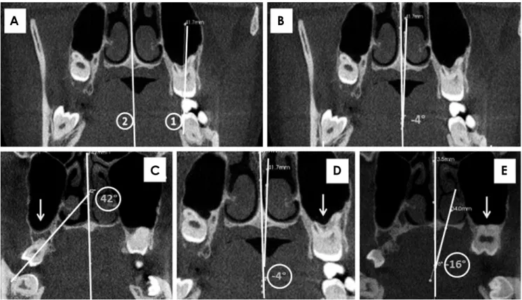
The position of the third molar on the buccolingual axis was assessed in a coronal section. The classification was made according to the angle formed between the refer- ence line(line 1, Fig. 1A) drawn on the third molar long axis(longitudinal axis of the tooth) and the reference line (line 2, Fig. 1A) in the midsagittal plane. To measure the angle formed between the reference lines 1 and 2, line 1 was shifted until it intersected with line 2, and the angle was then measured(Fig. 1B). The maxillary third molars were then classified as buccal when the angle formed was greater than +10°(Fig. 1C), central when the angle formed was between +10° and -10°(Fig. 1D), and lin- gual when it was less than -10°(Fig. 1E).
The classification of the mesial-distal position was made in the sagittal section. The angles formed by the in- tersection of the reference line drawn on the long axis of the third molar(longitudinal axis) and another line pass- ing through the occlusal plane of the maxillary first molar (line tangent to the cusps of the first molar) were ana- lyzed. The alignment was considered distoangular when the angle formed was less than or equal to 75°(Fig. 2A), vertical when the angle formed was between 76° and 104°
(Fig. 2B), and mesioangular when the angle was greater than or equal to 105°(Fig. 2C).
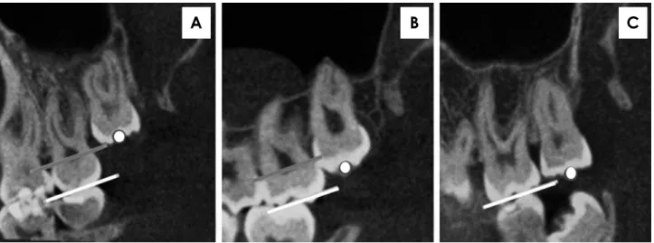
To assess the depth of impaction, the level of the occlu- sal plane of the maxillary third molar was analyzed. The tooth was classified according to the midpoint of its oc- clusal surface in relation to the occlusal plane of the max- illary second molar. The tooth was considered to be high when the midpoint of its occlusal plane was found coro- nally to the cementoenamel of the maxillary second mo- lar(Fig. 3A), medium when the midpoint of the occlusal plane appeared between the cementoenamel junction and the occlusal plane of the maxillary second molar(Fig. 3B), and low when the midpoint of the occlusal plane was found at the same level or caudally to the occlusal plane of the maxillary second molar(Fig. 3C).
Three hundred CBCT images of maxillary third molars were classified, and associations with gender and age were examined using analysis of variance with the Scheffé post-hoc test. To determine the associations among the 3 classifications, the chi-square test was used. Statistical analyses were conducted using SPSS version 13.0(SPSS Inc., Chicago, IL, USA), with a 5% significance level (P<.05).
Results
Among the 300 maxillary third molars, 151(50.3%) teeth were on the right side, and 149(49.7%) teeth were on the left. The age of patients ranged from 18 to 40 years, with a mean of 23.74±4.76 years. Regarding gender, 100 third molars belonged to male patients(33.3%), and 200 third molars belonged to female patients(66.7%).
The distribution of the 300 IMTMs according to the proposed classification is presented in Table 1. Table 2 shows the 3-dimensional classification data distributed by gender and age.
A significant association with age was found for the mesial-distal position(P=.005) because younger individ- uals exhibited an increased prevalence of the distoangular position, while there was a tendency toward verticaliza-
Fig. 1. Coronal cone-beam computed tomography sections. A. Line 1 passing through the long axis of the maxillary third molar and line 2 passing through the midsagittal plane. B. The intersection of lines 1 and 2 and formation of the buccolingual angle. Maxillary third molars (white arrows) classified according to the buccolingual position as buccal(C), central(D), or lingual(E).
A B
C D E
Fig. 2. Sagittal cone-beam computed tomography sections. The mesial-distal position of the maxillary third molar is classified as distoan- gular(A), vertical(B), or mesioangular(C).
A B C
tion with increasing age. In terms of the depth of impac- tion, the correlation analyses demonstrated that younger individuals exhibited a greater tendency toward medium and high depths of impaction, which tended to decrease with age(i.e., the frequency of a low depth of impaction increased; P=.0001).
The associations between gender and each of the clas- sifications of maxillary third molars were also evaluated, and none were significant(buccolingual position: P = .417; mesial-distal position: P=.052; and depth of im- paction: P=.738).
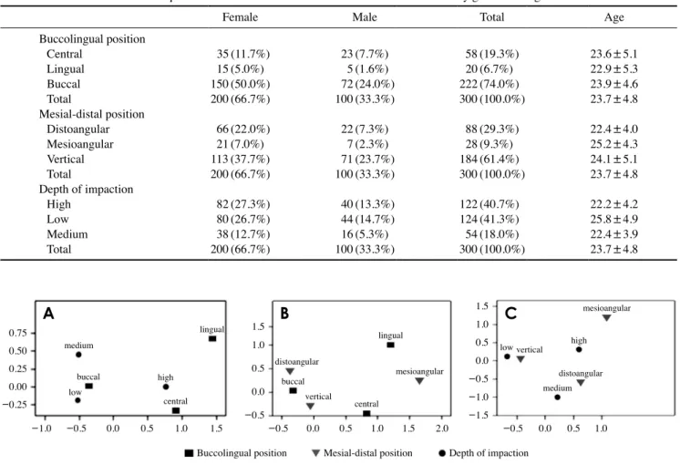
Significant associations were found between each of the classifications(buccolingual position with depth of im- paction, mesial-distal position with buccolingual position,
and mesial-distal position with depth of impaction). Fig- ure 4 illustrates the spatial distribution of the associations between the classifications.
Discussion
To our knowledge, this was the first study to assess the depth and angular position of IMTMs based on CBCT images, and most importantly, this study was the first to determine the buccolingual position of these teeth. The scientific literature on IMTM depth and angular position that has been published in databases(PubMed) contains results based on 2-dimensional images.1,3,4,8-13 Because of the nature of radiographs(i.e., 2-dimensional images),
Table 1. Distribution of third molar positions with respect to buccolingual position, mesial-distal position, and depth of impaction
Distoangular Mesioangular Vertical Total
Central HighLow Medium Lingual
HighLow Medium Buccal
HighLow Medium
8(2.6%) 6(2.0%)
- 2(0.6%) 6(2.0%) 5(1.6%)
- 1(0.3%) 74(24.6%) 3(10.3%) 19(6.3%) 24(8.0%)
12(4.0%) 11(3.6%) 1(0.3%) 6(2.0%)- 6(2.0%)
- 10(3.3%)- 6(2.0%) 4(1.3%)
-
38(12.6%) 23(7.6%) 13(4.3%) 2(0.6%) 8(2.6%) 6(2.0%) 1(0.3%) 1(0.3%) 138(46.0%)
28(9.3%) 86(28.6%)
24(8.0%)
58(19.3%) 40(13.3%) 14(4.6%) 4(1.3%) 20(6.7%) 17(5.6%) 1(0.3%) 2(0.6%) 222(74.0%) 65(21.6%) 109(36.3%) 48(16.0%)
Total 88(29.3%) 28(9.3%) 184(61.3%) 300(100%)
Fig. 3. Sagittal cone-beam computed tomography sections. The level of the occlusal plane of the maxillary third molar is classified as high when the midpoint of the maxillary third molar(white dot) is at or above the cementoenamel junction of the maxillary second molar(gray line)(A), as medium when the midpoint of the maxillary third molar(white dot) is between the occlusal plane of the maxillary second mo- lar(white line) and the cementoenamel junction(gray line)(B), and as low when the midpoint of the maxillary third molar(white dot) is at or below the occlusal plane of the maxillary second molar(white line)(C).
A B C
it has been impossible to determine the buccolingual po- sition. Indeed, none of the previous studies analyzed the buccolingual position of the IMTMs.1,3,4,8-13 Thus, the aims of this study were to present a new classification of the depth and angular position of IMTMs based on CBCT images and to determine the associations of gender, age, and depth with each angular position.
A preoperative CBCT examination is now considered an important assessment tool for planning third molar surgery and for predicting the risks related to surgical in- terventions.2 Some important findings that may affect the result of the removal of an IMTM can be obtained from CBCT images, but not from 2-dimensional images, such as the buccolingual position, the real proximity of the roots of the IMTM to the sinus floor,12 and the relation- ship of the IMTM with the second molar.14
In terms of the buccolingual position, 74.0% of IMTMs had a buccal position, 19.3% a central position, and 6.7%
a lingual position. Because this is a novel classification system, it is not possible to compare these findings with other reports. Neither clinical examinations nor 2-dimen- sional imaging techniques can identify when third molars are impacted at such angulations. Panoramic radiography is considered quite imprecise for determining the amount of overlying bone. Thus, surgical difficulty may be ex- pected when the tooth is in a deeper position inside the bone due to limited access to the surgical site and the need to remove a larger amount of bone. However, a CBCT examination can indicate the position of the tooth, the amount of overlying bone, and its relationships with the surrounding structures. Tomographic examination is thus an aid for diagnosis and therapy because it reveals the an- atomical situation with greater precision. The procedures performed during extraction are more predictable because of the better visualization of the anatomical obstacles and the 3-dimensional position of the third molar.7 Further
Table 2. Absolute and relative frequencies of the 3-dimensional classification of third molars by gender and age
Female Male Total Age
Buccolingual position Central
Lingual Buccal Total
Mesial-distal position Distoangular Mesioangular Vertical Total
Depth of impaction HighLow
Medium Total
35(11.7%) 15(5.0%) 150(50.0%) 200(66.7%) 66(22.0%) 21(7.0%) 113(37.7%) 200(66.7%) 82(27.3%) 80(26.7%) 38(12.7%) 200(66.7%)
23(7.7%) 5(1.6%) 72(24.0%) 100(33.3%) 22(7.3%) 7(2.3%) 71(23.7%) 100(33.3%) 40(13.3%) 44(14.7%) 16(5.3%) 100(33.3%)
58(19.3%) 20(6.7%) 222(74.0%) 300(100.0%)
88(29.3%) 28(9.3%) 184(61.4%) 300(100.0%) 122(40.7%) 124(41.3%) 54(18.0%) 300(100.0%)
23.6±5.1 22.9±5.3 23.9±4.6 23.7±4.8 22.4±4.0 25.2±4.3 24.1±5.1 23.7±4.8 22.2±4.2 25.8±4.9 22.4±3.9 23.7±4.8
Fig. 4. Spatial distribution of the associations between the buccolingual position and the depth of impaction(A), the mesial-distal position and the buccolingual position(B), and the mesial-distal position and the depth of impaction(C). The interpretation must be based on the distance between points; that is, the closer 2 points are, the stronger the association between them.
A B C
0.75 0.50 0.25 0.00 -0.25
1.0 0.5 0.0 -0.5
1.5 1.0 0.5 0.0 -0.5 -1.0 -1.5 1.5
medium
medium
buccal buccal
low
lingual lingual
high
low high
vertical
vertical
central central
distoangular
distoangular mesioangular
mesioangular
Buccolingual position Mesial-distal position Depth of impaction
-1.0 -0.5 0.0 0.5 1.0 1.5 -0.5 0.0 0.5 1.0 1.5 2.0 -0.5 0.0 0.5 1.0
studies should investigate whether buccolingual angula- tion predisposes surgical procedures to complications.
The most frequently observed IMTM position was ver- tical(61.3%), and this finding is consistent with those re- ported by other authors.3,8-12 However, Topkara and Sari1 reported that the distoangular position was the most fre- quent position and that in many studies, the angulation of IMTMs was usually determined based on visual impres- sions according to the Winter classification. Although var- ious authors have cited the Winter classification5 in their studies, the methodology used to evaluate the angles that determine the different positions is quite variable. Ventä et al.15 used the angle between the tooth’s occlusal surface and the occlusal plane of the first and second molar for IMTM classification, and classified teeth as vertical(-10°
to +10°), mesioangular(11° to 70°), distoangular(-11°
to -70°) or horizontal(>70°). In contrast, Sandhu and Kaur13 used the angle between the axis of the second mo- lar and that of the IMTM to classify IMTMs as vertical(0°
to 25°), mesioangular(26° to 75°), horizontal(>75°), or distoangular(<0°). Hassan10 used the angle between the long axis of the third molar and the long axis of the sec- ond molar to classify IMTMs as vertical(-10° to +10°), mesioangular(11° to 79°), horizontal(80° to 100°), dis- toangular(-11° to -79°), or buccolingual(crown over- lapping the root). De Carvalho et al.3 classified angles from 0° to 30° as horizontal, 31° to 60° as mesioangular, 61° to 90° as vertical, and >90° as distoangular. In the present study, the IMTMs were classified as follows: dis- toangular when the angle was ≤75°, vertical when the angle was 76° to 104°, and mesioangular when the angle was ≥105°. To establish these angle ranges, each third molar was classified visually using a modified version of the Winter classification.5 Next, the ranges of the angles were measured.
In our study, the most common IMTM impaction level was low(41.3%). In other studies, this classification has been performed using the nomenclature of class A, which corresponds to the low level; class B, which corresponds to the medium level; and class C, which corresponds to the high level. Our results are comparable to those report- ed by others because the same criteria were used.11,12 Has- san,10 de Carvalho et al.,3 and Topkara and Sari1 reported that the most frequent impaction level was B, which cor- responds to the medium level in our study. Evaluating the depth of impaction of an IMTM is important because depth is considered to be a predictor of surgical difficulty,3 and also serves as an indication of the potential for oroantral perforation.12 These authors stated that radiographs are
not a reliable method for predicting the possibility of oroantral perforation. Therefore, surgeons should obtain reliable tomographic information regarding the depth of impaction when planning to remove such teeth.12
The associations of age with depth of impaction and classification of the mesial-distal position were signifi- cant. In older patients, maxillary third molars tended to be classified as low and in the mesioangular and vertical positions. These associations are important because the incidence of radiographic lesions increases with age, and partially impacted mesioangular third molars exhibit a high incidence of caries or periodontal bone loss in the adjacent second molar.8 Moreover, mesioangular and hor- izontal third molars are more strongly associated with the external root resorption of second molars, and one study reported that a significantly greater number of cases were diagnosed based on CBCT images than panoramic radio- graphs.14 According to Oenning et al.,14 CBCT images provided diagnostic information in the sagittal, axial, and coronal planes without an overlap of structures, whereas panoramic radiographs did not have the third dimension.
According to the results obtained from the tests of the associations between IMTM classifications, strong asso- ciations of the low position with the vertical and buccal positions were observed, and these positions represent the lowest degree of extraction difficulty. Regarding the as- sociation between the buccolingual and the mesial-distal categories, a cyclical distribution was observed, in which all positions were equally closely associated. Position and depth alone may not indicate an extremely difficult surgi- cal procedure, but some combinations of these variables can elicit problems. Therefore, these variables should be evaluated in combination, not in isolation.
Until now, planning of third molar removal surgery was based on mesial-distal position and depth obtained from 2-dimensional images. With CBCT, in addition to these positions, it is possible to assess the buccolingual posi- tion. Two-dimensional radiographs provide information that can be useful for surgical planning; however, only CBCT examinations permit the evaluation of the real sit- uation of the tooth.
This study underscores the role of CBCT in the diag- nosis and treatment planning for the surgical removal of IMTMs. Although the expected surgical difficulty of IMTM removal is low, some cases may present unexpect- ed difficulties. CBCT is the primary tool for identifying variables that predict surgical complications, because it enables a 3-dimensional evaluation, which led to a new proposal for the classification of the position and depth of
impaction of IMTMs. A future study will investigate the relationships of the IMTM classifications with intraopera- tive complications and postoperative morbidity.
References
1. Topkara A, Sari Z. Investigation of third molar impaction in Turkish orthodontic patients: prevalence, depth and angular positions. Eur J Dent 2013; 7(Suppl 1): S94-8.
2. Nakamori K, Tomihara K, Noguchi M. Clinical significance of computed tomography assessment for third molar surgery.
World J Radiol 2014; 6: 417-23.
3. de Carvalho RW, de Araújo Filho RC, do Egito Vasconcelos BC. Assessment of factors associated with surgical difficulty during removal of impacted maxillary third molars. J Oral Maxillofac Surg 2013; 71: 839-45.
4. Carvalho RW, Araújo-Filho RC, Vasconcelos BC. Adverse events during the removal of impacted maxillary third molars.
Int J Oral Maxillofac Surg 2014; 43: 1142-7.
5. Winter GB. Principles of exodontia as applied to the impacted mandibular third molar; a complete treatise on the operative technic with clinical diagnoses and radiographic interpreta- tions. St. Louis: American Medical Book Co.; 1926.
6. Pell GJ, Gregory BT. Impacted third molars: classification and modified technique for removal. Dent Dig 1933; 39: 330-8.
7. Bouquet A, Coudert JL, Bourgeois D, Mazoyer JF, Bossard D.
Contributions of reformatted computed tomography and pan- oramic radiography in the localization of third molars relative to the maxillary sinus. Oral Surg Oral Med Oral Pathol Oral
Radiol Endod 2004; 98: 342-7.
8. Jung YH, Cho BH. Prevalence of missing and impacted third molars in adults aged 25 years and above. Imaging Sci Dent 2013; 43: 219-25.
9. Celikoglu M, Miloglu O, Kazanci F. Frequency of agenesis, impaction, angulation, and related pathologic changes of third molar teeth in orthodontic patients. J Oral Maxillofac Surg 2010; 68: 990-5.
10. Hassan AH. Pattern of third molar impaction in a Saudi popu- lation. Clin Cosmet Investig Dent 2010; 2: 109-13.
11. Hashemipour MA, Tahmasbi-Arashlow M, Fahimi-Hanzaei F.
Incidence of impacted mandibular and maxillary third molars:
a radiographic study in a Southeast Iran population. Med Oral Patol Oral Cir Bucal 2013; 18: e140-5.
12. Lim AA, Wong CW, Allen JC Jr. Maxillary third molar: pat- terns of impaction and their relation to oroantral perforation. J Oral Maxillofac Surg 2012; 70: 1035-9.
13. Sandhu S, Kaur T. Radiographic study of the positional chan- ges and eruption of impacted third molars in young adults of an Asian Indian population. J Oral Maxillofac Surg 2008; 66:
1617-24.
14. Oenning AC, Neves FS, Alencar PN, Prado RF, Groppo FC, Haiter-Neto F. External root resorption of the second molar associated with third molar impaction: comparison of pan- oramic radiography and cone beam computed tomography. J Oral Maxillofac Surg 2014; 72: 1444-55.
15. Ventä I, Turtola L, Ylipaavalniemi P. Radiographic follow-up of impacted third molars from age 20 to 32 years. Int J Oral Maxillofac Surg 2001; 30: 54-7.