Copyright © 2011 by Th e Korean Orthopaedic Association
Th is is an Open Access article distributed under the terms of the Creative Commons Attribution Non-Commercial License (http://creativecommons.org/licenses/by-nc/3.0) which permits unrestricted non-commercial use, distribution, and reproduction in any medium, provided the original work is properly cited.
Clinics in Orthopedic Surgery • pISSN 2005-291X eISSN 2005-4408
Long-term Outcomes of Ulnar Shortening Osteotomy for Idiopathic Ulnar Impaction
Syndrome: At Least 5-Years Follow-up
Goo Hyun Baek, MD, Hyuk Jin Lee, MD, Hyun Sik Gong, MD, Seung Hwan Rhee, MD, Jihyeung Kim, MD, Kang Wook Kim, MD, Bong Young Kong, MD, Won Seok Oh, MD
Department of Orthopedic Surgery, Seoul National University College of Medicine, Seoul, Korea
Received February 10, 2011; Accepted March 9, 2011 Correspondence to: Goo Hyun Baek, MD
Department of Orthopedic Surgery, Seoul National University College of Medicine, 101 Daehak-ro, Jongno-gu, Seoul 110-744, Korea
Tel: +82-2-2072-3787, Fax: +82-2-764-2718 E-mail: [email protected]
Ulnar impaction syndrome can cause medial wrist pain related to excessive weight bearing across the ulnar as- pect of the wrist.1) Idiopathic ulnar impaction syndrome is frequently seen radiographically in patients with static positive ulnar variance or dynamic positive ulnar variance
Background: There have been few outcomes studies with follow-up after performing ulnar shortening osteotomy for ulnar impac- tion syndrome. We investigated the long-term clinical and radiological outcomes of ulnar shortening osteotomy for the treatment of idiopathic ulnar impaction syndrome.
Methods: We retrospectively reviewed 36 patients who had undergone ulnar shortening osteotomy for idiopathic ulnar impaction syndrome for a mean follow-up of 79.1 months (range, 62 to 132 months). The modifi ed Gartland and Werley scores were mea- sured pre- and postoperatively. The radiographic parameters for the assessment of the distal radioulnar joint (DRUJ) as well as the relationship between these radiographic parameters and the clinical and radiological outcomes were determined.
Results: The average modifi ed Gartland and Werley wrist score improved from 65.5 ± 8.1 preoperatively to 93.4 ± 5.8 at the last follow-up visit. The average preoperative ulnar variance of 4.7 ± 2.0 mm was reduced to an average of −0.6 ± 1.4 mm postopera- tively. Osteoarthritic changes of the DRUJ were fi rst seen at 34.8 ± 11.1 months follow-up in 6 of 36 wrists (16.7%). Those who had osteoarthritic changes in the DRUJ had signifi cantly wider preoperative ulnar variance, a longer distal radioulnar distance and a greater length of ulnar shortening, but the wrist scores of the patients who had osteoarthritic changes in the DRUJ were compa- rable to those who did not have osteoarthritic changes in the DRUJ.
Conclusions: The clinical outcomes are satisfactory for even more than 5 years after ulnar shortening osteotomy for treating id- iopathic ulnar impaction syndrome despite the osteoarthritic changes of the DRUJ. The patients who need a larger degree of ulnar shortening may develop DRUJ arthritis.
Keywords: Idiopathic ulnar impaction syndrome, Ulnar shortening osteotomy, Long-term outcome, Osteoarthritis, Distal radioul- nar joint
with wrist pronation and a forceful grip.1,2)
Although ulnar shortening osteotomy has been successfully used for treating idiopathic ulnar impaction syndrome,3-5) several long term studies showed that 28- 38% of the patients had radiographic degenerative changes in the distal radioulnar joint (DRUJ) 2-6 years after this procedure.4,6,7) Koppel et al.6) suggested that such changes are due to joint remodeling and they are unlikely to lead to symptomatic osteoarthritis because these patients were symptom-free. However, Sagerman et al.8) reported that one of their patients who had undergone ulnar shorten-
ing osteotomy developed symptomatic distal radioulnar arthritis. Nishiwaki et al.9) showed that the greater the amount of ulnar shortening, the higher the peak pressure is at the DRUJ, suggesting that excessive pressure at the DRUJ can lead to osteoarthritis of this joint. Furthermore, morphological variations in the DRUJ may infl uence the congruity and the pressure at the DRUJ aft er ulnar short- ening osteotomy.8,10,11)
Th ere have been few reports with regard to the long term outcomes of ulnar shortening osteotomy. Here we hypothesize that the preoperative DRUJ types and short- ening length may aff ect the long term outcomes of ulnar shortening osteotomy. Th us, we evaluated the clinical and radiographic outcomes of ulnar shortening osteotomy in patients who have idiopathic ulnar impaction syndrome with a minimum of 5 years follow-up.
METHODS
Th is study was approved by the Institutional Review Board of Seoul National University Hospital and informed con- sent was obtained from all the enrolled patients.
Demographic and Surgical Data
Thirty six patients with idiopathic ulnar impaction syn- drome were treated with ulnar shortening osteotomy and they were followed for a minimum of five years (range, 62 to 132 months). Th e average duration of postoperative follow-up was 79.1 ± 15.7 months. Th ere were 12 men and 24 women with an average age of 45.8 ± 11.5 years. None of them had a history of wrist or forearm fracture or pre- mature growth arrest.
Th e diagnosis was made by the medical history and physical examination, including the use of a provocative maneuver such as the ulnocarpal stress test,1) and the plain radiographs, which showed static or dynamic positive ulnar variance12) with or without cystic change(s) of the lunate or triquetrum.
Th e inclusion criteria for the diagnosis were 1) the absence of trauma to the forearm and wrist; 2) symptoms of ulnar wrist pain that are aggravated by pronation and ulnar deviation of the wrist with activities such as open- ing a jar, squeezing a wet towel, typing or shift ing gears; 3) a positive provocation test (the ulnocarpal stress test); 4) positive ulnar variance with or without cystic change(s) of the carpus seen on plain X-rays; and 5) a follow-up period of more than 5 years.
Th e exclusion criteria were 1) radiologic evidence of old fracture of the forearm or wrist; 2) radiologic evidence of congenital anomalies such as a Madelung deformity;3)
and 3) less than 5 years follow-up.
A shortening transverse osteotomy was performed to obtain a final ulnar variance between 0 and −1 mm.
Th is was fi xed with a six or seven-hole 3.5 mm standard compression plate (25 cases) or a one-third tubular plate (Stryker Trauma, Selzach, Switzerland) when the ulna was too small for a standard compression plate (11 cases). A postoperative long arm splint was applied for the fi rst two weeks, and this was followed by a removable long arm splint for 4 to 8 weeks.13)
Clinical Evaluations
All 36 wrists of 36 patients were evaluated preoperatively using the Gartland and Werley evaluation scoring system, as modifi ed by Chun and Palmer.5) In this grading system, equal emphasis was placed on the subjective and objec- tive fi ndings and with giving 50 points to these two sets of fi ndings for a maximum score of 100 points. Th e subjec- tive variables were pain, function and motion and the ob- jective measurements were the range of motion, the grip and pinch strength, healing of the osteotomy and the pre- operative and postoperative ulnar variance. Th e result was excellent when the wrist score was 91 to 100, good when it was 81 to 90, fair when it was 66 to 80 and poor when it was 0 to 65. The patients were evaluated postoperatively every two to four weeks until radiologic union was con- fi rmed, and the wrist scores were obtained at six months and once yearly thereaft er.
Radiographic Evaluations
All the radiographic images were digitally acquired using a picture archiving and communication system (PACS, M- view, Infi nitt, Korea) and measurements were done using PACS soft ware. Th e radiographs of the wrist were evaluat- ed by two authors. Intra-observer reliability was evaluated by asking one examiner to repeat all the radiographical assessments aft er a period of 3 weeks. Inter-observer reli- ability was evaluated by asking another author to verify all the radiographical assessments.
Th e anteroposterior images of the wrist were taken with the shoulder in 90 degrees abduction, the elbow in 90 degrees flexion and the forearm in neutral rotation. The lateral images were taken with 0o shoulder abduction, and with the elbow and forearm maintaining the same amount of fl exion and rotation.
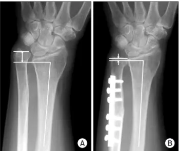
Ulnar variances were measured by the method of perpendiculars, whereby a line is drawn perpendicular to the longitudinal axis of the radius at its distal ulnar aspect, and the distance between this point and the line at the end of the ulna is measured (Fig. 1).14) Dorsal subluxation of
the DRUJ was quantifi ed preoperatively by measuring the radioulnar distance15) on a true lateral radiograph of the wrist, in which the pisoscaphoid distance was less than 3 mm (Fig. 2).
Th e types of the DRUJ have been classifi ed by Tolat et al.16) as follows (Fig. 3).
· Type I: Th e apposing joint surfaces are parallel to the lon- gitudinal axis of the ulna.
· Type II: Th e apposing joint surfaces are oblique (the sig- moid notch angle being positive).
· Type III: The apposing joint surfaces are in a rever se oblique (the sigmoid notch angle being negative).
We defined osteoarthritic changes of the DRUJ as
any osteophyte formation seen around the DRUJ on the plain radiography (Fig. 4).
Statistical Analysis
Th e wrist scores and ulnar variances were analyzed using paired Student’s t-tests. Correlation analyses were per- formed to assess for any associations between the ulnar
Fig. 1. (A) Measurement of ulnar variance (arrow) on the preoperative anteroposterior wrist radiograph. (B) Measurement of ulnar variance (arrows) on the postoperative anteroposterior wrist radiograph.
Fig. 2. (A) The preoperative lateral radiograph showed the measurement of the radioulnar distance (RU). The pisoscaphoid distance (PS) was used to define a true lateral radiograph of the wrist. (B) The postoperative lateral radiograph showed a decrease in the RU.
Fig. 3. Types of the distal radioulnar joint (DRUJ). (A) Type I: The apposing joint surfaces (b & c) are parallel to the long axis of the ulna (a). (B) Type II: The apposing joint surfaces (b & c) are oblique to the long axis of the ulna (a). The sigmoid notch and the ulnar seat angles are positive. (C) Type III:
The apposing joint surfaces (b & c) are reversely oblique to the long axis of the ulna (a). The sigmoid notch and the ulnar seat angles are negative.
variance and the radioulnar distance. Th e Mann-Whitney U-test was also used for comparisons between the group with DRUJ arthritis and those who did not have DRUJ arthritis. Cronbach’s α test was used to measure the intra- and interobserver reliability. A p-value of < 0.05 was con- sidered statistically signifi cant.
RESULTS
Clinical Evaluations
Th e average scores had improved from 65.5 ± 8.1 preop- eratively to 93.4 ± 5.8 at the last follow-up visit (p < 0.001, paired Student’s t-tests). Each component of the wrist score demonstrated statistically significant improvement (Table 1).
There were no nonunions or infections. Two pa- tients developed postoperative refl ex sympathetic dystro- phy, which improved at 9 months and 11 months, respec- tively, after the operation. Both the patients’ wrist scores were favorable at the last follow-up. One patient was diag- nosed with post-motor vehicle accident myofascial pain syndrome aft er surgery, and the fi nal wrist score was fair.
Radiographic Findings
The inter-observer reliabilities of the radiographic mea- surements (the type of DRUJ, the perioperative ulnar vari- ance and the radioulnar distance) were 0.95, 0.84, and 0.86, respectively (Cronbach α coeffi cient value). Th e intra-ob- server reliablities of the radiographic measurements were 0.97, 0.90 and 0.88, respectively. These results indicated that the measurements were reliable, as taken by a single
observer (one of the authors) and they were used in the analyses.
Th e preoperative ulnar variance was 4.7 ± 2.0 mm, and the postoperative ulnar variance was −0.6 ± 1.4 mm (p
< 0.001, the paired Student’s t-test). Th e shortened length was on average 5.3 ± 2.2 mm.
The mean radioulnar distance was 4.8 ± 1.7 mm preoperatively and 2.2 ± 2.2 mm at the fi nal follow-up (p <
0.001, the paired Student’s t-test) (Table 2).
Th e preoperative ulnar variance demonstrated weak positive correlation with the preoperative radioulnar dis- tance (R = 0.395, p = 0.014, Pearson’s correlation), indi- cating the more positive the ulnar variance, the larger the radioulnar distance with the ulnar translating dorsally.
Th e types of DRUJ were as follows.
· Type I: 1 patient (2.8%).
· Type II: 9 patients (25.0%).
· Type III: 26 patients (72.2%).
Osteoarthritis of the DRUJ
Radiographic changes consistent with osteoarthritis of the DRUJ were noted in 6 of 36 wrists (16.7%). Th ese changes were fi rst noted at a mean follow-up period of 34.8 months (range, 22 to 46 months) after surgery. This occurred in one of the type II (1/9) cases and in 5 of the type III (5/26) cases. We analyzed the cases with and without DRUJ osteoarthritis. We compared these two groups using the Mann-Whitney U-test (Table 3).
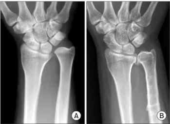
There was no difference in the clinical outcomes Fig. 4. (A) Positive ulnar variance was seen on the preoperative
anteroposterior wrist radiograph. No osteoarthritic changes of the distal radioulnar joint (DRUJ) were seen. (B) Osteoarthritic changes of the DRUJ after a follow-up period of 44 months.
Table 1. The Outcomes According to the Chun and Palmer Wrist Grading System
Preoperative Last follow-up p -value*
Wrist score 65.5 ± 8.1 93.4 ± 5.76 < 0.001 Subjective score 29.3 ± 6.7 45.4 ± 3.7 < 0.001 Objective score 36.2 ± 2.1 48.0 ± 2.6 < 0.001 Values are presented as mean ± SD.
*p using paired Student t-tests.
Table 2. Radiologic Findings
Preoperative Last follow-up p -value*
Ulnar variance (mm) 4.7 ± 2.0 −0.6 ± 1.4 < 0.001 Radioulnar distance (mm) 4.8 ± 1.7 2.2 ± 2.2 < 0.001 Values are presented as mean ± SD.
*p using paired Student t-tests.
between the two groups. Th e patients with osteoarthritic change of the DRUJ did not complain of pain or limita- tion of motion on the last follow-up visit, but one patient showed crepitus on forearm rotation while compressing the DRUJ.
Th ere was no statistically signifi cant diff erence in the wrist scores between the two groups preoperatively and at the last follow-up. Th ere was also no diff erence regardless of age and the follow-up duration.
However, there was a statistically signifi cance diff er- ence with regard to ulnar variance. Th e preoperative ulnar variance in those with osteoarthritis (OA) was 6.7 ± 1.4 mm and that in those patients without OA was 4.3 ± 1.9 mm (p = 0.008).
Th ere was a signifi cant diff erence in the shortening length between the two groups (those with OA, 7.0 ± 2.0 mm; those without OA, 4.8 ± 2.1 mm; p = 0.016).
In terms of the radioulnar distance, there was a sig- nifi cant diff erence between the two groups preoperatively (6.8 ± 2.0 mm in those with OA; 4.5 ± 1.4 mm in those without OA; p = 0.006) but there was no signifi cant diff er- ence postoperatively in those with OA (1.9 ± 3.9 mm) and those without OA (2.2 ± 1.8 mm, p = 0.279).
DISCUSSION
Th is study demonstrates that the clinical outcomes of ul- nar shortening osteotomy are satisfactory for more than 5 years postoperatively, despite the fact that 16% of the pa- tients had radiographic arthritic changes of the DRUJ that
appeared at a mean follow-up of 3 years aft er surgery.
We also found changes in the postoperative radio- graphs when compared to those done preoperatively. Th e radioulnar distance had signifi cantly decreased postopera- tively.
The factors associated with the observed osteoar- thritic changes of the DRUJ were analyzed. Based on the statistical date, we concluded that factors such as the ul- nar variance, the degree of shortening and the radioulnar distance were related to the postoperative osteoarthritic changes.
The clinical outcomes were excellent. The aver- age wrist score had improved from 65.5 to 93.4. Most of the patients were able to use their wrists without pain or discomfort even after 5 years. Our long term data dem- onstrated satisfactory results, which was consistent with previous follow-up reports of a shorter duration.3-5,17)
In our study, type III was the most common type of DRUJ followed by type II and then type I. Tolat et al.16) reported that in patients with a normal DRUJ, type II was the most common (50%) followed by type I (38%) and type III (12%). With regard to the declination angles, Mo- remans et al.17) reported that 8 had less than 85 degrees (similar to type III), 5 had more than 95 degrees (similar to type II), and 15 had between 85 and 95 degrees (simi- lar to type I), which was different from our results. We revealed that type III comprised the majority of cases of DRUJ in the patients with idiopathic ulnar impaction syn- drome among our study cohort. Studies focusing on the diff erent types of DRUJ in an asymptomatic cohort would be helpful.
There was some correlation among the ulnar vari- ance and the distal radioulnar distance. Th ere was a posi- tive correlation between ulnar variance and the distal radioulnar distance, which is consistent with our previous data.3)
We found radiographic changes consistent with osteoarthritis of the DRUJ in 6 of 38 wrists after ulnar shortening osteotomy. Regardless, there were no differ- ences in clinical outcome whether or not OA was present.
There were no significant differences with regard to the wrist scores either. Some authors maintain that problems may occur in the DRUJ aft er ulnar shortening at the last follow-up. Deshmukh et al.10) reported that the the DRUJ was incongruent after ulnar shortening in a cadaveric study. Miura et al.18) reported that the peak DRUJ pres- sure had increased in normal cadavers when the ulna was shortened, and especially in the type I and type III joints.
Tolat et al.16) suggested that the type III joint is diffi cult to shorten beyond 2 to 3 mm without disturbing the confi gu- Table 3. Comparison of Two Groups with/without Distal Radioulnar
Osteoarthritis (OA)
OA No OA p -value*
Age (yr) 46.2 ± 6.5 45.8 ± 12.3 1.000
Follow-up duration (mo) 70.2 ± 11.2 77.6 ± 17.1 0.356 Wrist score
Preoperative 58.0 ± 2.8 66.3 ± 8.1 0.190 Postoperative 91.3 ± 5.3 93.6 ± 5.9 0.610 Ulnar variance (mm)
Preoperative 6.7 ± 1.4 4.3 ± 1.9 0.008 Postoperative 0.5 ± 0.8 −0.77 ± 1.4 0.030 Shortening amount (mm) 7.0 ± 2.0 4.8 ± 2.1 0.016 Values are presented as mean ± SD.
*p-value using Mann-Whitney U-test.
ration of the DRUJ. Minami and Kato4) reported that the postoperative occurrence of degenerative changes of the DRUJ was seen in 28% of wrists aft er ulnar shortening os- teotomy. Koppel et al.6) also reported radiological changes of the DRUJ were seen in 38% of wrists aft er ulnar short- ening osteotomy. Nakamura et al.7) reported that postop- erative osteoarthrosis of the DRUJ was seen in 12 wrists (38%), but only 2 cases were symptomatic.
We conclude that the preoperative ulnar variance, the radioulnar distance and the degree of shortening could be attributed to the osteoarthritic changes of the DRUJ.
An increased distal radioulnar distance is seen with greater degrees of dorsal subluxation. In our study, the radioulnar distance was decreased on the postoperative ra- diographs, implying a decrease in dorsal subluxation aft er ulnar shortening. Th is should result in increased pressure at the DRUJ.
Thus, both the ulnar variance and the radioulnar distance should decrease aft er ulnar shortening osteotomy, and the resulting increase in pressure at the DRUJ may contribute to the arthritic changes of the DRUJ found in this study.
There are several limitations to be noted in this study. First, there were no CTs or MRIs performed in our study. Th erefore, we could not analyze the transverse sec- tion of the DRUJ or accurately measure the ulnar and sig-
moid notch angles or evaluate the ligament status around the ulnar wrist such as the triangular fi brocartilage com- plex. Second, functional evaluation such as DASH (disabil- ities of the arm, shoulder and hand) was not performed.
The general medical condition of the upper extremities was not assessed. Th ird, the number of cases with arthritic changes of the DRUJ was small and the number of type III DRUJs (which already harbor factors that increase the risk for subsequent problems at the DRUJ)8,16) comprised the majority of all the cases, as well as those with OA.
In conclusion, the clinical outcomes are satisfactory for even more than 5 years after ulnar shortening oste- otomy for treating idiopathic ulnar impaction syndrome despite of the osteoarthritic changes of the DRUJ. Howev- er, we must assess the osteoarthritic changes of the DRUJ with longer term follow-up.
CONFLICT OF INTEREST
No potential confl ict of interest relevant to this article was reported.
ACKNOWLEDGEMENTS
Th is study was supported by the Korean Human Technol- ogy Research Foundation (KOHTERF-2009-04).
REFERENCES
triangular fi brocartilage complex tear. J Jpn Soc Surg Hand.
2002;19(3):219-24.
8. Sagerman SD, Zogby RG, Palmer AK, Werner FW, Fortino MD. Relative articular inclination of the distal radioulnar joint: a radiographic study. J Hand Surg Am. 1995;20(4):597- 601.
9. Nishiwaki M, Nakamura T, Nakao Y, Nagura T, Toyama Y.
Ulnar shortening eff ect on distal radioulnar joint stability: a biomechanical study. J Hand Surg Am. 2005;30(4):719-26.
10. Deshmukh SC, Shanahan D, Coulthard D. Distal radioulnar joint incongruity aft er shortening of the ulna. J Hand Surg Br. 2000;25(5):434-8.
11. Tolat AR, Stanley JK, Trail IA. A cadaveric study of the anat- omy and stability of the distal radioulnar joint in the coronal and transverse planes. J Hand Surg Br. 1996;21(5):587-94.
12. Jung JM, Baek GH, Kim JH, Lee YH, Chung MS. Changes in ulnar variance in relation to forearm rotation and grip. J Bone Joint Surg Br. 2001;83(7):1029-33.
13. Baek GH, Chung MS, Lee YH, Gong HS, Lee S, Kim HH.
1. Friedman SL, Palmer AK, Short WH, Levinsohn EM, Hal- perin LS. The change in ulnar variance with grip. J Hand Surg Am. 1993;18(4):713-6.
2. Palmer AK, Glisson RR, Werner FW. Ulnar variance deter- mination. J Hand Surg Am. 1982;7(4):376-9.
3. Baek GH, Chung MS, Lee YH, Gong HS, Lee S, Kim HH.
Ulnar shortening osteotomy in idiopathic ulnar impaction syndrome. J Bone Joint Surg Am. 2005;87(12):2649-54.
4. Minami A, Kato H. Ulnar shortening for triangular fibro- cartilage complex tears associated with ulnar positive vari- ance. J Hand Surg Am. 1998;23(5):904-8.
5. Chun S, Palmer AK. The ulnar impaction syndrome:
follow-up of ulnar shortening osteotomy. J Hand Surg Am.
1993;18(1):46-53.
6. Koppel M, Hargreaves IC, Herbert TJ. Ulnar shortening os- teotomy for ulnar carpal instability and ulnar carpal impac- tion. J Hand Surg Br. 1997;22(4):451-6.
7. Nakamura T, Takayama S, Kikuchi Y, Osada N, Ono H.
Long-term results of the ulnar shortening procedure for
Ulnar shortening osteotomy in idiopathic ulnar impac- tion syndrome: surgical technique. J Bone Joint Surg Am.
2006;88 Suppl 1 Pt 2:212-20.
14. Coleman DA, Blair WF, Shurr D. Resection of the radial head for fracture of the radial head: long-term follow-up of seventeen cases. J Bone Joint Surg Am. 1987;69(3):385-92.
15. Nakamura R, Horii E, Imaeda T, Tsunoda K, Nakao E. Distal radioulnar joint subluxation and dislocation diagnosed by standard roentgenography. Skeletal Radiol. 1995;24(2):91-4.
16. Tolat AR, Sanderson PL, De Smet L, Stanley JK. Th e gym-
nast's wrist: acquired positive ulnar variance following chronic epiphyseal injury. J Hand Surg Br. 1992;17(6):678- 81.
17. Moermans A, Degreef I, De Smet L. Ulnar shortening os- teotomy for ulnar ideopathic impaction syndrome. Scand J Plast Reconstr Surg Hand Surg. 2007;41(6):310-4.
18. Miura T, Firoozbakhsh K, Cheema T, Moneim MS, Ed- munds M, Meltzer S. Dynamic effects of joint-leveling procedure on pressure at the distal radioulnar joint. J Hand Surg Am. 2005;30(4):711-8.