www.jkfas.org pISSN 1738-3757 eISSN 2288-8551 J Korean Foot Ankle Soc 2015;19(1):7-10 http://dx.doi.org/10.14193/jkfas.2015.19.1.7
OLT의 치료 방법은 다양하나 최근에는 관절경적으로 골연골 병변 을 변연 절제하고 골수를 자극하는 미세 골절술을 가장 선호한다.
개방성 절개 없이 작은 전방 삽입구 두 개나 추가 삽입구 한 개를 이용해 수술을 진행하고 수술 후 회복이 빠르며, 상처의 합병증도 적은 장점이 있다.4)
OLT에서 시행한 미세 골절술의 성공 여부는 수술 후 발목의 통 증 감소와 환자의 일상생활로의 복귀 및 발목의 불안감 등이 사라 져야 한다. 미세 골절술을 받은 후 약 70%의 환자가 평균 4개월에 서 수술 전의 생활로 복귀가 가능하다고 한다.4,5) 그러나, 많게는 30%의 환자가 일차 미세 골절술을 시행받고 단기적 혹은 장기적으 로 통증이 생겨 만족하지 못한 결과를 보인다. 일차 미세 골절술의 불만족한 경우를 문헌을 통해 고찰하고, 그 환자에 대한 2차 치료 로 어떤 술기를 적용할지 알아보고자 한다. 특히 자가 연골세포 이 식술(autologous chondrocyte implantation, ACI)의 효과를 중심으 로 다른 술기와 비교하고, 그 유용성에 대해서 알아보고자 하였다.
서 론
거골에 발생하는 골연골 병변(osteochondral lesion of talus, OLT)은 발목의 급성 또는 만성 통증을 유발하며, 발목의 불안감 혹은 간헐적 잠김 증상 등이 발생할 수 있다. 발목 내 병변의 위치 와 관계없이 통증의 위치가 다르거나 모호해서 진단이 힘든 경우 가 많다. OLT는 일반적으로 Berndt와 Harty1)의 방사선학적 분류에 낭종을 동반한 경우, 손상된 연골 주위 골부종 소견이 있는 경우를 추가하여 사용하고 있다. 병기 I, IIA의 경우 보존적 치료를 우선적 으로 하며, 이를 실패한 경우와 병기 IIB, III, IV와 같이 보존적 치 료의 실패 가능성이 높은 경우는 수술적 치료를 1차로 시행한다.2,3)
Received January 14, 2015 Revised January 29, 2015 Accepted February 9, 2015 Corresponding Author: Jin Su Kim
Surgery of Foot and Ankle, Eulji General Hospital, Eulji University College of Medicine, 68 Hangeulbiseok-ro, Nowon-gu, Seoul 139-711, Korea Tel: 82-2-970-8561, Fax: 82-2-974-8259, E-mail: [email protected] Financial support: None.
Conflict of interest: None.
Review Article
This is an Open Access article distributed under the terms of the Creative Commons Attribution Non-Commercial License (http://creativecommons.org/licenses/CC
by-nc/3.0) which permits unrestricted non-commercial use, distribution, and reproduction in any medium, provided the original work is properly cited.
Copyright 2015 Korean Foot and Ankle Society. All rights reserved.ⓒ
Microfracture as a reparative strategy is the treatment of choice for an osteochondral lesion of talus. Although the results of microfrac- ture are generally excellent, at least 30% of patients who received microfracture have acute or chronic ankle pain with several or un- known causes. The most important factor for unsatisfactory outcome after microfracture is the size of the lesion. For failed osteochon- dral lesion of talus, the second options are autologous osteochondral graft, autologous chondrocyte implantation, or re-microfracture. In this article, we present the autologous chondrocyte implantation as a second procedure for failed microfracture and compare its clinical outcome with other methods based on a literature review.
Key Words: Osteochondral lesion of talus, Microfracture, Autologous chondrocyte implantation
거골 골연골 병변에 대한 미세 골절술 실패 후 2차 치료로서 자가연골 세포 이식술
김진수
을지대학교 의과대학 을지병원 족부족관절정형외과
Autologous Chondrocyte Implantation as a Secondary Procedure after Failed Microfracture for Osteochondral Lesion of Talus
Jin Su Kim
Surgery of Foot and Ankle, Eulji General Hospital, Eulji University College of Medicine, Seoul, Korea
8 Vol. 19 No. 1, March 2015
다. 특히 손상된 연골의 크기가 150 mm2인 경우에는 미세 골절술 시행 후 80%에서 예후가 불량하므로6) 수술 전 환자에게 그 정보 를 제공해야 하며, 미세 골절술 이외의 이식 수술방식(replacement strategies)이 필요함을 알려야 한다.
미세 골절술의 기술적 실패요인으로는 골연골 병변의 불충분한 변연절제, 연골하골을 완전히 노출시키지 않은 경우나 골수가 나 오지 않는 얕은 천공 등을 들고 있다.13) 과도한 변연절제로 연골 병 변을 수술 전보다 크게 만들거나, 다발성 천공 시 견갑부(shoulder lesion)의 파손 혹은 연골하골의 추가 손상이 발생하는 경우도 있 다. 일차 미세 골절술 시 연골의 손상 부위가 확인되지 않아 부드 러워진 부위(softening)만을 천공하는 경우도 있으나, 많은 경우 OLT의 골연골 피판의 손상 부위(flap) 후방이 떨어져(posterior de- tachment) 전방 관절경으로 확인하기 힘든 경우가 있어 주의를 요 한다. 나이가 어린 환자에서 발생한 내측의 큰 OLT의 경우 연골은 손상이 없는 경우도 있기 때문에, 정상으로 보이는 연골 부위를 제 거하고 OLT를 완전히 변연절제를 시행하는 것이 좋다. 혹은 고정 을 시행하여 골편을 유합시켜야 한다.
이차적 수술의 방법으로는 ACI, 자가 골연골 이식술(osteo- chondral autograft transfer system, OATS), 동종 골연골 이식술 (osteochondral allograft), 콜라겐 유도 연골자가 재생법(autolo- gous collagen-induced chondrogenesis), 금속 임플란트(metallic implant), 연소자기 동종 연골절편 이식술(particulated juvenile articular cartilage allograft), 혈소판 풍부혈장(platelet-rich plasma), 골수세포 이식술(mesenchymal stem cell) 등의 다양한 방법이 존 재한다.11) 이 중 국내에서 가능한 이식수술 방법은 OATS, ACI, 콜 라겐 유도 연골자가 재생법, 혈소판 풍부혈장 이식술 및 골수세포 이식술이나, 보험의 적용을 받을 수 있는 것은 OLT의 크기가 150
본 론
1. 미세 골절술 실패의 정의
미세 골절술의 실패라 함은 1차로 수술 후, 추시관찰상 Ameri- can Orthopaedic Foot and Ankle Society (AOFAS) 발목-후족부점 수(ankle-hindfoot score)가 80점 이하인 경우를 말한다.6) 일반적으 로 1차 수술 후 6개월 이상 경과하고,7) 발목의 통증 및 증상이 수술 전과 동일하거나 더 증상의 빈도가 잦아진 경우를 말하며, 자각통 증이 매일 있다면 수술의 효과를 보지 못하였다고 할 것이다. 연골 손상은 회복되는 기간이 긴 특성상 수술 술기의 문제가 없다면 1년 이상 경과관찰을 하는 것이 좋을 것이다. 임상적 점수가 1년 이상 의 추시관찰에서 증상이 조금씩 회복되는 경우도 있기 때문이다.
여러 저자들의 보고에 따르면 1차 수술 후 통증이 지속되어 2차 수 술까지 걸리는 기간은 평균 16∼21개월로 알려져 있다.7,8)
2. 동반 손상의 확인
OLT의 증상은 모호한 경우가 많으며, 발목 염좌 혹은 골절 등의 외상력이 있다면 동반된 손상이 있을 가능성이 높다.9) 발목의 만 성 불안정성(내측, 외측 및 경비 간 이개)이 교정되지 않았거나 골 성 충돌증후군 혹은 연부조직 충돌에 의한 관절 내 병변이 있는 경 우로, 이는 동시 혹은 선교정이 이루어져야 한다. 1차 수술에서 이 런 병변이 동시에 교정되지 않았다면 추시관찰 중 회복되지 않는 통증의 원인을 감별해내기가 어렵다. 또한, 이런 동반 손상에 의한 증상과 증상 없이 발견되는 OLT가 동시에 존재할 수 있기 때문에 주의해야 한다.9) 수술 전, 수술 후 추시상 자기공명영상(magnetic resonance image, MRI)이 연골의 상태와 주변 관절 상태에 대해 많 은 정보를 제공한다. T2 강조영상에서 연골 손상 부위 주변으로 골 수부종을 보이거나, 충돌이 일어나는 부위의 강조영상과 활액낭염 이 있는 경우 통증과 연관성이 높다.10)
발목관절의 관절염이 이미 발생한 경우나 발목의 내반변형이 동 반된 경우에는 연골재생을 위한 수술보다 발목관절의 변형을 교정 하거나 관절염의 치료하는 방향으로 치료 목적을 수정해야 할 것 이다.
3. OLT에 대한 미세 골절술의 실패 원인 및 2차 치료의 결정 미세 골절술은 비수술적 치료에 반응하지 않는 OLT에 가장 많이 사용하는 골수 자극 치료방식(reparative strategy)이다. 연골하부에 구멍을 내어서 섬유연골을 재생시키기 위한 목적으로 시행하며, 단기적으로 임상적 회복을 보이나 장기적으로 갈수록 재생된 섬유 연골의 견고성이 떨어져 유효성이 감소하게 된다.11) OLT의 크기, 연골하 낭종, 위치, 환자의 나이, 신체질량지수(body mass index), 증상 지속 기간, 외상력 및 동반된 손상이 미세 골절술 이후 그 결 과를 불량하게 만들 수 있지만, Loveday 등12)의 체계적 고찰에 의 하면 OLT의 크기만이 치료방침을 결정할 수 있는 유일한 인자이
Failed OLT after microfracture
POD <1 year
Treatment plan change Yes
Wait and see
No
MRI follow-up/further evaluation Osteoarthritis and
or deformity?
Yes Arthroscopic re-microfracture
No No
Yes Technical error or concomitant lesions?
Size of OLT >100 mm ?2
Arthroscopic autologous chondrocyte implantation
Figure 1. Our flowchart for treatment of failed osteochondral lesion of talus. OLT: osteochondral lesion of talus, POD: postoperative days, MRI: magnetic resonance image.
www.jkfas.org 9 Jin Su Kim. ACI for Failed Microfracture
맞추기가 어렵고, 공여부에서 채취 및 이식하는 과정이 기술적으 로 어려운 단점이 있다. 공여해야 하는 사이즈가 큰 경우에는 무릎 연골을 주로 사용하여, 무릎 기능을 감소시킬 위험성이 있다. ACI 의 경우는 작은 크기의 연골편 혹은 손상된 거골의 연골편을 이용 하여 세포배양을 시행함으로써 OATS에 비해 타 관절에 입히는 해 가 적고, 최근에는 관절경적 ACI를 이용하여 내과절골술을 요하 지 않게 되어 성적이 더욱 우수해졌다.18) Niemeyer 등19)이 보고한 메타 분석에서도 ACI가 OLT에 효과적인 방법으로 보고되고 있다.
Minas 등20)이 무릎에서 시행한 연구에서는 일차적으로 미세 골절 술을 시행하고 실패한 OLT에 대해서 ACI를 시행할 경우 76%에서 만족하나, 1차 치료로서의 ACI에 비해 실패 가능성이 3배 높게 보 고되고 있다. 그러므로 사이즈가 큰 OLT의 경우에는 1차 수술로 ACI 혹은 OATS를 시행하는 것도 좋은 선택일 것이다.
결 론
OLT에 대해서 미세 골절술을 실패한 경우, 그 원인이 거골의 연 골 병변 사이즈가 큰 경우라면 ACI를 적용하는 것은 임상적으로 유용한 방법으로 생각된다. 하지만 아직까지 ACI와 다른 술기 간 의 직접 비교 연구가 없으므로 이후 더 많은 증례를 대상으로 한 장기간의 비교 연구 결과가 필요할 것이다.
REFERENCES
11 Berndt AL, Harty M. Transchondral fractures (osteochondritis dissecans) of the talus1 J Bone Joint Surg Am1 1959;41:988-10201 21 Lee KT, Young KW, Lee YK, Park SY, Jang MS. Results in conser- vative treatment of osteochondral lesion of talus1 J Korean Foot Ankle Soc1 2008;12:145-91
31 Anderson IF, Crichton KJ, Grattan-Smith T, Cooper RA, Brazier D. Osteochondral fractures of the dome of the talus1 J Bone Joint Surg Am1 1989;71:1143-521
41 Lee KB, Bai LB, Yoon TR, Jung ST, Seon JK. Second-look ar- throscopic findings and clinical outcomes after microfracture for osteochondral lesions of the talus1 Am J Sports Med1 2009;37 Suppl 1:63S-70S1
mm2 이상이거나, 1차 수술이 실패한 경우에 한해 OATS와 ACI만 가능하다.
일차 미세 골절술의 실패 이후 치료의 방침을 결정은 Dragoni 등14)이 고안한 순차도를 따라가면, OLT의 병변의 크기가 2 cm2보 다 작은 경우에는 OATS를 시행하고, 3 cm2보다 크기가 클 경우에 는 연골하골의 손상이 없다면 ACI를, 연골하골의 손상이 있는 경우 에는 OATS를 시행하도록 권고하고 있다. ACI가 실패한 경우에는 다시 OATS를 시행해 볼 수 있다. 저자의 경우에는 2차 수술 전 촬 영한 MRI 및 진찰소견상 기술적인 실패요인 혹은 동반 병변이 있 는 경우면서, MRI 시상면상 크기가 10 mm보다 작은 경우에는 동 반 병변을 교정하면서 완전한 변연절제 및 미세 골절술을 재시행 한다. 하지만 기술적 실패가 아니거나, 연골 손상의 크기가 10 mm 이상인 경우에는 연골하골의 손상 여부에 관계없이 ACI의 적응증 으로 삼고 있다(Fig. 1).
4. ACI의 결과 및 타 방법과의 비교
현재는 미세 골절술로 실패한 OLT의 치료 결과에 대한 level 4의 연구 결과는 있지만, 각각의 수술 방식을 직접 비교한 연구는 거의 없다. Yoon 등15)이 보고한 미세 골절술로 실패한 OLT에 대해, 미 세 골절술을 재시행한 군과 OATS를 시행한 군을 비교하여 OATS 가 더 우수한 결과를 보이는 것이 찾아 볼 수 있는 유일한 보고이 다. 이 보고에서도 OLT의 크기가 주요한 요인이며, 특히 미세 골절 술을 재시행할 때에는 실패율이 높아지므로, 150 mm2 이상의 재 발한 OLT는 OATS가 더 선호된다고 하였다. ACI의 경우, 미세 골 절술로 실패한 OLT만을 대상으로 연구한 문헌은 찾을 수 없었다.
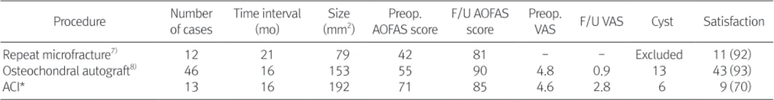
본 교실에서 시행된 ACI 중 1차 수술로 미세 골절술을 받고 실패한 증례를 후향적으로 분석해 보았을 때 총 13예, 평균 크기 192 mm2, 42개월 추시상 9명(70%)이 만족한 결과를 보였다. 타 연구와 단 순한 비교에서는 만족도 및 AOFAS 점수 결과가 떨어지는 것으로 보이나, 이는 평균 OLT의 크기가 크기 때문으로 생각된다(Table 1).7,8) 2차 수술로 추천되는 ACI 및 OATS는 연골 병변이 중심부와 후측에 위치할 경우 내과절골술을 시행하게 되고 이때 경골의 관 절면을 불규칙하게 만들어서 임상 결과를 감소시킨다.16,17) 그리고 OATS의 경우 손상된 부위가 원통형이 아니면 사이즈를 정확하게
Table 1. Simple Comparative Data between Several Procedures for Failed Osteochondral Lesion of Talus
Procedure Number
of cases
Time interval (mo)
Size (mm2)
Preop.
AOFAS score
F/U AOFAS score
Preop.
VAS F/U VAS Cyst Satisfaction Repeat microfracture7)
Osteochondral autograft8) ACI*
12 46 13
21 16 16
79 153 192
42 55 71
81 90 85
- 4.8 4.6
- 0.9 2.8
Excluded 13 6
11 (92) 43 (93) 9 (70) Values are presented as number or number (%).
Preop.: preoperative, AOFAS score: American Orthopaedic Foot and Ankle Society ankle-hindfoot score, F/U: follow-up, VAS: visual analog scale, -:
not checked, ACI: autologous chondrocyte implantation.
*Our data; not published.
10 Vol. 19 No. 1, March 2015
141 Dragoni M, Bonasia DE, Amendola A. Osteochondral talar al- lograft for large osteochondral defects: technique tip1 Foot Ankle Int1 2011;32:910-61
151 Yoon HS, Park YJ, Lee M, Choi WJ, Lee JW. Osteochondral autol- ogous transplantation is superior to repeat arthroscopy for the treatment of osteochondral lesions of the talus after failed pri- mary arthroscopic treatment1 Am J Sports Med1 2014;42:1896- 9031
161 Lee KT, Kim JS, Young KW, Lee YK, Park YU, Kim YH, et al. The use of fibrin matrix-mixed gel-type autologous chondrocyte implantation in the treatment for osteochondral lesions of the talus1 Knee Surg Sports Traumatol Arthrosc1 2013;21:1251-601 171 Kim YS, Park EH, Kim YC, Koh YG, Lee JW. Factors associated
with the clinical outcomes of the osteochondral autograft trans- fer system in osteochondral lesions of the talus: second-look arthroscopic evaluation1 Am J Sports Med1 2012;40:2709-191 181 Giannini S, Buda R, Ruffilli A, Cavallo M, Pagliazzi G, Bulzamini
MC, et al. Arthroscopic autologous chondrocyte implanta- tion in the ankle joint1 Knee Surg Sports Traumatol Arthrosc1 2014;22:1311-91
191 Niemeyer P, Salzmann G, Schmal H, Mayr H, Südkamp NP.
Autologous chondrocyte implantation for the treatment of chondral and osteochondral defects of the talus: a meta-analysis of available evidence1 Knee Surg Sports Traumatol Arthrosc1 2012;20:1696-7031
201 Minas T, Gomoll AH, Rosenberger R, Royce RO, Bryant T. In- creased failure rate of autologous chondrocyte implantation af- ter previous treatment with marrow stimulation techniques1 Am J Sports Med1 2009;37:902-81
51 Gobbi A, Francisco RA, Lubowitz JH, Allegra F, Canata G. Os- teochondral lesions of the talus: randomized controlled trial comparing chondroplasty, microfracture, and osteochondral autograft transplantation1 Arthroscopy1 2006;22:1085-921 61 Choi WJ, Park KK, Kim BS, Lee JW. Osteochondral lesion of
the talus: is there a critical defect size for poor outcome? Am J Sports Med1 2009;37:1974-801
71 Savva N, Jabur M, Davies M, Saxby T. Osteochondral lesions of the talus: results of repeat arthroscopic debridement1 Foot Ankle Int1 2007;28:669-731
81 Georgiannos D, Bisbinas I, Badekas A. Osteochondral transplan- tation of autologous graft for the treatment of osteochondral lesions of talus: 5- to 7-year follow-up1 Knee Surg Sports Trau- matol Arthrosc1 Published onlin October 19, 2014; doi: 1011007/
s00167-014-3389-31
91 McGahan PJ, Pinney SJ. Current concept review: osteochondral lesions of the talus1 Foot Ankle Int1 2010;31:90-1011
101 Robinson P, White LM. Soft-tissue and osseous impingement syndromes of the ankle: role of imaging in diagnosis and man- agement1 Radiographics1 2002;22:1457-69; discussion 1470-11 111 Hannon CP, Smyth NA, Murawski CD, Savage-Elliott I, Deyer
TW, Calder JD, et al. Osteochondral lesions of the talus: aspects of current management1 Bone Joint J1 2014;96-B:164-711 121 Loveday D, Clifton R, Robinson A. Interventions for treating
osteochondral defects of the talus in adults1 Cochrane Database Syst Rev1 2010;(8):CD0081041
131 Ogilvie-Harris DJ, Sarrosa EA. Arthroscopic treatment after pre- vious failed open surgery for osteochondritis dissecans of the talus1 Arthroscopy1 1999;15:809-121