유방에서 발견되는 인환세포암은 드문 질환으로 원발성과 전 이성으로 분류할 수 있다. 원발성 인환세포암은 침윤성 소엽암 의 변이체로서, 평균연령이 다른 유방암보다는 다소 높으며, 임 상진행이 빠르다 (1-2).
전이성 인환세포암은 위장관계에 생긴 인환세포암이 유방으 로 전이한 것으로 드물게 증례보고 되어있다 (3-5). 전이성 유 방암은 드물지만, 유방촬영술소견은 비교적 많이 보고되어 있다 (6-9). 그러나 초음파소견에 대한 보고는 드물다 (6, 10).
본 연구는 유방에서 발생한 인환세포암의 임상적 특징과 초음 파소견을 포함한 방사선학적 소견을 알아내어 다른 유방암과의 차이점 및 유사점을 알아보고자 하는데 있다.
대상과 방법
유방의 인환세포암으로 확진된 5명환자의 임상소견, 유방촬 영술소견, 그리고 초음파소견을 후향적으로 분석하였다. 환자는 23-49세로, 평균 37세였다. 모두 유방종괴를 호소하였지만 특
별한 위장관 증상은 없었다. 유방촬영은 Senographe 500 T (CGR, France) 또는 Senographe DMR (GE, Milwaukee, Wisconsin)을 이용하였고, 초음파기기는 ATL HDI 3000 (Advanced Technology Laboratories, Bothell, Wash., U.S.A.) 의 7.5 MHz 선형 탐촉자를 이용하였다. 유방병소는 16 G 바늘 을 이용하여 코어생검검사로 확진하였다. 생검 부위는 초음파 유도하에 종양이 있는 부위에서 시행하였고 (n = 2), 전반적인 피부비후만 있는 경우 (n = 3)는 촉진상 가장 두꺼운 부위에서 시행하였다. 유방병소의 확진후 원발병소를 찾기위해 모든 환자 는 위내시경을 시행하였다. 유방 및 위의 생검조직은 현미경학 적 병리조직검사와 난포호르몬 수용체(estrogen receptor, ER), 황체호르몬 수용체(progesterone receptor, PR)와 gross cystic disease fluid protein-15 (GCDFP-15)를 포함한 면역 조직화학적검사를 모두 시행하였다. 원발성과 전이성 유방암의 구분을 위해, 원발병소를 찾기위한 모든 검사를 시행하여, 4명 은 위의 인환세포암이 확진되어 전이성 인환세포 유방암으로, 1 명은 원발성 인환세포 유방암으로 진단하였다.
유방의 인환세포암: 임상적, 방사선학적 소견1
곽진영2・김은경・오기근・이용희3
목적: 유방 인환세포암의 임상적, 방사선학적인 소견을 알아보고자 하였다.
대상과 방법:유방의 인환세포암으로 확진된 5명의 환자 (연령분포 23-49세, 평균 37세)를 대상
으로 임상소견, 유방촬영술, 그리고 초음파소견을 후향적으로 분석하였다. 진단은 초음파 유도하 에 코어생검검사로 하였다. 모든 환자는 유방병변의 확진후 위내시경을 시행하여, 4명은 위의 인 환세포암이 발견되어 전이성 유방암으로, 1명은 원발성 유방암으로 확진하였다.
결과: 전이성 인환세포 유방암을 가진 4명 환자중 3명은 유방 동통과 종창을 호소하였다. 유방촬 영술상 음영증가와 피부비후가 있었고, 석회화는 없었다. 초음파상 미만성 피부비후, 림프관확장 소견, 그리고 액와림프절종대가 있었다. 3명 모두 유방촬영술이나 초음파상 종괴는 보이지 않았 다. 나머지 전이성 인환세포 유방암을 가진 한명은 점점 커지는 종괴가 만져졌다. 환자의 유방촬 영술은 고음영소견이었으나, 초음파상 경계가 불분명한 저에코의 종괴가 있었다. 원발성 인환세 포 유방암으로 확진된 1명은 만져지는 종괴를 호소했고, 방사선학적소견은 경계가 불분명한 종괴 를 보여 다른 원발성 유방암과의 차이점이 없었다.
결론:유방의 전이성 인환세포암은 염증성 유방암과 유사한 증상을 보이나, 비교적 나이가 젊고, 방사선학적소견은 미세 석회화나 종괴없이 미만성 음영증가와 피부비후로 보이는 것이 특징이었 다.
1연세대학교 의과대학 진단방사선학교실
2포천중문의과대학 분당차병원 진단방사선학교실
3포천중문의과대학 분당차병원 해부병리학교실
이 논문은 2000년 2월 3일 접수하여 2000년 7월 31일에 채택되었음.
결 과
원발성 인환세포암 1명과 전이성 인환세포암 4명의 임상 및 방사선학적 소견을 Table 1에 요약하였다. 치밀유방 4명, 지방
유방 1명이었다.
원발성 인환세포암
49세 여자로 만져지는 종괴를 주소로 하였으며, 유방촬영술 에서 경계가 불분명한 증가음영의 소견이 왼쪽 유방 상부에서
Table 1. Summary of Five Patients with Primary and Metastatic Signet Ring Cell Carcinoma of the Breast: Clinical and Radiologic Findings
No# Age Location Symptom Duration ER GCDFP Mammographic Sonographic
(months) /PR -15 Findings Findings
1 49 Lt. Palpable
2 + Asymmetric mass
mass /+ +
density
2 41 Lt. Breast
4 - Diffuse increased Diffuse skin thickening,
enlargement /- -
density increased SQ echogenecity
3 23 Rt. Breast
4 - Diffuse increased Diffuse skin thickening,
enlargement /- -
density increased SQ echogenecity
4 36 Lt. Breast
5 - Diffuse increased Diffuse skin thickening,
enlargement /- -
density increased SQ echogenecity
5 38 Lt. Palpable
3 - No abnormality mass
mass /- -
#1 : primary signet ring cell carcinoma, 2-5 : metastatic signet ring cell carcinoma from stomach, Lt : left, Rt : right, SQ = subcutaneous
A B
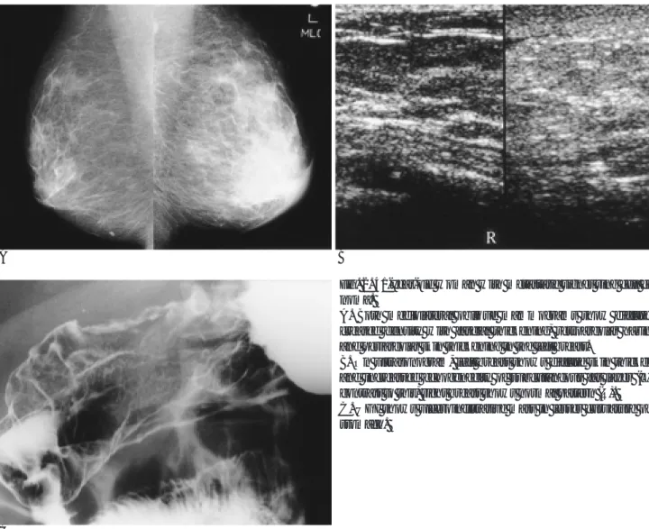
Fig. 1. 49-year-old woman with primary signet ring cell carcino- ma.
A. Both mediolateral oblique (MLO) mammograms show asym- metric increased density in the upper portion of the left breast (arrows) and lymph node enlargement (arrowheads) in the axil- la.
B. Ultrasound shows an 4×3 cm ill-defined hypoechoic mass with irregular margin and heterogenous posterior shadowing.
C. Photomicrographic findings of breast specimen (H & E, x 200) shows infiltration of signet ring cells (arrows) which has ec- centric, semilunar nuclei and cytoplasmic vacuoles. Endoscopic examination was negative.
보였으며, 액와부에는 내부 저음영이 소실되어 전이가 의심되는 임파절이 보였다 (Fig. 1A). 초음파검사에서는 경계가 불분명한 저에코음영의 종괴가 있었고, 크기는 4×3 cm이었으며, 불규칙 한 후방음영이 있었다 (Fig. 1B). 유방 종괴의 병리학적소견은 편심성 위치의 반월형 모양의 핵과, 세포질에 소포를 가진 인환 세포가 침윤하는 소견이었다 (Fig. 1C). 그리고 ER, PR, 그리고 GCDFP-15를 이용한 면역조직화학적검사에서 모두 양성이어 서, 유방에서 유래한 인환세포암임을 시사했다. 위내시경검사를 포함한 다른 검사에서 다른 원발성으로 고려할 수 있는 병변은 없었다.
전이성 인환세포암 임상증상
환자의 연령은 23-41세였고, 평균 34세로 비교적 젊은 연령 층이었다. 이중 3명은 유방종창, 미만성 유방부종, 유방동통, 그 리고 피부발적을 호소하였으며 나머지 1명은 4개월동안 점점 커지는 종괴를 호소하였다. 모든 환자에서 특별한 위장관 증상 은 없었다.
영상 소견
유방촬영술소견은 3명은 전반적 음영증가와 피부비후, 그리 고 근막의 비후가 있었고, 석회화는 없었으며 이들의 초음파소 견은 미만성 피부비후가 있었고, 종괴는 보이지 않았다 (Fig.
2A, B). 이중 1명에서는 복부 전산화단층촬영에서 양측 난소전 이가 의심되었다. 3명 모두 액와림프절이 커져있었고, 내부 고 에코의 소실이 있어, 전이성 림프절 종대로 생각하였다. 이중 1 명에서 액와림프절 생검을 시행하였고, 인환세포암세포가 있어 전이성 림프절 종대로 진단하였다.
나머지 1명은 위의 3명과는 방사선학적 소견이 달라, 유방촬 영술은 고음영소견이었고 (Fig. 3A), 초음파에서 경계가 불분명 한 저에코의 종괴가 있었고 후방음영은 없었다. 동측 액와에는 콤마모양의 경계가 분명한 림프절종대가 있었고, 이의 내부에는 중심성 고에코가 소실되어 있어, 전이성 임파절을 시사하는 소 견이었다 (Fig. 3B, C). 한달후 시행한 초음파검사에서 기존의 종괴는 1 cm에서 1.8 cm으로 커졌고, 종괴 주위에 경계가 불분 명한 5 mm 전후의 저에코의 결절이 2-3개 더 보였다 (Fig.
3D).
A
C
B
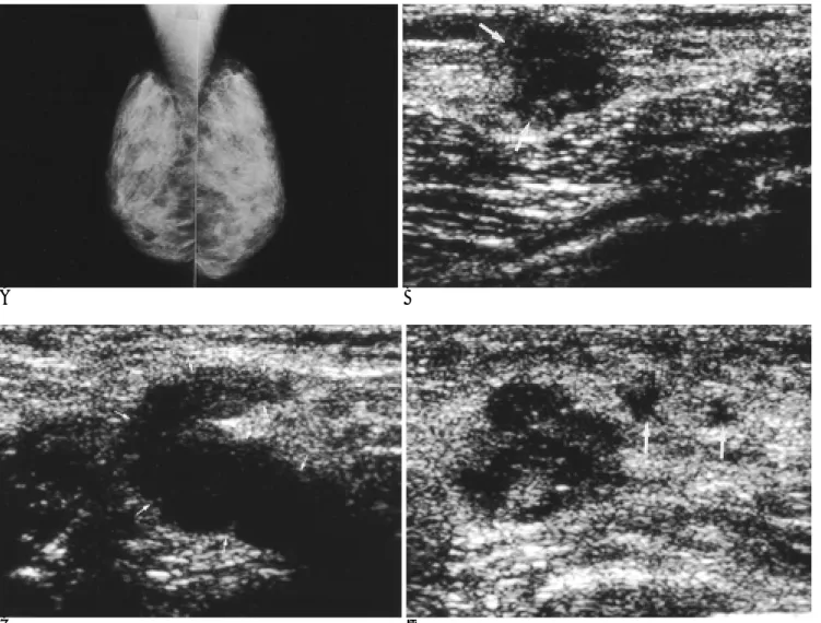
Fig. 2. 41-year-old woman with metastatic signet ring cell carci- noma.
A. Both mediolateral oblique mammograms show diffuse in- creased density with fascial thickening, retroareolar haziness, and periareolar skin thickening in the left breast.
B. On ultrasonogram, left breast shows diffuse skin thickening and increassed echogenecity of subcutaneous fat layer (L). In contrast to this, right breast shows normal pattern (R).
C. UGI shows ulceroinfiltrative mass in lesser curvature of the stomach.
위장 검사 및 병리 소견
4명 모두 위내시경에서 뚜렷한 점막병변이 있었고, 조직검사 상 인환세포암으로 진단되었다. 이 세포는 ER, PR및 GCDFP- 15염색에서 모두 음성이었다. 이중 상부위장조영술을 시행한 3 명에서 진행성 위암의 소견이 있었다 (Fig. 2C).
고 찰
유방의 인환세포암은 원발성과 전이성으로 분류된다. 원발성 유방 인환세포암은 침윤성 소엽암의 변이체로서, 전체 침윤성 소엽암의 8.7%를 차지하는 드문 종양중의 하나이다 (1, 11). 이 질환은 소포성의 mucicarminophilic 세포가 20%이상 포함되어 있으며, 임상적, 병리학적 소견이 점액성암과는 다른 유방종양 을 의미한다. 발견시 평균연령이 다른 유방암보다는 다소 높으 며, 임상진행이 빨라 예후가 나쁘다 (1-2). 유방의 원발성 인환
세포암은 여러장기로 전이할 수 있으며, 호발부위는 폐, 뼈, 림프 절, 간등으로 상대적으로 위장관으로의 전이는 드물다고하며, 전이시는 위장관 증상을 동반하지 않는 경우가 많다고 한다. 그 리고 유방의 원발성 인환세포암은 ER과 PR에 양성인 경우가 많 아서 호르몬 치료에 좋은 성적을 기대할 수 있다고 하였다 (12).
전이성 유방 인환세포암은 위장관계의 인환세포암이 유방으 로 전이된것으로, Cavazzini등 (3)은 위의 인환세포암에서 유방 으로 전이된 환자의 임상적, 방사선학적 소견을 보고하였다. 임 상소견은 염증성 유방암과 유사하였고, 방사선학적으로는 석회 화가 없는 경계가 좋지않은 종괴로 보여 악성을 시사하는 소견 을 보였다고 하였다. 박등 (10)의 보고는 인환세포암은 아니었 지만 위선암에서 전이된 유방병변이 염증성 유방암과 유사한 소 견을 보인 경우였다. 본 연구에서는 위 인환세포암에서 유방전 이를 한 4명 모두 특별한 위장관 증상이 없이 유방이 첫 병발병 소였으며, 그 중3명이 임상적으로 염증성 유방암과 유사한 소견
A B
C D
Fig. 3. 38-year-old woman with metastatic signet ring cell carcinoma.
A. Both mediolateral oblique mammograms show diffuse dense breast patterns with no definite evidence of mass.
B. Ultrasonogram of the left breast shows 1 cm ill-defined hypoechoic mass without posterior shadowing or enhancement (arrows).
을 보였다.
유방전이는 유방암중 1.2-6.6%의 빈도를 나타내는 드문 질 환이다 (6, 7). 유방전이에 관한 유방촬영술소견은 비교적 많이 보고되어 있지만 (6-9), 초음파소견의 보고는 드물다 (6, 10).
유방전이의 초음파소견은 경계가 좋은 저에코 종괴로 보이거나, 때로는 불규칙한 변연을 가지는 종괴로 보이며, 후방음영은 없 다고 보고하고 있다 (10). 본 연구에서는 전이성 인환세포암으 로 확진된 4명중 3명은 유방촬영술에서 전반적인 음영증가와 피부 비후, 그리고 근막비후의 소견이 있었지만, 내부에 미세석 회화의 소견은 보이지 않았고, 초음파검사에서도 종괴없이 미만 성 피부비후로만 보여, 염증성 유방암의 소견과는 달랐다. 나머 지 한명은 초음파검사에서 후방음영이 없는 경계가 불분명한 저 에코의 종괴로, 유방전이시 흔히 보는 경계가 좋은 종괴와는 다 른 소견이었다.
유방전이를 유발하는 인자에 관한 연구는 많이 있었지만, 명 백히 밝혀진 바 없다. 그러나 유방전이가 있었던 남성과 횡문근 육종에서 유방전이가 있었던 젊은 여성에서 호르몬이 한 인자로 작용한다는 보고가 있다 (13-14). 유방전이가 상대적으로 젊 은 연령층에서 나타난다는 것도, 유방의 생리학적 상태가 전이 에 영향을 미친다는것을 나타낸다 (15). 본 연구에서도 전이성 인환세포암으로 확진된 환자의 평균연령 34세로 비교적 젊은 연령이었다. 유방전이는 임상적으로 전신으로 진행되었음을 의 미하며, 예후가 아주 나빠 1년내에 80%이상의 사망률을 보인다 (16).
유방병변이 인환세포암인경우 원발성과 전이성 인환세포암 의 감별은 어려운 경우가 많으며, 이의 구분은 중요한데, 전이성 유방암일경우 불필요한 유방절제술을 피할 수 있고 (16), 원발 성인경우 항에스트로겐요법이 생존기간을 연장시킬 수 있기 때 문이다 (12). 방사선학적으로는 유방의 원발성 인환세포암이 위로 전이시, 장막을 침범하는 전이양상을 보여, 형성성 위조직 염(linitis plastica)형태로 보이는 경우가 흔하다 (12). 그리고 면역조직화학적검사에서, 원발성 유방암은 ER 양성을 나타내는 경우가 많다. 그러나 최근보고에 의하면 (17) 원발성 위종양의 23%에서도 ER 양성을 보인다고 하였다. 따라서 원발성 유방암 의 구분을 위해서는 GDCFP-15를 이용한 면역조직화학적 검 사가 유방에서 유래한 종양의 진단에 유용한 지표가 되므로 (12), 본 연구에서도 원발성과 전이성 유방의 감별을 위한 검사 로 포함하였다.
이 연구에서는 위 인환세포암에서 유방 전이로 진단된 4명은 위내시경에서 모두 뚜렷한 점막병변이 있었고, 이것은 유방의 원발성 인환세포암이 전이시 흔히 장막을 침범하는 소견과는 다 른점이라 할 수 있다. 또한 원발성에 비해 비교적 젊은 나이에 호발한다는 점도 구별점 중의 하나이다. 그리고 전이성 인환세 포암이 PR, ER, 그리고 GCDFP-15를 이용한 면역조직화학적 검사에서 음성으로 나와 유방에서 유래하지 않았음을 뒷받침해 주고있다.
결론적으로, 유방의 전이성 인환세포암은 염증성 유방암과 유 사한 증상을 보이나, 비교적 나이가 젊고, 방사선학적소견에서 미세 석회화나 종괴가 없는 것이 차이점이었다. 그리고 유방의 인환세포암은 위 인환세포암으로부터 전이된 경우가 대부분으 로, 유방병변이 인환세포암으로 확진된경우, 뚜렷한 위장증상이 없더라도 위내시경을 검사를 하여, 전이성 인환세포암을 감별하 여야 하겠다.
참 고 문 헌
1. Steinbrecher JS, Silverberg SG. Signet-ring cell carcinoma of the breast. The mucinous variant of infiltrating lobular carcinoma?
Cancer 1976;37:828-840
2. Fechner RE. Infiltrating lobular carcinoma without lobular carci- noma in-situ. Cancer 1972;29:1539-1545
3. Cavazzini G, Colpani F, Cantore M, Aitini E, et al. Breast metasta- sis from gastric ring cell carcinomas, mimicking inflammatory car- cinoma. A case report. Tumori 1993; 79:450-453
4. de la Cruz Mera A, Marino Cotelo A. Breast metastases. Acta Cytol 1998;42:1304-1306
5. Domanski HA. Metastases to the breast from extramammary neo- plasms : a report of six cases with diagnosis by fine needle aspira- tion cytology. Acta Cytol 1996;40:1293-1300
6. Derchi LE, Rizzatto G, Giugeppetti GM, Dini G, Garabenta A.
Metastatic tumors in the breast: sonographic findings. J Ultrasound Med 1985;4:69-74
7. Yamasaki H, Saw D, Zdanowitz, Faltz LL. Ovarian carcinoma metastasis to the breast case report and review of the literature.
Am J Surg Pathol 1993;17:193-197
8. Paulus DD, Libshitz HI. Metastasis to the breast. Radiol Clin North Am 1982;20:561-568
9. McCrea ES, Johnston C, Haney PY. Metastases to the breast. AJR Am J Roentgenol 1983;141:685-690
10. 박정미, 권진숙, 공경엽. 위암에서 전이된 유방암: 증례보고. 대한방 사선의학회지 1998;38:1139-1141
11. Eltorky M, Hall JC, Osborne PT, El Zeky F. Signet-ring cell variant of invasive lobular carcinoma of the breast. Arch Pathol Lab Med 1994;118(3):245-248
12. Yim H, Jin YM, Shim C, Park HB. Gastric metastasis of mammary signet ring cell carcinoma--a differential diagnosis with primary gas- tric signet ring cell carcinoma. J Korean Med Sci 1997;12(3):256-261 13. Toombs BD, Kalisher L. Metastatic disease to the breast : clinical,
pathologic, and radiographic features. AJR Am J Roentgenol 1977;
129:673-676
14. Howarth CB, Caces JN, Pratt CB. Breast metastasis in children with rhabdomyosarcoma. Cancer 1980;46:2520-2524
15. Alexander HR, Turnbull AD, Rosen PP. Isolated breast metastases from gastrointestinal carcinomas: report of two cases. J Surg Oncol 1989;42:264-266
16. Hajdu SI, Urban JA. Cancers metastatic to the breast. Cancer 1972;
29:1691-1696
17. Kojima O, Takahashi T, Kawakami S, Uehara Y, Matsui M. Locali- zation of estrogen receptors in gastric cancer using immunohisto- chemical staining of monoclonal antibody. Cancer 1991;67:2401-2406
J Korean Radiol Soc 2000;43:377-382
Address reprint requests to : Eun-Kyung Kim, M.D., Department of Diagnostic Radiology, Yonsei University College of Medicine 134 Shinchon-Dong, Seodaemun-gu, Seoul 120-752, Korea.
Tel. 82-2-361-5837 Fax. 82-2-393-3035 E-mail: [email protected]
Signet Ring Cell Carcinoma of the Breast:
Clinical and Radiologic findings
1Jin-Young Kwak, M.D.2, Eun-Kyung Kim, M.D., Ki Keun Oh, M.D., Yong Hee Lee, M.D.3
1Department of Diagnostic Radiology, Yonsei University College of Medicine
2Department of Diagnostic Radiology, Pundang CHA General Hospital, College of Medicine, Pochon CHA University
3Department of Pathology, Pundang CHA General Hospital, College of Medicine, Pochon CHA University
Purpose: To evaluate the clinical and imaging findings of signet ring cell carcinoma of the breast.
Materials and Methods: We retrospectively evaluated the clinical, mammographic and ultrasonographic (US) findings of five patients aged 23-49 (mean 37) years with signet ring cell carcinoma of the breast. Diagnosis in- volved US-guided core-needle biopsy. In all patients the stomach was evaluated endoscopically after confirma- tion of the breast lesion. Metastatic breast cancer was confirmed in four patients and primary breast cancer in one.
Results: Three of the four patients with metastatic signet ring cell carcinoma complained of breast pain and swelling or enlargement. Mammography indicated the presence showed of diffuse increased density and skin thickening, without calcifications, while US demonstrated diffuse marked skin thickening, lymphatic dilata- tion, and axillary lymph node enlargement. Neither modality revealed the presence of mass, however. In the remaining patient, an enlarged breast mass was observed; mammography showed no abnormality, but US re- vealed an ill-defined hypoechoic mass. Mammographic and US findings in the patient with primary signet ring cell carcinoma of the breast indicated an ill-defined spiculated mass, resembling other breast carcinomas.
Conclusion: Metastatic signet ring cell carcinoma of the breast showed clinical symptoms similar to these seen in inflammatory breast cancer, though the former condition occurred in younger women. Radiographs demonstrated diffuse increased density and skin thickening without associated microcalcifications or mass.
Index words :Breast neoplasm, diagnosis Breast neoplasm, US Breast neoplasm, metastases