• 검색 결과가 없습니다.

The Effect of Saphenous Vein Ablation on Combined Segmental Popliteal Vein Reflux

N/A
N/A
Protected

Academic year: 2021

Share "The Effect of Saphenous Vein Ablation on Combined Segmental Popliteal Vein Reflux"

Copied!
6
0
0

로드 중.... (전체 텍스트 보기)

전체 글

(1)

ISSN: 2233-601X (Print) ISSN: 2093-6516 (Online)

This study was presented at 2nd College of Phlebology international veins meeting, London, United Kingdom, March 14-16, 2018.

Received: August 2, 2018, Revised: August 15, 2018, Accepted: August 16, 2018, Published online: October 5, 2018

Corresponding author: Ki-Pyo Hong, Department of Thoracic and Cardiovascular Surgery, National Health Insurance Service Ilsan Hospital, 100 Ilsan-ro, Ilsandong-gu, Goyang 10444, Korea

(Tel) 82-31-900-0254 (Fax) 82-31-900-0343 (E-mail) kipyoh@nhimc.or.kr

© The Korean Society for Thoracic and Cardiovascular Surgery. 2018. All right reserved.

This is an open access article distributed under the terms of the Creative Commons Attribution Non-Commercial License (http://creativecommons.org/

licenses/by-nc/4.0) which permits unrestricted non-commercial use, distribution, and reproduction in any medium, provided the original work is properly cited.

The Effect of Saphenous Vein Ablation on Combined Segmental Popliteal Vein Reflux

Ki-Pyo Hong, M.D., Ph.D., Do-Kyun Kim, M.D., Ph.D.

Department of Thoracic and Cardiovascular Surgery, National Health Insurance Service Ilsan Hospital

Background: This study examined the role of superficial vein surgery in patients with combined superficial venous reflux and segmental popliteal vein reflux. Methods: We retrospectively reviewed 42 limbs in 38 pa- tients with combined superficial venous reflux and segmental popliteal vein reflux who underwent saphenous vein ablation between January 2014 and February 2017. Patients underwent outpatient follow-up duplex ul- trasonography at 3, 6, and 12 months postoperatively. Resolution of deep vein reflux was defined as re- versed blood flow in a popliteal segment for less than 1.0 second and a decrease in the reflux time of more than 20% of the preoperative reflux time. Results: The mean follow-up period was 9 months (range, 3–23 months). Saphenous vein ablations were performed by stripping in 24 limbs and radiofrequency ablation in 18 limbs. Preoperative segmental popliteal vein reflux resolved in 21 of the 42 limbs (50%). Conclusion: This study demonstrated that superficial venous surgery corrected segmental popliteal vein reflux in 50% of limbs with combined superficial venous reflux and segmental popliteal vein reflux. Other prospective studies are necessary to elucidate the etiology of the non-reversible cases.

Key words: 1. Popliteal vein 2. Saphenous vein 3. Duplex ultrasonography 4. Venous insufficiency

Introduction

Chronic venous disease (CVD) is caused by long-standing increased venous pressure. The etiol- ogy of increased venous pressure can be deep vein reflux (DVR), superficial vein reflux (SVR), and in- competent perforator veins, alone or in combination.

In the literature, SVR alone has been reported to be the main etiology of venous ulceration in up to 57%

of cases and to be combined with DVR in up to 32%

of cases [1]. However, McEnroe et al. [2] reported that deep venous insufficiency alone was the leading

cause of venous hypertension, accounting for 72% of patients with venous ulceration, while 13% had su- perficial venous insufficiency alone and the remaining 15% had both deep and superficial venous insufficiency. Chronic deep venous insufficiency has been shown to hasten the progression of venous dis- ease, leading to an increase in the rate of venous ul- ceration formation and, when coexisting with SVR, it has been found to be an additional factor influencing clinical status, as well as DVR [3]. Therefore, reso- lution of DVR along with SVR is important for pre- venting chronic venous insufficiency.

https://doi.org/10.5090/kjtcs.2018.51.5.338

(2)

Several pieces of evidence have shown that seg- mental DVR can be resolved after ablation of the combined incompetent greater saphenous vein (GSV) [4-7]. However, most studies have investigated the impact of ablation of the GSV on coexisting femoral vein reflux, but not on popliteal vein reflux (PVR).

The venous severity scoring system allots higher scores to reflux in popliteal veins, because it is thought to be more critical in the process of chronic venous in- sufficiency than DVR above the knee [8]. Further- more, PVR was reported to be strongly associated with delayed and failed healing of venous ulcers [9].

The aim of this study was to examine the effect of saphenous vein ablation on combined segmental PVR.

Methods

This study complied with the Declaration of Helsinki and was approved by the Institutional Review Board of National Health Insurance Service Ilsan Hospital (IRB approval no., 2015-04-011). Since this was a retrospective study, patients were not re- quired to provide informed consent.

Patients who underwent saphenous vein ablation between January 2014 and February 2017 with a di- agnosis of saphenous vein reflux combined with seg- mental PVR were included in this study. The follow- ing exclusion criteria were used: (1) limbs with deep axial reflux; (2) a history of previous varicose vein treatment in the ipsilateral limb, including surgery, endovenous thermal ablation, or sclerotherapy; and/or (3) a history of deep venous thrombosis.

During the physical examination, the clinical se- verity of venous disease was graded from C1 to C6, according to the Clinical Class, Etiology, Anatomy, and Pathophysiology Classification.

At baseline and at each follow-up visit, disease se- verity and disease-related quality of life were ap- praised by the Venous Clinical Severity Score (VCSS) and the Aberdeen Varicose Vein Questionnaire (AVVQ) score.

All patients scheduled to undergo varicose vein surgery underwent duplex ultrasound (DUS) before surgery. A LOGIQ5 PRO (5–12 MHz linear probe; GE HealthCare, Seoul, Korea) was used. Following rou- tine manual DUS, all patients underwent evaluation of the GSV, small saphenous vein (SSV), perforator vein, common femoral vein, and popliteal vein.

Patients were assessed in the standing position, and their non–weight-bearing limb was investigated. The superficial and deep venous systems were examined.

Venous reflux was defined as reversed blood flow for more than 0.5 seconds for the saphenous vein and more than 1.0 second for a deep vein on release of compression distal to the segment of the vein under examination. Segmental PVR was defined as an in- competent segment of deep vein below the knee with a competent common femoral vein.

The treatment options available to patients were either conventional surgery or radiofrequency abla- tion (RFA). Although RFA was considered preferable, conventional surgery was performed if the patient requested the treatment to be covered by insurance, as RFA was not covered by the National Health Insurance Service. In the conventional operations, high ligation and stripping of the GSV or SSV com- bined with stab avulsions of tributaries were performed. While stripping of the GSV was per- formed from groin to knee level, the procedure for the SSV was performed from the knee crease to the mid-calf level. RFA was performed from 2 cm distal to the saphenofemoral junction to the knee level for the GSV and from 2 cm distal to the saphenopopli- teal junction to the mid-calf level for the SSV, com- bined with stab avulsion phlebectomy or sclero- therapy of tributaries.

After conventional surgery or RFA, a compression bandage was applied for 1 day. Once the com- pression bandage was removed, the patient was re- quired to wear compression stockings 24 hours a day for 1 week and then during the daytime for the following week.

Patients underwent outpatient follow-up DUS at 3, 6, and 12 months postoperatively. Resolution of DVR was defined as reversed blood flow in a popliteal segment for less than 1.0 second and a decrease in the reflux time of more than 20% of the pre- operative reflux time. Aggravation of DVR was de- fined as reversed blood flow in a popliteal segment for more than 1.0 second and an increase of reflux time for more than 20% of the preoperative reflux time. Postoperative changes in the range of 20% of the preoperative reflux time were considered to be in the range of technical error.

The data were analyzed using IBM SPSS ver. 21.0

(IBM Corp., Armonk, NY, USA). The Student t-test,

(3)

Table 1. Demographic data and CEAP classification

Characteristic Value

No. of patients 38

No. of limbs 42

Duration of follow-up (mo) 11 (3–34) Sex

Male 18

Female 24

Age (yr) 58 (27–83)

CEAP classification

C1 1

C2 39

C4 1

C6 1

E

P

A

S

,

D

P

R

42

Incompetent saphenous vein

GSV 21

SSV 4

GSV and SSV 17

Values are presented as number or mean (range).

CEAP, Clinical Class, Etiology, Anatomy, and Pathophysiology Clas- sification; GSV, great saphenous vein; SSV, small saphenous vein.

Table 3. Correlations between clinical factors and the reso- lution of segmental PVR

Variable

Postoperative

resolution of PVR p-value

No Yes

Sex 0.03

Male 5 (24) 13 (62)

Female 16 (76) 8 (38)

Age (yr) 60.8±10.9 57.3±8.3 0.25

Follow-up (mo) 9.0±4.5 8.7±4.9 0.80

Preoperative duration of PVR (sec)

1.92±0.67 1.84±1.03 0.78 The ablated segment in the

superficial system

0.47

GSV 9 12

SSV 3 1

GSV and SSV 9 8

Truncal vein management 0.76

High ligation and stripping 13 (54) 11 (46) Radiofrequency ablation 8 (44) 10 (56)

Values are presented as number (%) or mean±standard deviation.

PVR, popliteal vein reflux; GSV, great saphenous vein; SSV, small saphenous vein.

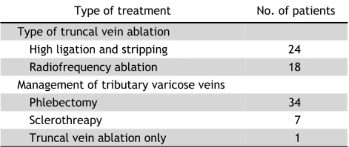
Table 2. Management of incompetent superficial veins (N=42) Type of treatment No. of patients Type of truncal vein ablation

High ligation and stripping 24

Radiofrequency ablation 18

Management of tributary varicose veins

Phlebectomy 34

Sclerothreapy 7

Truncal vein ablation only 1

chi-square test, and Fisher exact test were used to assess the statistical significance of differences be- tween groups. All p-values <0.05 were considered to indicate statistical significance.

Results

Overall, saphenous vein surgery was performed in 467 limbs in 303 patients during the study period.

Among them, 44 limbs from 40 patients fulfilled the inclusion criteria, and follow-up was performed in 38 patients (follow-up rate, 95%). Demographic data are listed in Table 1. The distribution of treatment op- tions is presented in Table 2.

The mean preoperative and postoperative reflux

times of the incompetent popliteal vein were 1.88 seconds (range, 1.01–5.24 seconds) and 1.23 seconds (range, 0–3.91 seconds), respectively. On postopera- tive DUS, resolution of segmental PVR was noted in 21 of 42 limbs (50%). In the remaining 21 limbs, 3 demonstrated improvement of PVR, and aggravation of PVR occurred in 6 (14%).

The correlations between several clinical factors

and postoperative resolution of the incompetent pop-

liteal segment are presented in Table 3. The reso-

lution of segmental PVR was statistically independent

of age, follow-up duration, the preoperative duration

of PVR, the ablated segment in the superficial sys-

tem, and treatment modality. Although preoperative

and postoperative VCSS scores were recorded for all

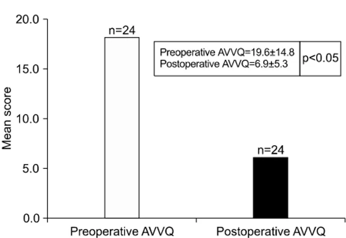
patients (n=42), postoperative AVVQ scores were on-

ly recorded for 24 patients, because the AVVQ was

not used during the early period of the study. The

postoperative VCSS and AVVQ scores demonstrated

significant improvements compared with the pre-

operative baseline scores (Figs. 1, 2). Based on a

comparison of the differences in the mean scores,

postoperative changes in the VCSS and AVVQ scores

showed no correlation with the resolution of PVR

(Table 4).

(4)

Fig. 1. Comparison of perioperative VCSS score. VCSS, Venous Clinical Severity Score.

Fig. 2. Comparison of perioperative AVVO score. AVVO, Aberdeen Varicose Vein Questionnaire.

Table 4. Perioperative changes in the VCSS and AVVQ according toresolution of segmental popliteal vein reflux

Variable

Postoperative resolution of popliteal

vein reflux p-value

No Yes

VCSS (N=42) 4.0±1.6 (n=21) 3.4±2.1 (n=21) 0.32 AVVQ (N=24) 13.3±11.5 (n=11) 9.8±8.8 (n=13) 0.40 Values are presented as mean±standard deviation.

VCSS, Venous Clinical Severity Score; AVVQ, Aberdeen Varicose Vein Questionnaire.

Discussion

Previous researchers have reported that SVR was present in approximately 90% of limbs with CVD, whereas DVR was detected in only 30% of limbs with CVD [10,11]. Labropoulos et al. [3] reported that the prevalence of DVR in limbs with combined superficial vein incompetence was 22%. While axial DVR is less likely to be abolished by superficial vein surgery, segmental DVR can be corrected after saphe- nous ablation in a substantial number of patients [4-7,12]. Puggioni et al. [7] reported complete dis- appearance of reflux in only 2 of 17 limbs (12%) with axial DVR after saphenous ablation. In addition, Maleti et al. [12] reported that preoperative axial DVR was not abolished in 81% of patients after sa- phenous ablation. In patients with segmental femoral vein reflux combined with SVR, Walsh et al. [4] and Sales et al. [5] reported that segmental femoral vein reflux was corrected after saphenous ablation in 27 of 29 limbs (93%) and 16 of 17 limbs (94%), respectively. Another study reported that nearly 50%

of limbs with segmental DVR were corrected after superficial venous surgery [6]. However, Puggioni et al. [7] demonstrated that segmental femoral vein re- flux was resolved in only 7 of 21 limbs (33%).

Despite the considerable variation in results, most studies demonstrated that at least one-third of seg- mental femoral vein reflux cases could be corrected by ablation of a combined incompetent GSV.

While most studies have focused on the resolution of segmental femoral vein reflux after the ablation of an incompetent GSV, we studied the resolution of

segmental PVR after the ablation of coexisting in-

competent saphenous veins. PVR is a significant risk

factor for venous ulceration [13], and the frequency

of PVR increases with the progression of clinical

symptoms [14]. Stuart et al. [15] also found that an

open or healed venous ulcer was strongly associated

with PVR. In the present study, after ablation of the

incompetent saphenous veins, segmental PVR was

corrected in 21 of 42 limbs (50%), regardless of the

treatment modality. This result demonstrates that a

substantial portion of segmental PVR cases can be

rectified after ablation of a coexisting incompetent

saphenous vein, as with segmental femoral vein re-

flux in previous studies [4-7,12]. The results of this

study could be explained through the hypothesis of

the ‘overload theory,’ according to which SVR might

cause increased venous return into the deep vein

system through the perforator veins, which causes a

volume overload in the deep veins, leading to an in-

creased caliber of the deep veins, followed by in-

competence of the valves of the deep veins. Two

possibilities might explain why only 50% of the cases

(5)

of segmental PVR were resolved. First, long-standing volume overload in the deep vein might cause irre- versible structural changes in the wall and valves of the deep vein. According to the overload theory, the diameter of the deep vein would decrease in patients with resolved DVR after saphenous ablation.

However, the literature contains no definitive evi- dence that saphenous vein ablation does indeed re- duce the diameter of the deep vein. We were not able to analyze perioperative changes in the diameter of the deep vein since we did not assess the width of the deep vein in routine DUS studies. Additionally, congenital abnormal anatomy of the valve could cause a non-reversible condition. Maleti et al. [12]

reported that there are 2 different kinds of valve morphology, with either symmetrical or asymmetrical cusps. After correction of volume overload in the deep vein system, valve competence could be re- stored in valves with symmetrical cusps, but not in those with asymmetrical cusps. The possible causes of asymmetrical valve cups of the deep vein could in- clude post-thrombotic changes or a congenital condition. An accurate preoperative evaluation of valve morphology is necessary to make a treatment plan based on the symmetricity of valve cusps.

Although Labropoulos et al. [3] reported that PVR was associated with SSV incompetence, we did not find such an anatomic association. In this study, the resolution of segmental PVR was not affected by the ablated segment in the superficial system. Nevertheless, the number of incompetent SSVs was too small to perform a meaningful analysis of their association with GSV incompetence. Hence, further study is re- quired before making a generalization.

1) Limitations of the study

This study has the limitation of being a retro- spective single-center study. In addition, although DVR is known to be closely associated with chronic venous insufficiency, as manifested by conditions such as venous ulcers, the majority of the patients in the study (93%) were classified as having C2 disease.

Therefore, we were not able to assess the prognosis of venous ulcers according to whether PVR was resolved.

2) Conclusion

This study demonstrated that saphenous vein abla-

tion corrected segmental PVR in 50% of limbs with combined SVR and segmental PVR, regardless of the treatment modality. Other prospective studies are necessary to elucidate the etiology of the non-rever- sible cases.

Conflict of interest

No potential conflict of interest relevant to this ar- ticle was reported.

Acknowledgments

This work was supported by the National Health Insurance Service Ilsan Hospital Research Grant (2015-25).

References

1. Scriven JM, Hartshorne T, Bell PR, Naylor AR, London NJ.

Single-visit venous ulcer assessment clinic: the first year.

Br J Surg 1997;84:334-6.

2. McEnroe CS, O’Donnell TF Jr, Mackey WC. Correlation of clinical findings with venous hemodynamics in 386 pa- tients with chronic venous insufficiency. Am J Surg 1988;156:148-52.

3. Labropoulos N, Tassiopoulos AK, Kang SS, Mansour MA, Littooy FN, Baker WH. Prevalence of deep venous reflux in patients with primary superficial vein incompetence. J Vasc Surg 2000;32:663-8.

4. Walsh JC, Bergan JJ, Beeman S, Comer TP. Femoral venous reflux abolished by greater saphenous vein stripping. Ann Vasc Surg 1994;8:566-70.

5. Sales CM, Bilof ML, Petrillo KA, Luka NL. Correction of low- er extremity deep venous incompetence by ablation of su- perficial venous reflux. Ann Vasc Surg 1996;10:186-9.

6. Adam DJ, Bello M, Hartshorne T, London NJ. Role of super- ficial venous surgery in patients with combined superficial and segmental deep venous reflux. Eur J Vasc Endovasc Surg 2003;25:469-72.

7. Puggioni A, Lurie F, Kistner RL, Eklof B. How often is deep venous reflux eliminated after saphenous vein ablation? J Vasc Surg 2003;38:517-21.

8. Danielsson G, Eklof B, Grandinetti A, Lurie F, Kistner RL.

Deep axial reflux, an important contributor to skin changes or ulcer in chronic venous disease. J Vasc Surg 2003;38:1336-41.

9. Brittenden J, Bradbury AW, Allan PL, Prescott RJ, Harper DR, Ruckley CV. Popliteal vein reflux reduces the healing of chronic venous ulcer. Br J Surg 1998;85:60-2.

10. Kistner RL, Eklof B, Masuda EM. Diagnosis of chronic ve-

nous disease of the lower extremities: the “CEAP”

(6)

classification. Mayo Clin Proc 1996;71:338-45.

11. Labropoulos N. CEAP in clinical practice. Vasc Surg 1997;31:224-5.

12. Maleti O, Lugli M, Perrin M. After superficial ablation for superficial reflux associated with primary deep axial re- flux, can variable outcomes be caused by deep venous valve anomalies? Eur J Vasc Endovasc Surg 2017;53:229-36.

13. Robertson L, Lee AJ, Gallagher K, et al. Risk factors for chronic ulceration in patients with varicose veins: a case control study. J Vasc Surg 2009;49:1490-8.

14. Welch HJ, Young CM, Semegran AB, Iafrati MD, Mackey WC, O’Donnell TF Jr. Duplex assessment of venous reflux and chronic venous insufficiency: the significance of deep ve- nous reflux. J Vasc Surg 1996;24:755-62.

15. Stuart WP, Adam DJ, Allan PL, Ruckley CV, Bradbury AW.

The relationship between the number, competence, and

diameter of medial calf perforating veins and the clinical

status in healthy subjects and patients with lower-limb

venous disease. J Vasc Surg 2000;32:138-43.

수치

Table 2. Management of incompetent superficial veins (N=42) Type of treatment No. of patients Type of truncal vein ablation
Fig. 1. Comparison of perioperative VCSS score. VCSS, Venous  Clinical Severity Score.

참조

관련 문서

Post-surgical use of radioiodine (131I) in patients with papillary and follicular thyroid cancer and the issue of remnant ablation: a consensus

After first field tests, we expect electric passenger drones or eVTOL aircraft (short for electric vertical take-off and landing) to start providing commercial mobility

1 John Owen, Justification by Faith Alone, in The Works of John Owen, ed. John Bolt, trans. Scott Clark, "Do This and Live: Christ's Active Obedience as the

(c) the same mask pattern can result in a substantial degree of undercutting using substantial degree of undercutting using an etchant with a fast convex undercut rate such

As a result of the study, it was confirmed that only human resource competency had a positive(+) effect on the intercompany cooperation method of IT SMEs,

The effect of Combined Exercise program on Body composition, Physical fitness and thickness of subcutaneous fat in. Obese

Thermal balloon ablation versus endometrial resection for the treatment of abnormal uterine bleeding. Thermal balloon ablation in myoma-induced menorrhagia

The result suggested that a taekwon aerobics program had a positive effect on the increase of growth hormone in blood lipid in middle school girls.. The