Korean J Thorac Cardiovasc Surg 2014;47:145-148 □ Case Report □ http://dx.doi.org/10.5090/kjtcs.2014.47.2.145 ISSN: 2233-601X (Print) ISSN: 2093-6516 (Online)
− 145 −
Departments of 1Thoracic and Cardiovascular Surgery, 2Radiology, and 3Pathology, Konkuk University School of Medicine Received: July 25, 2013, Revised: October 11, 2013, Accepted: October 14, 2013
Corresponding author: Je Kyoun Shin, Konkuk University Medical Center, Konkuk University School of Medicine, 120 Neungdong-ro, Gwangjin-gu, Seoul 143-729, Korea
(Tel) 82-2-2030-5042 (Fax) 82-2-2030-5009 (E-mail) [email protected]
C The Korean Society for Thoracic and Cardiovascular Surgery. 2014. All right reserved.
CC This is an open access article distributed under the terms of the Creative Commons Attribution Non-Commercial License (http://creative- commons.org/licenses/by-nc/3.0) which permits unrestricted non-commercial use, distribution, and reproduction in any medium, provided the original work is properly cited.
Cardiac Parasitic Infection in Trichinellosis Associated with Right Ventricle Outflow Tract Obstruction
Seung Ho Bang, M.D.
1, Jae Bum Park, M.D.
1, Hyun Keun Chee, M.D.
1, Jun Seok Kim, M.D.
1, Sung Min Ko, M.D.
2, Wan Seop Kim, M.D.
3, Je Kyoun Shin, M.D.
1Here, we present a rare case of cardiac parasitic infection found in an adult female patient who had the symp- toms of dyspnea upon exertion. She was diagnosed with a double-chambered right ventricle due to infundibular hy- pertrophy confirmed by transthoracic echocardiography and cardiac computed tomography. We performed surgery of infundibulectomy around the pulmonary valve. In the end, histopathological findings of the resected infundibular muscle demonstrated trichinellosis, a type of roundworm infection.
Key words: 1. Parasite infection, heart 2. Trichinellosis
3. Right ventricle
CASE REPORT
A 60-year-old woman had been diagnosed with hyper- tension and cardiomegaly in 2003 but was doing well without any specific medication or treatment. She visited the depart- ment of cardiovascular internal medicine at Konkuk Universi- ty Medical Center with a complaint of aggravating dyspnea on exertion, functional impairment class 3 (New York Heart Association) in December 2010. Then, she was referred to our department of cardiovascular surgery upon the diagnosis of a double-chambered right ventricle with pulmonary in- fundibular hypertrophy by two-dimensional transthoracic echocardiography (TTE). Upon physical examination, she was found to have mild hypertension (135/92 mmHg) with a reg- ular heartbeat of 67 beats/ min and a body temperature of 36oC. Cardiac auscultation revealed grade III systolic ejection murmur at the left lower parasternal border. The patient’s
electrocardiogram showed a sinus rhythm with a right bundle branch block, right axis deviation, and right ventricular hypertrophy. Chest radiography revealed mild cardiomegaly with a computed tomography (CT) ratio of 60%. A cardiac CT revealed a perimembranous septal aneurysm (size:
5.9×10.1 mm) without thrombus and rupture, right ventricular outflow tract obstruction (RVOTO), and bronchiectasis with atelectasis at the medial segment of right middle lobe.
Transesophageal echocardiography revealed a high-velocity systolic jet (4.8 m/sec, peak pressure gradient=93 mmHg) through the right ventricular outflow track due to a discrete and round muscular obstruction, but remarkable tricuspid re- gurgitation or leakage through a ventricular septal defect was not noted. Magnetic resonance imaging verified thickened muscular bands between the thin-walled infundibulum and the inflow segment of the right ventricle causing the dou- ble-chambered right ventricle (Fig. 1). There were no other
Seung Ho Bang, et al
− 146 −
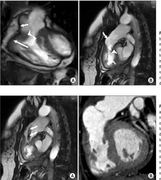
Fig. 1. (A) Coronal and (B) sagittal balanced steady-state free-precision magnetic resonance images of the right ventricular outflow tract of the heart during diastole show thickened muscular bands (arrowheads) between the thin-walled infundibulum (short ar- rows) and the inflow segment of the right ventricle (long arrow) causing a double-chambered right ventricle.
Fig. 2. (A) Sagittal balanced steady-st- ate free precession magnetic reso- nance image of the right ventricular outflow tract of the heart during sys- tole shows a jet phenomenon (arrows) due to the pressure gradient distal to the stenosis. (B) Sagittal multi-pla- nar reconstruction computed tomog- raphy image of the right ventricular outflow tract of the heart during dia- stole after surgery shows the re- moval of the thickened muscular ba- nds and the widened right ventricular outflow tract.
specific abnormalities. The patient underwent median sternotomy. After cardiopulmonary bypass (CPB) was ini- tiated as usual, fibrillatory arrest was performed while avoid- ing deep hypothermia. We first incised the main pulmonary artery and then, proceeded to reconstruct the right ventricular outflow tract with infundibulectomy of the hypertrophied muscle. Whitish fibrotic thickening was noted in the resected infundibular muscle, so we sent it to the pathology laboratory. The CPB time was 42 minutes, and the CPB was weaned without difficulty. Mechanical ventilation was applied for 1 day postoperatively. The patient was transferred to the general ward from the intensive care unit 3 days after the operation. A peak pressure gradient in the right ventricular
outflow tract estimated on the basis of TTE was 12 mmHg in the 7 days after surgery; this was a remarkable decrease from the peak pressure gradient of 93 mmHg obtained by pre- operative echocardiography. A postoperative follow-up CT al- so revealed satisfactory results displaying no significant RVOTO or other abnormalities in the right ventricle or the pulmonary valve and the arteries (Fig. 2). Meanwhile, histo- pathological findings of the resected infundibular muscle in the right ventricle demonstrated trichinellosis, a type of roundworm infection (Fig. 3). We asked the patient again for other information about her dietary habits for the past few years. We noted that she had eaten undercooked wild boar and birds for quite a long time approximately 20 years
Cardiac Parasitic Infection in Trichinellosis Associated with Right Ventricle Outflow Tract Obstruction
− 147 −
Fig. 3. The larvae of Trichinella spiralis are seen in the fibromuscular tissue of the right ventricle. (A) H&E, ×40. (B) H&E, ×200.
earlier. The patient recovered uneventfully and was dis- charged on postoperative day 14 after taking some doses of albendazole (400 mg) daily postoperatively. Since the surgery, in the last 28 months, she has visited the outpatient clinic pe- riodically and has been found to be in good condition.
DISCUSSION
Trichinellosis, also known as trichinosis, is a food-borne parasitic infection widely dispersed in various regions all over the world. Further, the disease is endemic in many areas of Asia, Eastern Europe, and Latin America. Humans are ex- posed to the infection by ingestion of undercooked or raw meat of animals such as pigs, wild boars, and horses con- taminated with the Trichinella spiralis larvae [1]. Reported in 55 countries across the world, human trichinellosis has be- come an important public health problem. There have also been quite a few outbreaks of trichinellosis in Asia, partic- ularly in China and Thailand. Including the first outbreak in 1997, 34 cases of human trichinellosis were reported in Korea until 2010 [2].
A parasitic cycle can be divided into two phases: the gas- trointestinal (GI; enteral) phase and the muscular (parenteral or systemic) phase, which may coexist for a certain period of time lasting from a few days to weeks. After ingestion of the contaminated meat, the Trichinella spiralis larvae are released in the stomach in the GI phase; these then penetrate the mu-
cosa of the small intestine, where they mature into adult worms 4 to 5 days after infection. After copulation in the in- testine, the female worms shed newborn larvae into the lym- phatic vessels. In the muscular phase, the larvae released in the GI mucosa migrate to the blood vessels by spreading throughout the body. The penetration and persistent presence of these larvae in the cells of the striated skeletal muscles lead to three major cell modifications: the disappearance of sarcomere myofibrils, the encapsulation of the larvae, and the development of a capillary network around the infected cells [3]. The length of the incubation period varies from 1 to 4 weeks, depending on the severity of the disease. The in- cubation period is generally shorter in the case of more se- vere forms of trichinellosis.
In most cases, the predominant symptoms are GI problems such as vomiting, diarrhea, and abdominal pain, as well as fever, periorbital edema, and myalgia. Other complications such as encephalitis, ocular disease, pneumonia, and pleuritis can also be observed in some severe cases. Trichinellosis is a common infective disease, with cardiovascular complications occurring in 10% to 60% of all patients; further, most of the myocardial damage occurs during the invasive infective stage [4]. The abovementioned cardiovascular complications include myocarditis, thromboembolism, pericarditis, and Takotsubo cardiomyopathy. These cardiovascular problems can occur in moderate-to-severe cases of trichinellosis, usually later in the infection. Among them, myocarditis develops in 5% to 20%
Seung Ho Bang, et al
− 148 − of all the infected patients. However, death from trichinellosis is rare. Twenty fatalities out of 10,030 cases were reported in a worldwide survey conducted by the International Commis- sion on Trichinellosis between January 1995 and June 1997 [3]. At the time of detection, trichinellosis infection and leu- kocytosis with eosinophilic predominance is usually noticed.
Eosinophilia, the earliest and the most common laboratory finding, is present in almost all cases. The serum levels of cardiac enzymes such as creatine kinase or troponin-I usually increase. Electrocardiography may display sinus bradycardia or tachycardia, right bundle branch block, atrial fibrillation, first-degree atrioventricular block, and supraventricular pre- mature beats.
Trichinellosis is definitively diagnosed by revealing the en- cysted larvae through a muscle biopsy, but this method can- not be applied to all patients. Thus, serologic tests may be helpful for the diagnosis, and an enzyme-linked immuno- sorbent assay is the most commonly used assay with 99%
sensitivity and 91% to 96% specificity [5]. Finally, the diag- nosis of an infection depends on the correlation of various clinical symptoms and the relevant laboratory results, as well as a carefully taken anamnesis. Although medical treatment during the early stages of infection has been shown to be ef- fective, the treatment of trichinellosis with drugs has been de- bated for years. The medications used to treat trichinellosis include anthelmintics and glucocorticosteroids. There are sev- eral known anthelmintics such as mebendazole, albendazole, and thiabendazole. Among them, a certain study revealed that thiabendazole has certain side effects of intolerable dizziness, urticaria, generalized maculopapular rash, and tinnitus; hence, the use of thiabendazole to treat trichinellosis has been discontinued. Drug therapy is effective if it starts within 1 week (early stage) after infection, but it is difficult to dis-
criminate whether the patient is infected or not until the oc- currence of specific symptoms. Therefore, the patient is usu- ally recommended to take medications within 4 to 6 weeks after infection, and 48 hours after ingestion of undercooked or raw meat, particularly that of wild boars or bears. The ste- roid treatment of glucocorticoids should be combined with mebendazole or albendazole for the protection of immedi- ate-type hypersensitivity reactions. Additionally, albendazole should be administered with care because the administration of dexamethasone might increase the serum level of albendazole.
CONFLICT OF INTEREST
No potential conflict of interest relevant to this article was reported.
REFERENCES
1. Neghina R, Neghina AM, Marincu I. Reviews on trichi- nellosis (III): cardiovascular involvement. Foodborne Pathog Dis 2011;8:853-60.
2. Rhee JY, Hong ST, Lee HJ, Seo M, Kim SB. The fifth out- break of trichinosis in Korea. Korean J Parasitol 2011;49:
405-8.
3. Dupouy-Camet J, Kociecka W, Bruschi F, Bolas-Fernandez F, Pozio E. Opinion on the diagnosis and treatment of hu- man trichinellosis. Expert Opin Pharmacother 2002;3:1117-30.
4. Tint D, Cocuz ME, Ortan OF, Niculescu MD, Radoi M.
Cardiac involvement in trichinellosis: a case of left ven- tricular thrombosis. Am J Trop Med Hyg 2009;81:313-6.
5. Gamble HR, Pozio E, Bruschi F, Nockler K, Kapel CM, Gajadhar AA. International Commission on Trichinellosis:
recommendations on the use of serological tests for the de- tection of Trichinella infection in animals and man. Parasite 2004;11:3-13.