ORIGINAL ARTICLE
과형성 위용종의 내시경적 제거 후 헬리코박터 파일로리 감염 상태에 따른 위용종 재발률
강규호, 황수현, 김동우, 김대하, 김승영, 현종진, 정성우, 구자설, 정영걸, 임형준, 이상우
고려대학교 안산병원 소화기내과
The Effect of Helicobacter pylori Infection on Recurrence of Gastric Hyperplastic Polyp after Endoscopic Removal
Kyu Ho Kang, Su Hyun Hwang, Dongwoo Kim, Dae-Ha Kim, Seung Young Kim, Jong Jin Hyun, Sung Woo Jung, Ja Seol Koo, Young Kul Jung, Hyung Joon Yim and Sang Woo Lee
Division of Gastroenterology, Department of Internal Medicine, Korea University Ansan Hospital, Ansan, Korea
Background/Aims: Several previous studies suggest that eradication of Helicobacter pylori (H. pylori) leads to the disappearance of gastric hyperplastic polyps. However, little is known about the effect of H. pylori status and eradication on the recurrence of gastric polyps after endoscopic removal. Here, we investigated the recurrence of gastric polyps according to the final H. pylori status in pa- tients who underwent endoscopic removal of gastric hyperplastic polyps.
Methods: Between January 2011 and December 2016, patients who underwent endoscopic removal of gastric hyperplastic polyps and were followed-up for more than two months were enrolled. The success of H. pylori eradication was assessed by histology and rapid urease test or urea breath test, at least 4 weeks after the completion of eradication treatment. At follow-up, the recurrence of gastric polyp was evaluated via esophagogastroduodenoscopy.
Results: Seventy-nine patients were enrolled. During the mean follow-up period of 16.4 months, the recurrence rate of gastric polyp was 25.3%. Among those who received H. pylori eradication therapy, the H. pylori persistent group showed a higher recurrence of polyp than the H. pylori eradicated group; but there was no statistical significance (42.9% vs. 21.7%, p=0.269). Regarding the final H. pylori infection status, the recurrence rate of gastric polyps was significantly higher in the H. pylori positive group than in the H.
pylori negative group (42.9% vs. 18.9%, p=0.031). In multivariate analysis, the final H. pylori infection status was a significant risk factor for gastric polyp recurrence after endoscopic removal.
Conclusions: The final positive H. pylori infection status is significantly associated with higher recurrence of gastric hyperplastic polyps after endoscopic removal. (Korean J Gastroenterol 2018;71:213-218)
Key Words: Stomach neoplasms; Helicobacter pylori; Therapeutics; Recurrence
Received December 19, 2017. Revised January 11, 2018. Accepted January 26, 2018.
CC This is an open access article distributed under the terms of the Creative Commons Attribution Non-Commercial License (http://creativecommons.org/licenses/
by-nc/4.0) which permits unrestricted non-commercial use, distribution, and reproduction in any medium, provided the original work is properly cited.
Copyright © 2018. Korean Society of Gastroenterology.
교신저자: 이상우, 15355, 안산시 단원구 적금로 123, 고려대학교 안산병원 소화기내과
Correspondence to: Sang Woo Lee, Division of Gastroenterology, Department of Internal Medicine, Korea University Ansan Hospital, 123 Jeokgeum-ro, Danwon-gu, Ansan 15355, Korea. Tel: +82-31-412-5580, Fax: +82-31-8099-6373, E-mail: [email protected]
Financial support: None. Conflict of interest: None.
서 론
국가 암 검진 사업의 일환으로 상부위장관 내시경 검사가 보편화되어 위용종이 발견되는 경우가 많아지고 있다. 광의적
개념의 용종이란 위장관내의 안쪽으로 돌출된 병변을 의미하 나, 보다 정확하게는 위의 점막층에서 발생된 종양성 또는 과 형성 병변으로 정의할 수 있다.1위용종은 크게 상피성 용종과 비점막성 벽내용종으로 분류할 수 있으며, 상피성 용종에는
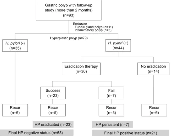
Fig. 1. Flow chart of this study.
H. pylori, Helicobacter pylori;
HP, Helicobacter pylori.
과형성 용종(hyperplastic polyp), 위저선 용종(fundic gland polyp), 위선종(gastric adenoma) 등이 있다.2 위용종은 대개 무증상으로, 상부위장관 내시경 검사에서 우연히 발견되는 경 우가 대부분이다.
위용종 환자를 대상으로 한 몇몇 연구들에서 헬리코박터 파일로리(
Helicobacter pylori
,H. pylori
) 제균 후 과형성 위 용종의 소실이 보고되었고,3-6H. pylori
양성인 과형성 위용종 환 자에서H. pylori
제균 요법을 시도해 볼 수 있다고 하였다.1-3,7,8 한편, 내시경으로 과형성 위용종이 절제된 경우,H. pylori
감염 상태가 과형성 용종의 재발에 영향을 미치는지에 대해서 는 잘 알려져 있지 않다. 따라서, 본 연구는 내시경으로 과형 성 위용종을 제거한 이후H. pylori
감염 및 제균 여부에 따른 위용종의 재발률에 대해 비교 분석해 보고자 하였다.대상 및 방법
1. 대상
2011년 1월부터 2016년 12월까지 위용종으로 진단받은 18세 이상의 성인을 대상으로 후향 연구를 시행하였다. 위용종을 조직생검 겸자나 내시경 용종 절제술 또는 내시경 점막 절제 술(endoscopic mucosal resection)로 제거하고, 조직 검사 결과 과형성 용종으로 확인된 예 중에서 최소 2개월 이상 추 적된 환자를 대상으로 하였다. 이 연구는 고려대학교 안산병
원 연구윤리심의위원회의 승인을 받았다(IRB No. AS17120-001).
2. 방법
1) 위용종의 진단 및 재발 확인
상부위장관 내시경 검사에서 위용종이 발견되면, 용종의 크 기에 따라 조직생검 겸자로 제거하거나, 용종 절제술 또는 내 시경 점막 절제술을 시행하였고, 병리조직 검사를 통해 과형 성 용종을 진단하였다. 위용종을 제거하고 최소 2개월 이후 시행한 상부위장관 내시경 검사에서 용종의 재발 여부를 평가 하였다.
2) H. pylori 감염
H. pylori
감염 여부는 상부위장관 내시경 검사를 통하여 전정부와 위체부에서 얻은 생검조직으로 신속요소분해효소 검사(campylobacter-like organism, CLO test) 또는 조직 검사를 시행하였으며, 그중 하나의 검사에서 양성이면H. py- lori
감염 양성으로 판정하였다. 조직 검사에서는 크레실 바이올 렛(cresyl-violet) 염색을 사용하였다.3) H. pylori 제균 치료와 효과 판정
H. pylori
감염 양성으로 진단된 환자는 기본 삼제 요법 (amoxicillin 1.0 g bid, clarithromycin 0.5 g bid, 양성자Table 1. Baseline Clinical Characteristics of Study Population according to H. pylori Infection
H. pylori (-) (n=35)
H. pylori (+)
(n=44) p-value
Age (years) 64.7±12.7 60.4±11.5 0.073
Sex Male Female
11 (31.4) 24 (68.6)
17 (38.6) 27 (61.4)
0.512
Diabetes mellitus 6 (17.1) 11 (25.0) 0.405
Hypertension 14 (40.0) 17 (38.6) 0.903
Alcohol 8 (22.9) 15 (34.1) 0.281
Smoking 4 (11.4) 7 (15.9) 0.574
Values are presented as mean±standard deviation or n (%).
H. pylori, Helicobacter pylori.
Table 2. Characteristics of Gastric Hyperplastic Polyps H. pylori (-)
(n=35)
H. pylori (+)
(n=44) p-value
Size (mm) 11.5±6.0 12.7±7.2 0.503
Location 0.172
Antrum 11 (31.4) 19 (43.2)
Body 15 (42.9) 19 (43.2)
Cardia/fundus 9 (25.7) 6 (13.6)
Number 0.076
1 21 (60.0) 32 (72.7)
≥2 14 (40.0) 12 (27.3)
Values are presented as mean±standard deviation or n (%).
H. pylori, Helicobacter pylori.
Fig. 2. Comparison of gastric hyperplastic polyp recurrence according to HP eradication status (HP eradicated vs. HP persistent). HP, Helicobacter pylori.
펌프 억제제 표준 용량 bid) 또는 non-bismuth 사제 요법인 동시 요법이나 순차 요법으로 제균 치료를 1-2주간 시행하였 다. 치료 종료 최소 4주 이후 상부위장관 내시경 검사를 통한 조직 검사와 CLO test 또는 요소호기 검사를 시행하였으며, 어느 한 검사에서라도 양성으로 확인되면
H. pylori
제균 실 패로 판정하였다.3. 통계분석
통계분석은 SPSS Statistics ver. 20.0 (IBM Co., Armonk, NY, USA)을 이용하였으며, 범주형 자료들은 chi-square test, 연속형 자료들은 t-test를 이용하여 비교하였다. p값이 0.05 미만인 경우 통계학적으로 유의한 것으로 정의하였다.
결 과
1. 환자 및 위용종의 특성
2011년부터 2016년까지 비선종성 용종을 절제하고, 2개월 이상 추적 관찰한 환자 93명 중에서 위저선 용종 11명과 염증 성 용종 3명을 제외하고, 과형성 용종 환자 79명을 분석하였다.
대상 환자 79명 중에서 35명은
H. pylori
음성, 44명은 양성이었고, 평균 추적 기간은 16.4개월(2-57개월)이었다.
H. pylori
감염이 양성인 44명 중 30명에서 제균 치료를 시행하였다.이 중 23명(76.7%)에서 제균 치료가 성공하였고, 7명(23.3%) 에서 제균 치료가 실패하였다(Fig. 1). 두 군의 평균 연령은
H. pylori
음성군에서 64.7±12.7세,H. pylori
양성군에서 60.4±11.5세로 양군 간에 유의한 차이를 보이지 않았고 (p=0.073), 성별이나 당뇨, 고혈압의 과거력, 흡연 및 음주력 에서도 두 군 간에 유의한 차이는 없었다(Table 1). 또한, 용 종의 크기나 위치, 개수에 있어서도 유의한 차이는 없었다 (Table 2).2. H. pylori 감염 및 제균 여부에 따른 위용종의 재발 대상 환자 79명 중 20명(25.3%)에서 추적 상부위장관 내시 경 검사 결과 용종 재발이 관찰되었다. 처음
H. pylori
감염이 음성인 35명 중에서는 6명(17.1%), 처음H. pylori
감염이 양 성으로 확인된 44명 중에서는 14명(31.8%)에서 용종이 재발 하였으나,H. pylori
감염 상태에 따른 용종 재발률은 유의한 차이가 없었다(p=0.136).H. pylori
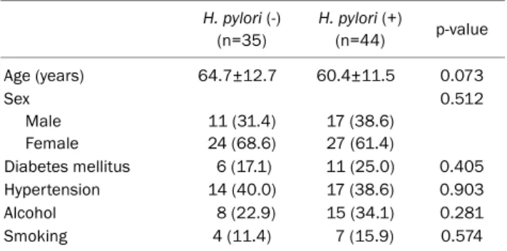
감염이 양성으로 확인되어 제균 치료를 시행한 30명 의 환자 중에서 제균 치료를 성공한 23명을 HP (Helicobacter
pylori
) eradicated group, 제균 치료가 실패한 7명을 HP persistent group으로 분류하여 과형성 위용종의 재발률을 비교하였을 때, 재발률은 HP eradicated group에서 21.7%, HP persistent group에서 42.9%로, HP persistent group에Table 3. Risk Factors of Gastric Hyperplastic Polyp Recurrence (Univariate and Multivariate Logistic Regression Analysis)
Univariate analysis Multivariate analysis
OR (95% CI) p-value OR (95% CI) p-value
Age (≥50) 1.63 (0.48-5.53) 0.430 1.51 (0.37-6.13) 0.562
Sex (male) 0.72 (0.24-2.15) 0.557 1.31 (0.30-5.61) 0.721
Diabetes mellitus 1.13 (0.32-3.97) 0.848
Hypertension 1.72 (0.58-5.09) 0.330 1.40 (0.42-4.63) 0.581
Alcohol 1.90 (0.56-6.46) 0.304 2.15 (0.45-10.2) 0.336
Smoking 0.89 (0.21-3.74) 0.872
Polyp size (≥10 mm) 0.56 (0.20-1.59) 0.275 1.32 (0.40-4.30) 0.650
Location of polyps
Non-antrum 1 1
Antrum 0.52 (0.17-1.63) 0.263 0.49 (0.13-1.84) 2.292
Polyp numbers
1 1
2-4 0.77 (0.24-2.48) 0.666
≥5 1.39 (0.12-16.6) 0.793
Initial HP positive status 2.26 (0.76-6.67) 0.141 1.66 (0.42-6.62) 0.474
Final HP positive status 0.31 (0.11-0.92) 0.035 0.28 (0.09-0.90) 0.033
OR, odds ratio; CI, confidence interval; HP, Helicobacter pylori.
Fig. 3. Comparison of gastric hyperplastic polyp recurrence according to final HP status (HP negative status vs. HP positive status). HP, Helicobacter pylori.
서 재발이 더 많았으나, 통계적 유의성은 없었다(p=0.269) (Fig. 2).
추적 상부위장관 내시경 검사 결과 위용종이 재발한 20명 에서 재발한 용종의 개수는 1-4개, 크기는 3-11 mm였으며, 조직생검 겸자 또는 용종 절제술로 제거하였다. 위용종이 재 발한 20명 중 17명은 재발 용종의 위치가 처음 발견된 용종과
같은 전정부, 위체부, 위저부에서 재발하였고, 처음
H. pylori
감염이 음성이었다가 재발한 6명 중에서 1명은 위저부에서 전정부로,H. pylori
제균 치료에 실패한 후 재발한 3명 중 1명과 처음H. pylori
양성이었으나 제균 치료를 하지 않고 재발한 6명 중 1명 등 2명은 전정부에서 위체부로 재발 용종 의 위치가 바뀌었다.3. 최종 H. pylori 감염 여부에 따른 위용종의 재발
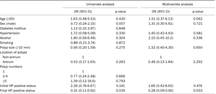
H. pylori
감염이 양성으로 확인되었으나 치료를 받지 않았 던 14명과 제균 치료를 시행하였으나 치료 실패한 7명을 최종H. pylori
감염이 양성으로(final HP positive group), 처음H. pylori
감염이 음성인 35명과 제균 치료가 성공한 23명을 최종H. pylori
감염 음성으로(final HP negative group) 구 분하였고, 각각H. pylori
양성인 그룹은 21명(26.6%),H. py- lori
음성인 그룹은 58명(73.4%)이었다(Fig. 1). 두 군의 위용 종의 재발률을 비교하였을 때, 최종H. pylori
양성군 중 9명 (42.9%)에서 과형성 위용종이 재발하였고, 최종H. pylori
음 성군에서 11명(18.9%)이 재발하여, 최종H. pylori
양성군에 서 재발률이 유의하게 더 높았다(p=0.031) (Fig. 3).4. 위용종의 재발에 영향을 주는 요인
위용종의 재발에 영향을 주는 요인에 대해 단변량 분석을 시행하고, 단변량 분석 결과 및 임상적으로 의미를 가질 수 있는 가능성이 있는 변수(나이, 성별, 처음 및 최종
H. pylori
감염 상태 등)를 가지고 이분형 로지스틱 회귀분석을 이용하 여 다변량 분석을 시행하였다. 그 결과 최종H. pylori
감염이 양성인 경우에서만 과형성 위용종의 재발률에 영향을 미치는것으로 확인되었다(Table 3).
고 찰
일반적으로 위용종은 0.4-2.0% 정도의 빈도로 발견되며,9,10 20대에서 위용종의 발견 빈도는 0.8%이나 70대의 노령에서 는 7.2%로 높은 빈도를 보이는 것과 같이, 연령이나 환자 개 개인의 특성에 따라 발견 빈도가 다르게 보고되기도 한다.11-13 위용종은 위점막의 정상적인 반응에 의해 발생될 수도 있 고, 염증성 반응에 의해서도 발생할 수 있다. 염증성 반응의 하나인
H. pylori
감염과 위용종의 발생과의 사이에도 관련성 이 있는 것으로 보고되고 있는데,14특히,H. pylori
가 위점막 의 손상을 일으켜 염증 반응의 매개체를 분비하여, inter- luekin-1β나 간세포성장인자(hepatocyte growth factor) 등 의 신호자극 물질을 활성화시켜 위축성 위염, 장상피화생, mucosa-associated lymphoid tissue lymphoma 및 위암 등을 발생시킬 수 있는 것과 마찬가지로, 이러한 신호자극 물 질들이 상피세포의 증식과 소와 증식(foveolar hyperplasia) 을 일으켜 과형성 위용종을 발생시킨다고 보고된다.15,16국내 연구에서 가장 흔하게 발견되는 위용종은 과형성 용 종으로 알려져 있으며, 다음으로 위저선 용종과 선종성 용종 의 순으로 많은 것으로 나타났지만, 서구에서는 위저선 용종 이 가장 흔한 것으로 나타났다.13,17,18 과형성 위용종은 여성에 서 높은 발생 빈도를 보이고, 대부분 크기는 1-2 cm 이하이 며, 유경성, 무경성 등 다양한 내시경 소견을 보인다.11,19 과형 성 위용종은 대부분 양성 경과를 보이며, 악성 종양은 약 0.4% 정도로 알려져 있다. 최근 국내외에서 과형성 용종의 개수, 크기 및 분엽화 등 용종의 특성과 악성 변화의 상관관계 에 관한 연구가 보고되었고, 크기가 1 cm를 초과하는 용종은 악성 변화의 위험성이 높아 용종 절제술을 권장하고 있다.20,21 크기 1 cm 이하의 작은 용종의 치료에 대해서는 정립된 개념이 없으나, 일반적으로 작은 용종은 조직 생검을 통해 제거하거나 조직 검사 결과를 바탕으로 내시경 추적 관찰을 하게 된다.22 또 한, 위용종 환자들에서
H. pylori
제균 치료를 하였을 때, 용종이 소실된다는 결과들이 보고되고 있어, 작은 용종에서는H. pylori
제균 치료를 먼저 하는 것이 권고되기도 한다.1-3,6-8본 연구에서 과형성 용종을 내시경으로 제거하고 최소 2개 월 이상 추적한 환자들을 대상으로
H. pylori
제균 여부와 최 종H. pylori
감염 여부에 따른 재발률을 비교하였다. 국내에 서는 아직 과형성 용종의H. pylori
제균 치료가 보험 급여가 되지 않아 소화성 궤양 반흔 등의 다른 적응증이 있거나 환자 가 원할 경우 제균 치료를 시행하였고, HP eradicated group 이 HP persistent group과 비교하여 재발률이 낮았으나, 대 상 환자가 상대적으로 적어 통계적 유의성을 확보하지 못한것으로 판단된다. 최종
H. pylori
감염 여부에 따라 비교하였 을 때는, 내시경으로 위용종을 제거한 이후에도H. pylori
감 염이 있는 것이H. pylori
감염 음성인 군보다 과형성 위용종 의 재발을 높일 수 있다는 것을 확인하였다.본 연구는 후향적 연구로서 선택 편향이라는 한계점과 단 일기관의 다소 적은 대상의 환자 연구라는 한계점이 있다. 그 러나 내시경으로 과형성 위용종을 제거한 이후
H. pylori
감 염 상태와 제균 여부에 따른 과형성 위용종의 재발에 관한 비교 연구는 처음이라는 점에 있어서 의의를 둘 수 있겠다.이에 따라 추후 많은 환자들을 대상으로 한 대단위 연구가 필요하며, 위용종의 병태생리 중 악성 변화에 미치는 영향이 보고된20 p16 하향조절, cyclin D1, Ki-67 이외의 분자 생물학 적 변수를 찾는 연구 및
H. pylori
제균 치료 후 위용종의 소실 에 관여하는 인자에 대한 연구가 더 필요할 것으로 생각한다.결론적으로, 위용종을 내시경으로 제거한 후에도
H. pylori
감염 양성인 군이H. pylori
감염 음성인 군보다 과형성 위용 종의 재발 빈도가 높은 경향을 보였다. 또한, 단변량 및 다변 량 분석 결과 최종H. pylori
감염 양성은 과형성 위용종의 재발에 영향을 미치는 유의한 위험인자로 확인되었다.요 약
목적: 본 연구는 과형성 위용종을 내시경으로 제거한 이후 최 종
H. pylori
감염 상태와H. pylori
제균 여부에 따른 과형성 위용종의 재발률에 대해 알아보고자 하였다.대상 및 방법: 2011년 1월부터 2016년 12월까지 과형성 위용 종을 내시경으로 제거하고, 상부위장관 내시경 추적 검사를 시행한 79명의 환자를 대상으로 진료기록을 후향적으로 분석 하였다.
H. pylori
감염과 성공적인 제균 여부는 내시경 생검 조직의 cresyl violet 특수염색과 CLO test, 또는 요소호기 검사를 통해 평가하였다. 상부위장관 내시경 추적 검사에서 과형성 위용종의 재발을 조사하고, 최종H. pylori
감염 상태 와 제균 여부에 따른 차이가 있는지 비교하였다.결과: 평균 16.4개월의 대상 환자 추적 기간 동안, HP pos- itive status group이 HP negative status group보다 과형 성 위용종의 재발률이 유의하게 더 높았다. HP eradicated group과 HP persistent group과의 비교에서는 HP persis- tent group에서 재발률이 더 높았으나, 통계적 유의성은 없었 다. 위용종의 재발에 영향을 미치는 인자에 대한 단변량 및 다변량 분석결과, 최종
H. pylori
감염 상태가 재발률에 유의 한 영향이 있었다.결론: 최종
H. pylori
감염 상태는 과형성 위용종을 내시경으 로 제거한 이후 위용종의 재발에 유의한 영향이 있었으나,H.
pylori
제균 여부에 따른 위용종의 재발률은 통계적 유의성이없었다.
색인단어: 위종양; 헬리코박터 파일로리; 치료법; 재발
REFERENCES
1. Park DY, Lauwers GY. Gastric polyps: classification and manage- ment. Arch Pathol Lab Med 2008;132:633-640.
2. Goddard AF, Badreldin R, Pritchard DM, Walker MM, Warren B;
British Society of Gastroenterology. The management of gastric polyps. Gut 2010;59:1270-1276.
3. Nam SY, Park BJ, Ryu KH, Nam JH. Effect of Helicobacter pylori infection and its eradication on the fate of gastric polyps. Eur J Gastroenterol 2016;28:449-454.
4. Kume K, Hirakoba M, Murata I, Yoshikawa I, Otsuki M.
Disappearance of both MALT lymphoma and hyperplastic polyps in the stomach after eradication of Helicobacter pylori. Am J Gastroenterol 2001;96:2796-2797.
5. Saccá N. Hyperplastic gastric polyps and Helicobacter pylori.
Scand J Gastroenterol 2003;38:904.
6. Ohkusa T, Miwa H, Hojo M, et al. Endoscopic, histological and se- rologic findings of gastric hyperplastic polyps after eradication of Helicobacter pylori: comparison between responder and non-responder cases. Digestion 2003;68:57-62.
7. Abraham SC, Nobukawa B, Giardiello FM, Hamilton SR, Wu TT.
Sporadic fundic gland polyps: common gastric polyps arising through activating mutations in the β-catenin gene. Am J Pathol 2001;158:1005-1010.
8. Malfertheiner P, Megraud F, O'Morain CA, et al. Management of Helicobacter pylori infection-the Maastricht V/Florence con- sensus report. Gut 2017;66:6-30.
9. Davis GR. Gastric polyps. In: Sleisenger MH, Fordtran JS, eds.
Gastrointestinal disease. 10th ed. Philadelphia: WB Saunders, 2015:763-764.
10. Carmack SW, Genta RM, Schuler CM, Saboorian MH. The current spectrum of gastric polyps: a 1-year national study of over 120,000 patients. Am J Gastroenterol 2009;104:1524-1532.
11. Kamiya T, Morishita T, Asakura H, Munakata Y, Miura S, Tsuchiya
M. Histoclinical long-standing follow-up study of hyperplastic pol- yps of the stomach. Am J Gastroenterol 1981;75:275-281.
12. Fan NN, Yang J, Sun G, et al. Changes in the spectrum of gastric polyps in the Chinese population. World J Gastroenterol 2015;21:9758-9764.
13. Morais DJ, Yamanaka A, Zeitune JM, Andreollo NA. Gastric pol- yps: a retrospective analysis of 26,000 digestive endoscopies.
Arq Gastroenterol 2007;44:14-17.
14. Yasunaga Y, Shinomura Y, Kanayama S, et al. Increased pro- duction of interleukin 1 beta and hepatocyte growth factor may contribute to foveolar hyperplasia in enlarged fold gastritis. Gut 1996;39:787-794.
15. Lipkin M, Enker WE, Winawer SJ. Tritiated-thymidine labeling of rectal epithelial cells in ‘non-prep’ biopsies of individuals at in- creased risk for colonic neoplasia. Cancer Lett 1987;37:153-161.
16. Bechi P, Balzi M, Becciolini A, et al. Helicobacter pylori and cell proliferation of the gastric mucosa: possible implications for gas- tric carcinogenesis. Am J Gastroenterol 1996;91:271-276.
17. Archimandritis A, Spiliadis C, Tzivras M, et al. Gastric epithelial polyps: a retrospective endoscopic study of 12974 symptomatic patients. Ital J Gastroenterol 1996;28:387-390.
18. Jalving M, Koornstra JJ, Wesseling J, Boezen H, DE Jong S, Kleibeuker JH. Increased risk of fundic gland polyps during long‐
term proton pump inhibitor therapy. Aliment Pharmacol Ther 2006;24:1341-1348.
19. Oberhuber G, Stolte M. Gastric polyps: an update of their pathol- ogy and biological significance. Virchows Arch 2000;437:581-590.
20. Ahn JY, Son DH, Choi KD, et al. Neoplasms arising in large gastric hyperplastic polyps: endoscopic and pathologic features.
Gastrointest Endosc 2014;80:1005-1013.e2.
21. Anjiki H, Mukaisho K, Kadomoto Y, et al. Adenocarcinoma arising in multiple hyperplastic polyps in a patient with Helicobacter py- lori infection and hypergastrinemia during long-term proton pump inhibitor therapy. Clin J Gastroenterol 2017;10:128-136.
22. Ji F, Wang ZW, Ning JW, Wang QY, Chen JY, Li YM. Effect of drug treatment on hyperplastic gastric polyps infected with Helicobacter pylori: a randomized, controlled trial. World J Gastroenterol 2006;
12:1770-1773.