大혐放射練醫學會誌 第 22 卷 第3 號 pp. 332 - 338, 1986 Journal 01 Korean Radiological Society, Vo1.22, No.3, 1986
Magnetic Resonance (MR) Imaging in Delayed Encephalopathy of Acute Carbon Monoxide Poisoning *
〈국문초록〉
- Cornparison with CT -
Kee Hyun Chang, M.D., Chang Hae Suh, M.D., In Wool
‘
Choo, M.D.Departmenl 01 Radiology, College 01 Medicine, Seoμ1 Natioηal U,ηwerslty
急性-嚴化밟素中毒後遺효에서의 磁氣共嗚影像*
-
電算化斷層播影과의 비교
-서울大學校 醫科大學 放射線科學敎室 張基賢· 徐昌海 · 朱仁옆
急性 -顧化뚫素中毒後追tJE을 보이는 11 例에 서 磁氣共嗚影像(MRI)과 電算化斷層擬影(CT) 을 시행 하여 각각 그 所見을 관찰 분석하고, 융?짧I的 않{直을 81교 검토하였 다.
MRI는 0.15 Tesla 常電향型을 이용하여 部分館和回復, T2 행調스판에코 또는 反轉回復등의 마
%택f 펄스시권스로 촬영하였다. 白質의 脫廣質을 암시하는 白質의 이 상소견이
MRI에서 는 4 例에 서 관찰펀 반면, CT에서는 2 例에서만 관찰되었다. 白質의 이상소견은 특히 T2 彈調스판에코像 과 反轉며復像에서 對照、度가 가장 뚜렷하였 으며 部分짧和回復像파 CT像에서는 對照度가 낮았마
淡蒼球病짧을 보인 1例와 腦쫓縮을 보인 3 例는 MRI와 CT에서 모두 함께 판찰되었다.
白質病變의 발견율은 CT보다 MRI 가 높으므로 -醒化뚫素中毒後道tJE으호 白質病變이 의심될 때
는
MRI를 먼저 시행하는 것 이 바랑직하다고 생각된다.
Eleven magnetic resonance (MR) and computed tomographic (CT) imagings were performed in nine patients with mild to moderate degree of delayed neuropsychiatric symptoms lollowing acute carbon monoxide (CO) poisoning, to evaluate the capability 01 MR in demonstrating any additional linding to CT. The MR images were
。 btainedusing 0.15 Tesla resistive system with various combination 01 three pulsle sequences, including partial saturation recovery, T2-weighted spin echo and inversion recovery
Bilateral white matter abnormalities suggesting demyelination were demonstrated in 4 patients with MR and in only 2 patients with CT. The contrast discrimination between normal and abnormal white matter proved to be better with T2-weighted spin echo and inversion recovery than with partial saturation recovery and CT
But necrosis 01 the globus pallidus (1 patient) and diffuse atrophy (3 patients) were equally demonstrated on both MR and CT
It is suggested that MR be used as a initial imaging method in the evaluation of the delayed encephalopathy following acute CO poisoning, especially for the detection of the possible white matter lesions
* 본 논문은 1985 년도 서울대학병원 특진연구비의 보조로 이루어진 것 임. Received April 23, 1986; accepted May 31, 1986
- 332-
Kee Hyun Chang, et al.: MRI in Delayecl Encephalopathy of Acute Carbon Monoxicle Poisoning -
Acute carbon monoxide (CO) poisoning produces Nine palienls were aged 1‘rOI11 201068 ycars (5 l11en hypoxia by c1isplacing oxygen from hemoglobin and and 4 wOlllen). Onc palienl (casc 1) had 3 sludies; thc preventing its release from hemoglobin in tissues, often first study in lhe 2 weeks, lhe scιond inlhc 1 ι’ monlh resulting in fetal event.' and the last in lhe 5 Illonths al‘tcr thc aιcidcn t. F ou J
Victims who survive acute CO poisoning may have patients wcre Sllldicd 2 to 3 J110nths aflCI. lhc accidcnts.
various delayed symptoms and signs. Occasionally, an In the 이her 4 palicnl 、 1 hc "ιIJlS IVCïC ohl‘lillCd 7 10 apparent recovery is foll。、vedwithin two days to three 48 months after the aιιidcnts.
weeks by a sudden neurological deterioration.' The AlI the MR studies wcre perforlllcd u 、 ing a ()‘ 15 degree of neuropsychiatric symptoms clepends upon Tesla resistive magnet cleveloped by Korca ^dvanccd the extent and severity 01' the pathologic changes in Institute of Science & Technology, Seoul, Korca‘ wilh
the brain. a aperture of 25cl11 head ιoi l.
The pathologic effects of CO poisoning are pre- Using a two-cIimensional Fourier transform mClhod sent in almost all organs of patients. However, the with 256 phase-encocIing data lines, various combina most important changes occur in lhe brain, which con- tions 01' following three pulse sequences were lIscd;
sist of necrosis of the globus pallidus and reticular zone partial saturation recovery (spin echo TE 30, TR of the substantia nigra, and the degeneration of the 二 500ms), inversion recovery (TE 30, TI 400, cerebral white matter.' . J TR 1,400ms) ancl T2 weightecl spin echo (TE
The cliagnostic superiority of magnetic resonance 60, TR 1 ,000ms) techniques. A total of 8-12 axial (MR) over CT has already been shown for various slices of about 13mm slice thickness 、vereoblained in neurological disease.4-7 But we could not find any MR each pulse sequence.
description on CO poisoning in the literatures, even In all patients the X-ray CT scans were obtained though the computed tomography (CT) findings and within 1 day to 1 week of the MR study. The CT scans their correlation with neuropathologic findings in CO were macle with an G E CT /T 8,800 ancl incluclecl poisoning have previously been described. 8." precontrast ancl postcontrast enhancecl-scans. The CT To evaluate the capability of M R in demonstrating scanner was operated at 120k Vp with contiguou any additional finding to CT in delayed 10mm sections
encephalopathy of CO poisoning, we studied M R in nine patients and compared the MR with CT results.
Subjects and Methods
Nine patients with a cliagnosis of delayed encephalopathy of acute CO poisoning were studied.
The diagnosis of acute CO poisoning was reaclily made by overwhelming circumstantial evidence. In Korea, many people have a unique, traditional home heating system using coal briquette as a fuel. CO-containing coal briquette gas can accidentally leak into the room through the cracks, resulting in acute CO poisoning There are so many victims in the winter season. Our nine patients who survived acute CO poisoning has had mild to moderate neuropsychiatric symptoms and signs, after recovery from the initial coma state. There is no patient with grave symptoms such as vegetative or quaclriplegic pat ients in our series
Results
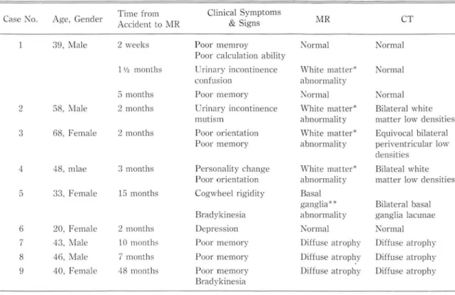
The clinical, MR and CT findings were summariz- ed in Table 1.
Four of 5 patients examined within 3 month after the accidents showed symmetrical bilateral abnor- malities in the white matter of the periventricular area and centrum ovale. These white matter abnormalities are seen as bright signal intensities on T2 weighted spin echo image and dark, low signal intensities on Tl weighted inversion recovery images, representing long T2 and Tl relaxation times, retrospectively.
In one patient with 3 times follow-up studies (case 1), there were no abnormalities on the initial M R studied iri the complete recovery state (lucicl internal), 2 weeks after the acciclent. But on the second M R studied 1 씨!eek after the delayed symptoms of urinary incontinence ancI confusion appeared, symmetrical
- The JOllrnal of the Korean Radiological Society, Vol. 22, No. 3, 1986 -
Table 1. SlIl1lmary of Clinical, MR ancl CT Finclings in Oelayed Encephalopathy of Acute CO Poisoning
Case No Age, Genclel Til1le frolll Clinical Sylllptollls
MR CT
Acciclent to MR & Signs
39, Male 2 weeks Poor memroy Normal Norl1lal
Poor calclllation ability
11; ' Illonths Urinary incontinence White Illatter* Normal
confllsion abnormality
5 months Poor memory Normal Normal
2 58. Male 2 months Urinary incontinence White matter* Bilateral 、、lhite
mlltlsm abnormality Illatter low c1ensities 3 68, Female 2 months Poor orientation White Illatter* Eqllivocal bilateral
Poor Illelllory abnorlllality periventriclllar low c1ensities
4 48, mlae 3 months Personality change White Illatter* Bilateal white Poor orientation abnormality Illatter low c1ensities 5 33. Felllale 15 months Cog、.vheel rigiclity Basal
ganglia** Bilateral basal Braclykinesia abnormality ganglia lacllnae
6 20, Felllale 2 months Oepression Normal Norlllal
7 43, Male 10 11l0nlh
,
POOr melllory Oiffllse atrophy Oiffuse atrophy 8 46, Male 7 months POOr melllory Oiffllse atrophy Oiffllse atrophy 40, Felllale 48 11l0nths Poor memory Oiffllse atrophy Oiffllse atrophyBradykinesia
White matter abnormality on MR represents bilaterallow signal intensities in inversion recovery illlages ancl/or bilateral high signal intensities in T2-weightecl spin echo ill1ages in the white matter of c1eep periventriclllar area ancl centrum ovale
* * Basal ganglia abnormality on M R represents bilateral focallo\V signal intensitips in globlls pallicllls on inversion recovery
bilateral white matter abnorll1alities were were changes of attenuation on CT. Subtle low den- demonstrated. The white ll1atter abnorll1alities disap- sities on CT were easily dell10nstrated on MR peared on the 3rd MR studied 5 1l10nth after the acci- (Fig. 2).
dent (Fig. 1). Only one patient who was examined 15 1l10nths
In general, the T2 weighted spin-echo and inver- after the accident dell10nstrated bilatera1 symll1etrical sion recovery techniques were useful in detecting the 10w densities in the globus pallidus, which was seen white matter abnorma1ity itself. Although partial on the both CT and inversion recovery images of MR saturation recovery ill1ages provided excellent (Fig. 3).
anatomic definition, they were not helpful in Three patients examined 7 months or more after separating the lesion froll1 norll1al surrounding the accidents dell10nstrated dilatation of the lateral
structures. ventricles and widening of the sulci, indicating diffuse
On the CT scans, only two of 4 patients with the atrophy.
white ll1atter abnormalities on MR showed diffuse
periventricular white matter low densities. The other Discussion two patient showed norll1al to subtle low densities on
CT, which is very difficult to differentiate the norll1al The neuropathologic changes including necrosis of from the abnormal white matter. the basal ganglia and degeneration of the white mat-
Contrast discrimination between normal and ab- ter in CO poisoning have been well documented in normal white matter was more apparent on MR than autopsy specill1ens and experill1ental animals. The le-
- Kee Hyun Chang, et al.: MRI in Delayed Encephalopathy of Acute Carbon Monoxide Poisoning -
A
C
E
B
D
Fig. 1. Case 1.
- 335-
A. 2 weeks after accident. SE 1000/60‘ No abnor- mality. A black dot in periventricular area of left parietal lobe is an artifact.
B,C,D. 10 month after accident.
B. SE 1000/60. Bilateral symmetrical areas of in- creased signal intensity in white matter C. IR 1400/400/30. Bilateral symmetrical areas of
decreased signal intensity in white matter‘ D. Constrast enhanced CT. No apparent abnor-
mality.
E. 5 months after accident. SE 1000/60. Bilateral white matter abnormalities disappeared. A black dot in left periventricular area is an ar- tifact.
- The JOl1rnal of the Korean Racliological Society, Vol. 22, No‘ 3, 1986
A
Fig. 2. Cäse ::J. 2 l1lonlhs after acciclent
A
A. SE 1000/(
,
0. ßiläteräl sYJll1llelricäl areas of increasecl signal intensity in white ll1atter. A 、vhitec10t in lefl lateral venlricle is an artifact.B. Contrasl-enhäncecl CT. Eql1ivocal white matter lesions showing bilateral periventric111ar low c1ensities
Fig. 3. Case ii. 1:ï months afLer acciclent A. IR I.JOO/4ll0/::JOO. ßilateral äreas of
focal 1 。、vclensity in globl1s pallidl1s ß. Non-contrasl CT. ßilaleral globl1s
pallicll1s lacunae
- 336-
B
B
- Kee Hyun Chang. et al.: MRI in Delayed Encephalopathy of Acute Carbon Monoxide Poisoning
sions are symmetrical, and involve the globus pallidus and the deep white matter of the periventricular cen trum semiovale and the corpus callosum. In acute state of CO poisoning the pathological alteration is characteristic of the hypoxic change producing aciclotic
이igemic edema, which progresses to incomplete
necrosis causing demyelinalion and gliosis, 01' com- plete necrosis terminating in cyslic lesions in latel state.2
‘
AII the abnormal M R findings in our cases were attributed 10 the lale palhologic change; the white matter abnormalities in our 4 cases were probably due to the demyelination, but not to the acute edematolls change. The demyelination may be reversible. In OUI case 1 with 3 times follow-up MR, the white matter abnormalities shown only on the 2nd MR (101 month) highly suggest reversible. incomplete demyelination.The superior sensitivity of MR to CT has been reported previously in a variety of demyelinating
with mild neuropsychiatric symptoms and signs, like those in our patients.
Most of the patients showing prominent abnormaì CT features reported in the literat 비 e had grave neurologic sequelae.8
With exception of the white malter lesions, the M R finclings appearecl to be.generally correlatecl with the CT fïnclings. The basal ganglia lacunae ancl c1ifTuse atrophy could be equally demonstrated in both M R and CT.
The M R is suggestecl to be used as a initial imag- ing methocl in the evaluation of the delayecl encephalopathy following acute CO poisoning, especially for the cletection of the possible white mat- ter lesions.
REFERENCES
disease including multiple sclerosis, subcortical 1. Lilienthal JL: Carbon monoxlde. Pharmacol Rev 2:32 .. -.35 .. , arteriosclerotic encephalopathy, progressive multifocal 7950
leukoencephalopathy, myelinolysis, methotrexate 2. Lapresle J, Fardeau M: The central nervous Sy5tPIll and encephalopathy and anoxic change."-7 The carbonmonoxidepoisoning. 11. Anatolllicalstuclyofbrain demyelinating process usually demonstrated prolonged lesiα15 following intoxication with carbon monoλ id, ‘ μ2 Tl and T2 on the MR images." cases). Prog 8rain Res 2 .. :31-75, 1967
In our two patients with mild clinical symptoms 3. Ginsberg MD. Myers RE. McDonagh BF: Experimel1tal car- (case 1 & case 3) the M R could detect the white mat- bon monoxide encephalopathy inthe primate. 11. Clinica!
ter lesions which were not evidently demonstrated on aspects. neuropathology, and physiological correlation the CT. Bilateral symmetrical myelinopathic lesions Arch Neurol 30:209-276, 197.J
as a delayed change of CO poisoning correspond with 4. Bydder Grv\, Steiner RE, Young Ir et al: Clinical N!\;IR im.lg the areas of low signal intensity in IR image (prolongecl ing of the brain; 7.J0 cases. AjR 73921S-2.36, 1982 Tl) and of high signal intensity in SE images (prolong- 5. Brant-Zaν.vadzki M. Davis PL. Crooks LE et al: NMR ed T2) in our cases. clemonstration of cerebral abnormalities: Comparison with
Our cases proved that MR is more sensitive ancl CT AjNR FI77-12 .. , 198.1
better than CT in the contrast discrimination between 6. Bradley WC, Waluch V, Yadley RA, Wycoff RR: Com the normal and abnormal white matter of delayed parison oi CT and MR il1 .. 00 patients suspectecl clisease encephalopathy of CO poisoning. of the brain ancl cervical spinal corcl. Racliology
Although the characteristic CT findings of CO '1.52.'695-702, 198 ..
poisoning inclucling bilateral symmetric areas of low 7. Zimmerman RA, Bilaniuk LT. Goldberg HI et al: Cerebral c1ensity in the basal ganglia ancl/or diffuse low c1ensi- NMR imaging' Early results with a 0.72 T Resistive Sysl;tem ty in the white matter has been previously reportecl AjNR 5.'1-7, 198 ..
in the many literatures,8'" it seems to be sometimes dif- 8. Kill1 KSκ Weinbergn PE. Suh JH, Ho SU: Acute carbon ficult to iclentify the subtle changes of the white mat- mnonoxide poisoníng: Computecl tomogl강phy uf the ter clegeneration on lhe CT scan which may show braín. Aji\/R 1-]99-.. 02, 1980
apparently normal 01' equivocal low density in the 9. Kabayashi K, Isaki K. Fukutani Y, et al: CT fíndíngs of the periventricular white matter, especially for the patients int'elval form of c"rbon monoxíde poísoníng comparecl
- 337-
- The Journal of the Korean Radiological Society, Vol. 22, No. 3, 1986 -
with neuropathological findings. Eur NellroI23 :34-43, 1984 10. Nardizzi LR: Compllterized tomographic correlate of car- bon monoxide poisoning. Arc!1 Nellrol.36:38-39, 1919 11. Sawada Y, Sakamoto T, Nishide K et al: Correlation of
pathological findings with computed tomographic findings after acute carbon monoxide poisoning. New Engl J Med 308ρ1):1296, 1983