Received:July 2, 2015, Revised (1st) July 23, 2015, (2nd) August 1, 2015, Accepted:August 1, 2015
Corresponding to:Sang Tae Choi, Division of Rheumatology, Department of Internal Medicine, Chung-Ang University College of Medicine, 84 Heukseok-ro, Dongjak-gu, Seoul 06974, Korea. E-mail:[email protected]
pISSN: 2093-940X, eISSN: 2233-4718
Copyright ⓒ 2016 by The Korean College of Rheumatology. All rights reserved.
This is a Free Access article, which permits unrestricted non-commerical use, distribution, and reproduction in any medium, provided the original work is properly cited.
통풍 환자의 혈청 호모시스테인 변화와 요신 및 지질, 신기능 변화의 연관성: 2년 추적 관찰 연구
박은혜ㆍ박지호ㆍ송정수ㆍ최상태
중앙대학교 의과대학 내과학교실 류마티스내과
Two-year Follow-up Study of the Relationship between the Changes of Serum Homocysteine and Those of Serum Uric Acid Levels, Lipid Profiles and Renal Function in Gout Patients
Eun Hye Park, Ji Ho Park, Jung-Soo Song, Sang Tae Choi
Division of Rheumatology, Department of Internal Medicine, Chung-Ang University College of Medicine, Seoul, Korea
Objective. Gout is known to be associated with cardiovascular disease (CVD), and hyperhomocysteinemia is one of the risk factors for CVD. We investigated the associations between the change of serum homocysteine (Hcy) level and those of the other parameters including serum uric acid level, renal function, and cholesterol profiles in chronic gout patients with longitudinal follow-up data. Methods. Ninety-one male patients with chronic gout and 97 age-matched healthy male control subjects were included in the previous study. Among them, 33 patients with gout and 39 healthy control subjects underwent follow-up tests for Hcy levels with an average of 24.00±9.12 months in this study. Results. The follow-up data showed that gout patients had significantly higher levels of Hcy in serum than control subjects (16.75±5.43 μmol/L vs. 13.17±3.83 μmol/L, p=0.002). In gout patients, the change of serum Hcy level after follow up showed positive correlation with the change of creatinine (γ=0.442, p=0.009) and negative correlation with estimated glomerular filtration rate (eGFR; γ=−0.528, p<0.001).
However, the change of serum Hcy level did not show correlation with the changes of uric acid level or the lipid profiles.
Conclusion. Serum Hcy level was elevated in gout patients compared with control subjects. The change of serum Hcy level showed negative correlation with the change of eGFR. Hyperhomocysteinemia in gout patients was associated with decreased renal function, but not with serum uric acid or lipid profiles. (J Rheum Dis 2016;23:30-36)
Key Words. Gout, Homocysteine, Uric acid, Estimated glomerular filtration rate
서 론
통풍은 관절이나 조직에 요산이 침착되어 급성 관절염, 통풍결절, 콩팥 돌증 등의 다양한 증상을 나타내는 질병군 이다. 통풍의 주된 증상은 관절염이지만, 통풍 환자에서 비만, 고혈압, 당뇨병, 고지혈증 등을 포함하는 대사 증후
군의 비율은 62.8%, 대사 증후군 환자 중에서 고요산혈증 및 통풍의 유병률은 70.7%으로 보고되는 등, 최근에는 관 절 증상 이외에도 대사 증후군이나 심혈관계 질환과의 연 관성에 대해서 많이 밝혀지고 있다[1]. 더욱이 통풍에서 심혈관계 질환의 위험도가 증가하여, 이로 인한 사망 누적 위험함수 비율이 1.97 (95% 신뢰구간 1.08∼3.59)로 유의
하게 높다고 보고되었다[2]. 통풍 환자에서 고요산혈증과 심혈관계 질환의 연관성을 이해하는 기전으로, 고요산혈 증이 혈관 내피세포의 기능을 저하시키고, 통풍의 급성 혹 은 만성 염증 반응이 심혈관 질환의 발생에 관여하는 것과 같은 몇 가지 기전들이 제시되고 있으나[3,4], 통풍과 심 혈관 질환과의 직접적인 연관성에 대해서는 아직도 명확 히 알려져 있지 않다.
호모시스테인(homocysteine)은 필수 아미노산인 메티오 닌(methionine)이 시스테인(cysteine)으로 분해될 때 생 성되는 중간 대사산물이다. 혈청 호모시스테인의 농도는 methylene tetrahydrofolate reductase의 변형과 같이 호 모시스테인의 대사에 관여하는 효소의 유전적 결함이 있 거나[5], 엽산 및 비타민 B6, 비타민 B12 등의 비타민이 결핍될 때 상승할 수 있다[6]. 또한 만성콩팥질환 환자에 서도 콩팥 배설기능 저하 및 대사 이상 등으로 혈중 호모 시스테인 농도가 증가하며[7], 흡연도 혈중 호모시스테인 의 농도 증가에 영향을 준다[8]. 이렇게 발생한 고호모시 스테인혈증은 활성산소의 생성을 촉진하고 혈관 내막의 비후 및 내탄성판의 파괴, 평활근의 비후, 과도한 혈소판 응집, 혈전 생성 등의 기전에 의해 동맥 내피세포를 손상 시킨다. 고호모시스테인혈증은 관상동맥질환, 뇌졸중 및 말초혈관질환 등의 주된 위험인자 중 하나로도 알려져 있 다[9].
최근 통풍에서 혈중 호모시스테인의 농도가 상승되어 있 다는 연구 결과들이 보고되고 있다[10,11]. 저자들은 국내 의 통풍 환자를 대상으로 한 연구에서 혈청 호모시스테인 이 혈중 요산의 농도가 잘 조절되는 환자들에게서도 높게 나타났으며, 이는 콩팥 기능의 감소와 연관이 있는 것으로 보고하기도 하였다[12]. 이러한 연구들은 통풍 환자에서 심혈관 질환이 증가하는 하나의 기전에 호모시스테인이 있음을 암시한다. 그러나 이들 연구들은 모두 단면적 연구 였다. 따라서 이번 연구에서는 저자들의 연구를 2년 동안 추적 관찰한 자료를 바탕으로 하여, 통풍 환자에서 호모시 스테인의 농도 변화가 요산 및 신기능, 콜레스테롤 등의 다른 변수들의 변화들과 어떠한 연관성이 있는지에 대해 알아보고자 한다.
대상 및 방법
대상
이 연구는 저자들이 보고한 연구[12]에 등록된 환자들에 대한 전향적이고 종적인 관찰 연구이다. 중앙대학교병원에 서 American College of Rheumatology (ACR) 진단 기준 [13]에 의거하여 통풍으로 진단받은 환자를 대상으로 시행 되었다. 처음 연구에 등록된 총 91명의 통풍 환자 중에서 33명, 97명의 건강한 대조군 중에서 39명에 대하여, 2011 년 12월부터 2014년 4월까지 혈중 호모시스테인 농도를 추 적하여 검사하였다(승인번호: IRB No. C2013170 (1130)).
방법
나이, 성별, 과거력, 통풍의 임상증상 등을 기록하였다.
임상 자료는 환자들의 혈액 채취와 동일한 날에 수집하였 다. 혈중 호모시스테인, 혈당, 요산, 혈액요소질소, 크레아 티닌, 콜레스테롤 및 기타 검사 결과들을 취합하였다. 혈 중 호모시스테인은 직접 화학 발광 방법을 이용한 경쟁적 면역 분석법(ADVIA Centaur HCY; Siemens Centaur Immunoassay Systems, Las Vegas, NV, USA)으로 검사 하였다. 사구체 여과율(estimated glomerular filtration rate, eGFR) (mL/min/1.73 m2)은 Modification of Diet in Renal Disease (MDRD) 공식: eGFR (mL/min/1.73 m2)=175×Serum creatinine (exp(−1.154))×Age (exp (−0.203))×(0.742 if female)×(1.212 if black)으로 계산 하였다[14].
자료분석
모든 자료는 평균±표준편차로 표기하였다. 평균값의 비 교를 위해 independent t-test를 시행하였고, 상관 분석은 Pearson’s χ2 test나 Spearman test를 이용하였다. p값이 0.05 미만인 경우 통계적인 유의성이 있는 것으로 간주하 였다. 자료의 통계 처리는 PASW Statistics ver. 18.0 (IBM Co., Armonk, NY, USA)을 이용하였다.
결 과
환자군 특성
추적 검사를 진행한 통풍 환자 33명, 대조군 39명의 추적 검사 당시의 평균 나이는 54.30±14.59세, 57.51±13.67 세(p=0.339)였고, 평균 추적 기간은 24.00±9.12개월 (p=0.963)이었다. 처음 혈중 호모시스테인을 검사했을 당 시, 통풍 환자들의 평균 유병 기간은 3.20±1.40년이었으 며, 고혈압 및 당뇨병이 동반된 환자는 각각 16명(48.5%), 3명(9.1%)이었다. 39명의 대조군 중 고혈압 및 당뇨병이 동반된 사람은 각각 6명(15.4%), 3명(7.7%)이었으며, 추 적 기간 동안 고혈압이 새로 발생한 사람이 2명(5.1%) 있 었으나, 당뇨병 및 협심증, 심근경색, 뇌졸중이 새로 발생 한 사람은 없었다.
추적 기간 동안 통풍 환자들이 복용한 요산 저하제의 종 류는 allopurinol (n=2, 6.1%), febuxostat (n=22, 66.7%), benzbromarone (n=8, 24.2%) 단독 요법 및 allo- purinol과 benzbromarone 병합 요법(n=1, 3.03%)이었으 며, 추적 기간 동안 사용한 요산 저하제의 평균 용량은 하 루 allopuriol 166.7±115.5 mg, febuxostat 78.2±8.5 mg, benzbromarone 61.1±33.3 mg이었다. 항고혈압약제는 칼슘통로차단제(10/33, 30.3%), 안지오텐신 II 수용체 길 항제(10/33, 30.3%), 베타차단제(3/33, 9.1%), 이뇨제 (1/33, 3.0%)였으며, 아스피린을 복용하고 있는 환자는 15.1% (5/33)였다. 추적 기간 동안 급성 통풍 발작이 발생
Table 1. Follow-up data of other parameters in gout and control groups
Parameter Gout (n=33) Control (n=39)
Initial Follow-up p-value Initial Follow-up p-value Age (yr)
Uric acid (mg/dL) BUN (mg/dL) Cr (mg/dL) eGFR (mL/min) Glucose (mg/dL) AST (IU/mL) ALT (IU/mL) TC (mg/dL) LDL (mg/dL) HDL (mg/dL) TG (mg/dL)
54.30±14.59 6.00±2.39 17.12±5.57 1.19±0.25 70.75±16.09 100.06±10.13 26.45±8.01 30.95±23.95 198.20±33.83 120.30±29.57 45.23±8.91 190.06±79.77
4.45±1.59 19.39±7.00 1.17±0.29 69.61±19.24 103.58±18.45 26.97±9.98 27.27±15.80 182.53±45.48 119.60±38.46 46.40±8.72 134.58±68.51
0.007 0.017 NS NS NS NS NS 0.045
NS NS 0.001
57.51±13.67 6.01±1.25 14.67±3.47 1.19±0.72 79.93±14.68 98.38±12.96 27.03±10.84 27.51±11.49 204.61±30.55 116.43±21.86 47.83±9.98 149.36±68.13
6.07±1.63 14.44±4.46 1.60±3.69 95.55±17.68 96.51±14.61 26.26±9.02 31.07±32.38 192.44±45.56 107.71±43.32 49.31±14.83 141.83±116.36
NS NS NS
<0.001 NS NS NS NS NS NS NS Values are presented as only mean±standard deviation. ALT: alanine aminotransferase, AST: aspartate aminotransferase, BUN:
blood urea nitrogen, Cr: creatinine, eGFR: estimated glomerular filtration rate, HDL: high density lipoprotein, LDL: low density lipoprotein, NS: not significant, TC: total cholesterol, TG: triglyceride.
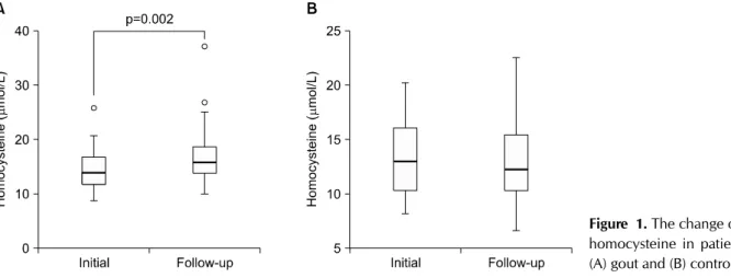
Figure 1. The change of serum homocysteine in patients with (A) gout and (B) control.
하였던 환자는 총 7명(21.2%)으로, 평균 1.29회의 통풍 발작이 발생하였고, 아스피린과 이뇨제를 사용한 환자에 서 통풍 발작의 증가 소견은 관찰되지 않았다.
통풍 환자 중 고혈압 및 당뇨병으로 투약 중인 환자는 16 명(48.5%) 및 3명(9.1%)으로 추적 후에도 변화가 없었으 나, 내당능장애가 있는 환자는 12명(36.4%)에서 17명 (51.5%)으로 추적 후 증가하였다(p=0.267). 혈청 고비중 지단백 수치가 낮은(남자 40 mg/dL 미만, 여자 50 mg/dL 미만) 환자는 10명(30.3%)에서 8명(24.2%)으로 감소하였 으며(p=0.774), 혈청 중성지방 수치가 150 mg/dL 이상으 로 높거나 혈청 중성지방 수치를 낮추기 위하여 콜레스테 롤 저하 약제를 복용 중인 환자도 26명(78.8%)에서 19명 (57.6%)으로 감소하였다(p=0.065). 다만 이들 변화들 가 운데 통계적인 유의성은 관찰되지 않았다. 추적 기간 동안 협심증, 심근경색, 뇌졸중이 새로 발생한 환자는 없었다.
혈청 호모시스테인 농도의 변화
추적 관찰이 가능했던 환자들을 대상으로 했을 때, 처음 연구 진행 당시 통풍 환자와 대조군의 혈청 호모시스테인 값은 14.38±3.74 μmol/L vs. 13.43±3.76 μmol/L (p=0.029)로 통풍 환자에서 유의하게 더 높았으며, 추적 검사에서도 16.75±5.43 μmol/L vs. 13.17±3.83 μmol/L (p=0.002) 로 대조군에 비하여 통풍 환자에게서 유의하게 높았다. 통 풍 환자에서 추적 전후의 혈청 호모시스테인 농도의 차이 는 2.37±4.10 μmol/L로 추적 후에 유의하게 높은 값을 보 였으며(p=0.002) (Figure 1A), 대조군에서는 −0.26±4.21 μmol/L로 통계적 유의성이 관찰되지 않았다(p=0.710) (Figure 1B). 통풍 환자에서 allopurinol, benzbromarone, febuxostat 등의 요산 저하제의 사용에 따른 혈청 호모시 스테인 농도 변화에 차이는 없었다. 통풍 환자와 대조군에 서 호모시스테인 외에 다른 변수들을 추적한 결과는 Table 1 에 제시하였다.
Figure 2. Correlations between the change of serum homo- cysteine level and the changes of (A) blood urea nitrogen (BUN), (B) serum creatinine (Cr), and (C) estimated glomer- ular filtration rate (eGFR). The change of serum homo- cysteine level was positively correlated with the change of Cr, and negatively correlated with the change of serum eGFR.
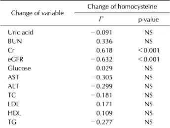
Table 2. Correlations between the change of serum homo- cysteine level and the change of other parameters
Change of variable Change of homocysteine Γ p-value Uric acid
BUN Cr eGFR Glucose AST ALT TC LDL HDL TG
−0.091 0.336 0.618
−0.632 0.029
−0.305
−0.299
−0.181 0.171 0.109
−0.277
NS NS
<0.001
<0.001 NS NS NS NS NS NS NS ALT: alanine aminotransferase, AST: aspartate aminotrans- ferase, BUN: blood urea nitrogen, Cr: creatinine, eGFR:
estimated glomerular filtration rate, HDL: high density lipoprotein, LDL: low density lipoprotein, NS: not significant, TC: total cholesterol, TG: triglyceride.
호모시스테인 수치 변화와 다른 변수들의 변화의 연관성
통풍 환자에서 추적한 혈청 호모시스테인 수치의 변화는 혈청요소단백의 변화와는 통계적으로 유의한 상관관계를 나타내지 않았으나(γ=0.183, p=0.300), 혈청 크레아티 닌의 변화와는 통계적으로 유의한 양의 상관관계(γ=0.442, p=0.009), eGFR의 변화와는 음의 상관관계를 보였다(γ
=−0.528, p<0.001) (Figure 2). 하지만 호모시스테인 수 치의 변화는 혈청 요산이나 지질의 변화와는 연관성이 없 었다(Table 2).
고 찰
이 연구에서 혈청 호모시스테인은 통풍으로 치료를 받으 며 혈청 요산 수치가 잘 조절되는 통풍 환자에서도 대조군 보다 높은 값을 나타냈다(p=0.002). 통풍 환자에서 요산 저하제의 사용에 따른 혈청 호모시스테인 농도 변화에 차 이는 없었으며, 호모시스테인 농도의 변화는 혈중 요산 및 콜레스테롤 농도의 변화와는 상관관계가 없었으나, 크레 아티닌 및 eGFR의 변화와는 유의한 연관성을 보였다(γ
=0.442, p=0.009; γ=−0.528, p<0.001, respectively).
이는 혈청 호모시스테인의 변화가 콩팥 기능의 감소와 연 관이 있음을 보여준다. 호모시스테인 농도의 변화와 혈액 요소질소의 변화는 연관성이 없었는데(γ=0.183, p=
0.300), 이는 혈액요소질소가 콩팥 기능뿐 아니라, 단백질 식이, 근육량, 체액량 등과 같은 다른 요소들에 의해서도 영향을 받기 때문이라고 생각된다. 2년 동안의 추적관찰 기간에 새롭게 발생한 당뇨병, 고혈압, 관상동맥 질환이나 뇌혈관 질환 환자는 없었으나, 내당능장애가 있는 환자는 36.4%에서 51.5%로 증가하였다.
통풍 환자에서 심혈관 질환의 유병률과 그로 인한 사망 률이 증가한다는 연구 결과들이 많이 보고되고 있다.
Framingham 연구 결과 통풍 환자에서 관상동맥질환이 60% 더 많이 발생하였고[15], 대만 연구에서는 통풍 발작 의 빈도가 심근경색의 발생과 승산비 1.18로 관련이 있다 고 보고되었다[16]. Full 등[17]의 연구에서는 통풍 환자 에서 급성 심근경색의 위험률이 26% 증가한다고 보고하 였고, 심혈관계 질환으로 인한 사망 누적위험함수 비율이 1.21 [18], 건강전문기관의 추적검사 결과에서는 통풍 환 자에서 심혈관 질환으로 인한 사망률의 상대위험도가 1.38 [19]로 보고되었다. 따라서 최근 통풍의 치료는 단순 히 관절 통증을 조절하는 것을 넘어서서, 이들 합병증을 예방하고 조절하는 것을 매우 강조되고 있다. 그러나 통풍 환자에서 심혈관 질환이 증가하는 기전에 대해서는 아직 도 명확히 밝혀지지 않고 있다. 특히 통풍 환자에서는 항 산화제 역할을 하는 요산의 농도가 높음에도 불구하고 오 히려 심혈관 질환이 증가하기에, 그 기전을 밝히기 위해 많은 연구들이 수행되어 왔다.
가장 대표적으로 밝혀지고 있는 것이 통풍과 대사 증후군 과의 연관성이다. 최근 연구들에서 통풍 환자의 대사 증후 군의 유병률은 대조군에 비해 유의하게 높은 것이 보고되 고 있다. 4,053명의 성인을 대상으로 한 단면적 연구 결과 고요산혈증과 대사 증후군, 즉 고인슐린혈증, 고혈압, 고지 혈증, 비만 등과 밀접한 연관성이 있었으며[20], 미국의 국 립건강 영양 검진 조사(1984∼1994년)의 데이터(8,807 명) 분석 결과, 통풍 환자에게서 대사 증후군의 유병률이 62.8%로(vs. 25.4%) 유의하게 더 높았다[21]. 환자군-대 조군 다기관 연구에서도, 통풍 환자에게서 대사 증후군의 유병률이 43.6%로 대조군(5.2%)과 비교하여 유의하게 높 게 보고되었다[22]. 이와 같이 통풍 환자에서 대사 증후군 이 증가하는 기전으로는 먼저 혈관 내피세포의 기능 이상 을 생각해 볼 수 있다. 고요산혈증이 있는 환자에게서 allo- purinol로 치료를 한 결과, 혈관 내피세포의 기능이 향상되 었음이 보고되었는데, 이는 고요산혈증이 혈관 내피세포 의 기능 이상을 초래할 수 있음을 암시한다[23]. 또한 요산 에 의해 지방 세포의 염증 변화 및 산화가 유발되어 대사 증후군이 발생하는 기전도 제시되고 있다[24].
다음으로 생각해 볼 수 있는 것은 통풍 환자의 급성 및 만성 염증 때에 발생하는 염증 물질들이 심혈관 질환의 진
행에 관여하는 것이다[4]. C-반응단백은 죽상경화반에서 빈번하게 확인되고, 산화되지 않은 저밀도 리포단백질에 결합하여 보체계 활성화를 촉진하는 등 혈관 손상에 직접 적으로 기여한다고 제시되고 있다[25]. 이러한 혈청 C-반 응단백이 상승되어 있는 군에서 심혈관 질환 발생의 승산 비가 2.13으로 높게 확인되었다[4].
셋째로 통풍 환자에서 높게 유지되고 있는 혈청 요산이 죽상경화반 내의 지질단백질을 산화시킴으로써 심혈관 질 환을 유발할 수 있다[26]. 산화 유리기의 생성을 촉진하는 동맥경화성 환경에서는 항산화 성향이 있는 요산이 역설 적으로 활성산소의 생성을 촉진하며, 죽상경화반 내의 지 질단백을 산화시키는 방식으로 요산염 산화환원 반응이 일어나게 된다.
통풍 환자들에게서 심혈관계 질환의 위험이 증가하는 또 다른 기전으로 생각해 볼 수 있는 것 중의 하나가 바로 호 모시스테인이다. 저자들은 이전 연구에서 통풍 환자들의 혈청 호모시스테인 농도가 증가되어 있음을 보고하였는데 [12], 평균 2년의 기간 동안 종적으로 추적한 이번 연구에 서도 비슷한 결과를 나타내고 있다. 호모시스테인은 동맥 경화 및 혈전 형성의 위험 인자 중 하나로 알려져 있다.
고호모시스테인혈증은 심근경색이나 다른 급성 관상동맥 증후군 및 심부전의 발생과 유의한 연관성이 있으며, 뇌졸 증의 발생과도 명확한 연관성이 있음이 보고되고 있다 [27,28]. 호모시스테인은 단구주화성인자(monocyte che- moattractant protein-1)와 인터루킨-8의 발현 및 분비를 촉진시킴으로써 백혈구의 모집을 촉진시키고, 평활근 세 포의 증식과 콜라겐 생성을 증진시킴으로써 혈관손상을 유발한다[9]. 또한 호모시스테인의 대사물인 티오락톤 (thiolactone)은 저밀도지질단백질과 결합하여 동맥의 내 막 내 대식세포에 의해 포식되고, 이러한 거품세포는 지질 을 죽상경화반으로 방출하게 된다[29]. 그 외에도 내피세 포의 tissue plasminogen activator 결합부의 쇠약, factor VIIa, V의 활성화, protein C와 heparin sulfate의 억제, fi- brinopeptide A와 prothrombin fragments I & II의 증가, 혈액의 점성도 증가, thrombomodulin의 기능 변화로 인 한 내피세포의 항혈전 활동의 감소 등의 기능을 한다 [30-32].
통풍 환자에게서 만성적인 고요산혈증으로 인하여 발생 하는 두 가지 주요 콩팥 관련 합병증으로는 요산 결석과 만성 요산 콩팥병이다. 통풍 환자에서 요산으로 인한 콩팥 돌증의 유병률은 20% 정도로, 통풍이 진단되지 않은 사람 들과 비교하여 수백 배 더 높으며, 통풍 환자의 부검 결과 79%∼99%에서 요산 침착으로 인한 만성 염증 및 섬유화 등을 보이는 콩팥병증이 발견되었다는 보고들이 있다 [33-35]. 종합해 보면, 통풍 환자들의 콩팥 기능이 감소하 면서, 콩팥에서의 호모시스테인의 배출이 감소하고, 엽산 이나 비타민 B6, 비타민 B12 등이 결핍되어 호모시스테인 의 대사가 저하됨으로 인해, 결국 혈청 호모시스테인의 농
도가 상승하여 심혈관 질환의 발생률이 증가하는 기전을 제시해 볼 수 있다. 특히 이번 연구에서는 요산 억제 치료 를 통하여 요산 농도가 잘 조절되고 있음에도 불구하고 (4.45±1.59 mg/dL), 통풍 환자의 혈청 호모시스테인의 농 도는 대조군에 비해 오히려 상승되어 있었다(16.75±5.43 μmol/L vs. 13.17±3.83 μmol/L, p=0.002). 이는 통풍 환자에서 심혈관계 질환의 위험도가 증가하는 이유가 단 지 고요산혈증에 있지 않음을 암시한다. 더불어 약물 치료 를 통하여 혈청 요산 농도가 잘 조절되고 있는 환자에서도, 콩팥 기능의 감소 여부 및 호모시스테인 농도를 관찰하여 심혈관계 질환의 위험성이 높은 환자에 대해 주의를 기울 일 필요가 있음을 의미한다.
이 연구에서 몇 가지 제한점을 고려해 볼 수 있다. 우선 환자의 수가 적었다는 점이다. 2년 동안의 추적 관찰 연구 기간 동안 탈락된 환자들이 많았던 점에 아쉬움이 있다.
다음으로는 2년간의 추적 관찰 사이에 곡선하면적을 이용 하여 값을 구했더라면 더 정확한 값을 얻을 수 있었을 가 능성이 있다. 셋째로는 실제 심혈관 질환의 발생률을 전향 적으로 보지 못했다는 점이다. 이 연구에서 2년 동안 추적 관찰하는 사이에 심혈관계 질환이 새롭게 발생한 환자는 발견되지 않았는데, 이에 대해 더 많은 수의 환자들을 대 상으로 장기간에 걸쳐 추적 관찰할 필요가 있다. 마지막으 로 혈중 호모시스테인 수치에 영향을 주는 혈중 엽산, 비 타민 B12, 비타민 B6 의 수치 및 흡연이나 카페인 과복용 여부, 환자의 체중 및 음식 섭취량, 운동 상태, 유전적 요 인 등에 대해 측정을 하지 못하였다는 점이 있다.
결 론
약물 치료를 통하여 혈청 요산 농도를 잘 조절하고 있음 에도 불구하고, 통풍 환자의 혈청 호모시스테인의 농도는 대조군보다 더 높았다. 추적한 호모시스테인의 변화는 사 구체 여과율 변화와 역의 상관관계를 보였다. 통풍 환자에 서 고호모시스테인혈증은 콩팥 기능의 감소와 연관성을 보였으며, 혈청 요산 및 콜레스테롤 농도와는 연관성이 없 었다.
CONFLICT OF INTEREST
No potential conflict of interest relevant to this article was reported.
REFERENCES
1. Puig JG, Martínez MA. Hyperuricemia, gout and the meta- bolic syndrome. Curr Opin Rheumatol 2008;20:187-91.
2. Kuo CF, See LC, Luo SF, Ko YS, Lin YS, Hwang JS, et al.
Gout: an independent risk factor for all-cause and car- diovascular mortality. Rheumatology (Oxford) 2010;49:
141-6.
3. Khosla UM, Zharikov S, Finch JL, Nakagawa T, Roncal C, Mu W, et al. Hyperuricemia induces endothelial dys- function. Kidney Int 2005;67:1739-42.
4. Danesh J, Whincup P, Walker M, Lennon L, Thomson A, Appleby P, et al. Low grade inflammation and coronary heart disease: prospective study and updated meta-analyses.
BMJ 2000;321:199-204.
5. Frosst P, Blom HJ, Milos R, Goyette P, Sheppard CA, Matthews RG, et al. A candidate genetic risk factor for vas- cular disease: a common mutation in methylenetetrahy- drofolate reductase. Nat Genet 1995;10:111-3.
6. Kang SS. Critical points for determining moderate hyper- homocyst(e)inaemia. Eur J Clin Invest 1995;25:806-8.
7. van Guldener C, Stam F, Stehouwer CD. Hyperhomocys- teinaemia in chronic kidney disease: focus on transme- thylation. Clin Chem Lab Med 2005;43:1026-31.
8. Bazzano LA, He J, Muntner P, Vupputuri S, Whelton PK.
Relationship between cigarette smoking and novel risk fac- tors for cardiovascular disease in the United States. Ann Intern Med 2003;138:891-7.
9. Majors A, Ehrhart LA, Pezacka EH. Homocysteine as a risk factor for vascular disease. Enhanced collagen production and accumulation by smooth muscle cells. Arterioscler Thromb Vasc Biol 1997;17:2074-81.
10. Slot O. Homocysteine, a marker of cardiovascular disease risk, is markedly elevated in patients with gout. Ann Rheum Dis 2013;72:457.
11. Cheng TT, Lai HM, Chang HW, Luo SF. Elevated serum ho- mocysteine levels for gouty patients. Clin Rheumatol 2005;24:103-6.
12. Choi ST, Kim JS, Song JS. Elevated serum homocysteine lev- els were not correlated with serum uric acid levels, but with decreased renal function in gouty patients. J Korean Med Sci 2014;29:788-92.
13. Wallace SL, Robinson H, Masi AT, Decker JL, McCarty DJ, Yü TF. Preliminary criteria for the classification of the acute arthritis of primary gout. Arthritis Rheum 1977;20:895- 900.
14. Levey AS, Bosch JP, Lewis JB, Greene T, Rogers N, Roth D.
A more accurate method to estimate glomerular filtration rate from serum creatinine: a new prediction equation.
Modification of Diet in Renal Disease Study Group. Ann Intern Med 1999;130:461-70.
15. Abbott RD, Brand FN, Kannel WB, Castelli WP. Gout and coronary heart disease: the Framingham study. J Clin Epidemiol 1988;41:237-42.
16. Chen SY, Chen CL, Shen ML. Severity of gouty arthritis is associated with Q-wave myocardial infarction: a large-scale, cross-sectional study. Clin Rheumatol 2007;26:308-13.
17. Full LE, Ruisanchez C, Monaco C. The inextricable link be- tween atherosclerosis and prototypical inflammatory dis- eases rheumatoid arthritis and systemic lupus erythe- matosus. Arthritis Res Ther 2009;11:217.
18. Krishnan E, Svendsen K, Neaton JD, Grandits G, Kuller LH;
MRFIT Research Group. Long-term cardiovascular mortal- ity among middle-aged men with gout. Arch Intern Med 2008;168:1104-10.
19. Choi HK, Curhan G. Independent impact of gout on mortal- ity and risk for coronary heart disease. Circulation 2007;116:894-900.
20. Rathmann W, Funkhouser E, Dyer AR, Roseman JM.
Relations of hyperuricemia with the various components of the insulin resistance syndrome in young black and white adults: the CARDIA study. Coronary artery risk develop- ment in young adults. Ann Epidemiol 1998;8:250-61.
21. Choi HK, Ford ES, Li C, Curhan G. Prevalence of the meta- bolic syndrome in patients with gout: the third National Health and Nutrition examination survey. Arthritis Rheum 2007;57:109-15.
22. Rho YH, Choi SJ, Lee YH, Ji JD, Choi KM, Baik SH, et al. The prevalence of metabolic syndrome in patients with gout: a multicenter study. J Korean Med Sci 2005;20:1029-33.
23. Feig DI, Kang DH, Johnson RJ. Uric acid and cardiovascular risk. N Engl J Med 2008;359:1811-21.
24. Sautin YY, Nakagawa T, Zharikov S, Johnson RJ. Adverse ef- fects of the classic antioxidant uric acid in adipocytes:
NADPH oxidase-mediated oxidative/nitrosative stress. Am J Physiol Cell Physiol 2007;293:C584-96.
25. Bhakdi S, Torzewski M, Klouche M, Hemmes M. Comple- ment and atherogenesis: binding of CRP to degraded, non- oxidized LDL enhances complement activation. Arterioscler Thromb Vasc Biol 1999;19:2348-54.
26. Hayden MR, Tyagi SC. Uric acid: a new look at an old risk marker for cardiovascular disease, metabolic syndrome, and type 2 diabetes mellitus: The urate redox shuttle. Nutr Metab (Lond) 2004;1:10.
27. Kelly PJ, Rosand J, Kistler JP, Shih VE, Silveira S, Plomaritoglou A, et al. Homocysteine, MTHFR 677C→T
polymorphism, and risk of ischemic stroke: results of a meta-analysis. Neurology 2002;59:529-36.
28. Iso H, Moriyama Y, Sato S, Kitamura A, Tanigawa T, Yamagishi K, et al. Serum total homocysteine concen- trations and risk of stroke and its subtypes in Japanese.
Circulation 2004;109:2766-72.
29. McCully KS. Homocysteine and vascular disease. Nat Med 1996;2:386-9.
30. Hayashi T, Honda G, Suzuki K. An atherogenic stimulus ho- mocysteine inhibits cofactor activity of thrombomodulin and enhances thrombomodulin expression in human um- bilical vein endothelial cells. Blood 1992;79:2930-6.
31. Hajjar KA. Homocysteine-induced modulation of tissue plasminogen activator binding to its endothelial cell mem- brane receptor. J Clin Invest 1993;91:2873-9.
32. Lentz SR, Sadler JE. Inhibition of thrombomodulin surface expression and protein C activation by the thrombogenic agent homocysteine. J Clin Invest 1991;88:1906-14.
33. Craswell PW, Price J, Boyle PD, Heazlewood VJ, Baddeley H, Lloyd HM, et al. Chronic renal failure with gout: a marker of chronic lead poisoning. Kidney Int 1984;26:319-23.
34. Batuman V, Maesaka JK, Haddad B, Tepper E, Landy E, Wedeen RP. The role of lead in gout nephropathy. N Engl J Med 1981;304:520-3.
35. Avram Z, Krishnan E. Hyperuricaemia−where nephrology meets rheumatology. Rheumatology (Oxford) 2008;47:
960-4.