J Korean Surg Soc 2013;84:292-297 http://dx.doi.org/10.4174/jkss.2013.84.5.292
ORIGINAL ARTICLE
JKSS JKSS JKSS
Journal of the Korean Surgical Society pISSN 2233-7903ㆍeISSN 2093-0488
Received October 26, 2012, Revised January 26, 2013, Accepted February 12, 2013 Correspondence to: Jae Hoon Lee
Division of Vascular Surgery, Department of Surgery, Yeungnam University College of Medicine, 170 Hyeonchung-ro, Nam-gu, Daegu 705-717, Korea
Tel: +82-53-620-4032, Fax: +82-53-624-1213, E-mail: [email protected]
cc Journal of the Korean Surgical Society is an Open Access Journal. All articles are distributed under the terms of the Creative Commons Attribution Non-Commercial License (http://creativecommons.org/licenses/by-nc/3.0/) which permits unrestricted non-commercial use, distribution, and reproduction in any medium, provided the original work is properly cited.
The results of aspiration thrombecomy in the
endovascular treatment for iliofemoral deep vein thrombosis
Jae Hoon Lee, Woo Hyung Kwun, Bo Yang Suh
Division of Vascular Surgery, Department of Surgery, Yeungnam University College of Medicine, Daegu, Korea
Purpose: The aim of this study is to evaluate the results of aspiration thrombectomy (AT) in the endovascular treatment for iliofemoral deep vein thrombosis (DVT) through the comparison of catheter directed thrombolysis (CDT) alone group and CDT with AT group. Methods: From November 2001 to April 2011, 100 patients received endovascular treatment with CDT alone or CDT with AT for iliofemoral DVT at Yeungnam University Medical Center. We compared procedure, clinical out- comes and complications between the two groups. Results: The mean age of patients was 60.48 ± 14.57 years. The patients consisted of 41 men and 59 women. CDT alone and CDT with AT were performed in 29 and 71 patients, respectively. The mean procedural time of the CDT-alone group was longer than the CDT with AT group (P < 0.001) and dose of urokinase used during the procedure significantly decreased in the CDT with AT group (P < 0.001). There were no statistically sig- nificant differences in clinical outcomes between the two groups. Cases of pulmonary embolism was not noted in each group in our series, but entrapped thrombus during procedure was noted in 6 of 37 in the CDT with AT group and 0 of 9 in the CDT-alone group among 46 patients with prophylactic inferior vena cava (IVC) filter insertion. Conclusion: In conclusion, CDT with AT is safe and effective for the treatment of an acute iliofemoral DVT. In AT treatment, prophylactic IVC filter in- sertion should be considered for the prevention of pulmonary embolism by floating thrombi.
Key Words: Deep vein thrombosis, Thrombolysis, Catheters, Mechanical thrombectomy, Vena cava filters
INTRODUCTION
Venous thromboembolism (VTE), which contains deep vein thrombosis (DVT) and pulmonary embolism (PE), is not an uncommon medical problem [1]. The reported age standardized incidence of first time VTE was 1.92 per 1,000 person-years in the United States [2]. In Korea, the in-
cidence of DVT was 5.31 per 100,000 in 2008 and is increas- ing with time [3]. VTE is a significant cause for morbidity and mortality in the general population, the socio- economic burden due to VTE is substantial [4]. Especially, iliofemoral DVT accounts for 20–25% of lower limb DVT and has the highest risk for post thrombotic syndrome (PTS) and post phlebitic syndrome [5]. Thus, patients with
iliofemoral DVT are more prone to lifelong complications.
Systemic anticoagulation therapy is a classic treatment modality for VTE. However, it does not actively dissolve thrombus and has no effect on PTS prevention [6].
Therefore, the various methods for effective thrombus re- moval that reduce complications such as post thrombotic morbidity have been introduced [5,7].
Catheter directed thrombolysis (CDT) for iliofemoral DVT was presented in the early 1990s as an alternative to anticoagulation, systemic thrombolysis, or surgical thrombectomy [8-11]. CDT is an image-guided technique involving infusion of thrombolytic agents through a mul- tiside hole infusion catheter or wire placed directly into a venous thrombus through a remote puncture site [12]. The main purpose of CDT is to restore the venous lumen and preserve the function of venous valves [13]. CDT im- proved long-term outcome after iliofemoral DVT by re- ducing PTS compared with conventional treatment with anticoagulation [14]. But the results of published studies demonstrated that the patients who underwent CDT needed long hospitalization and intensive care monitoring. And CDT therapy has a risk of hemorrhage, particularly in elderly patients [15]. Thus, CDT therapy may be used for patients with extensive acute proximal DVT (iliofemoral DVT, symptoms for ≤14 days, good functional status, life expectancy >1 year) who have a low risk of bleeding (grade 2B) according to the latest American College of Chest Physicians guidelines [16].
Manual aspiration thrombectomy (AT) was first re- ported in 1984 [17]. AT restores venous patency and re- lieves acute symptoms quickly as well as reduces the risk of hemorrhagic complication [18]. But AT also has several defects such as incomplete thrombus removal, the poten- tial for vein or valve injury and embolization risks [19].
This study was conducted to evaluate the results of AT in the endovascular treatment for iliofemoral DVT through a comparison of CDT-alone group and CDT with AT group, including the amount of thrombolytic agent, procedure duration, completion of thrombus removal, symptom resolve, patency rate, etc.
METHODS
From November 2001 to April 2011, 100 patients re- ceived endovascular treatment with CDT for iliofemoral DVT at Yeungnam University Medical Center. Demo- graphic characteristics and clinical outcomes were ana- lyzed retrospectively. The diagnosis of DVT was estab- lished using color Doppler ultrasonography (duplex scan) and 3-dimentional venous computed tomography (3D ve- nous CT). DVT was confirmed by venography performed during endovascular treatment in all patients. We have performed CDT alone in early period (∼2004) and CDT with AT in late period (∼2011). For complete clot dis- solution, we used urokinase in all patients. We analyzed total patients divided into two groups: 71 patients treated by CDT with AT and 29 patients treated by CDT alone.
Exclusion criteria were iliofemoral DVT more than 14 days from symptom onset, patients who could not use ur- okinase and anticoagulation, terminally ill patients, and the presence of PE. End-points of study were recurrent symptomatic venous thrombosis, development of PE, ma- jor hemorrhage needing transfusion, and death. All pa- tients were followed up every 6 months and duplex scans or 3D venous CT were checked.
In prone position on the angiographic table, the venog- raphy was done and the topography of the thrombus was established. After local anesthesia, an infusion catheter was introduced under ultrasonographic guidance, prefer- entially into the popliteal vein. A multiside hole catheter was located across the thrombosed venous segment and attached to a low dose infusion of a thrombolytic drug.
Urokinase was used as the thrombolytic agent in all cases.
Urokinase was given in a loading dose of 120,000–250,000 IU/hr for 2 hours followed by infusion of 30,000–60,000 IU/hr for 22 hours through the main route of catheter; the rate used depended on the patient’s condition, amount of thrombus, and the patient’s weight. Heparin was ad- ministered by infusion of 5,000 IU for 24 hours through the side route of catheter. Lysis progress was monitored at venography. The starting and ending times of thrombol- ysis, as well as the concentration and total amount of ur- okinase administered, were recorded. After infusion of the lytic agent, a 9 Fr VISTA guide catheter (Cordis, Miami
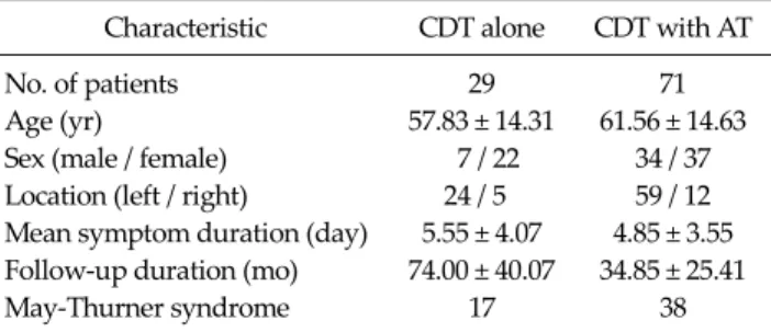
Table 1. The characteristics of the patients with acute deep vein thrombosis treated with CDT
Characteristic CDT alone CDT with AT
No. of patients 29 71
Age (yr) 57.83 ± 14.31 61.56 ± 14.63
Sex (male / female) 7 / 22 34 / 37
Location (left / right) 24 / 5 59 / 12 Mean symptom duration (day) 5.55 ± 4.07 4.85 ± 3.55 Follow-up duration (mo) 74.00 ± 40.07 34.85 ± 25.41
May-Thurner syndrome 17 38
Values are presented as mean ± standard deviation.
CDT, catheter directed thrombolysis; AT, aspiration thrombectomy.
Table 2.Thrombolytic agent amount and thrombolytic infusion time during the procedure
CDT alone group
CDT with
AT group P-value Urokinase amount
(million international unit)
2.48 ± 0.82 1.61 ± 0.44 <0.001a)
Duration (hr) 40.93 ± 15.59 22.01 ± 7.10 <0.001a) Values are presented as mean ± standard deviation.
CDT, catheter directed thrombolysis; AT, aspiration thrombectomy.
a)P < 0.05.
Lakes, FL, USA) was inserted. Using a simple plastic sy- ringe, aspiration of remnant thrombus was done gently.
Aspirations were performed from the caudal to cranial ends. While pulling the catheter back, negative pressure was applied. Advancing the catheter to the more proximal site, the procedures were repeated several times. The most cranial part was aspirated last to prevent embolization of a thrombus fragment. When thromboemboli were not seen at venography, aspiration was stopped. Fluoroscopic im- ages were used to help find residual thrombus. Percuta- neous transluminal angioplasty (PTA) and stent place- ment were used to treat underlying severe stenoses or to treat complications. If there was significant compression of the left common iliac vein with intraluminal webs con- sistent with iliac vein compression syndrome (May- Thurner syndrome), or if there was more than 50% steno- sis due to residual thrombi that could not be removed with CDT with/without AT in the iliac veins or the IVC, self-ex- panding stents were placed and dilated with a balloon catheter; Wall (Boston Scientific, Natick, MA, USA) in 10 cases, Nitis (Taewoong Medical, Seoul, Korea) in 13 cases, Zilver (Cook Ireland Ltd., Limerick, Ireland) in 4 cases, Hercules (S&G Biotech Inc., Seoul, Korea) in 5 cases and Smart (Cordis). A prophylactic retrievable IVC filter was inserted before or after the lytic infusion in 46 patients hav- ing a free floating thrombus at the IVC or common iliac vein. After recanalization of the major venous systems, in- cluding from common iliac vein to IVC junction, was ach- ieved, a final IVC venogram was performed to search for trapped thrombus in the IVC filter. Anticoagulation ther- apy with warfarin sodium was initiated and continued for
a minimum of 6 months. Low-molecular-weight heparin was overlapped with warfarin sodium until the interna- tional normalized radio (INR) was in the therapeutic range. Therapy was adjusted to attain an INR in the range of 2–3.
Statistical analysis was performed using the IBM SPSS ver. 18.0 (IBM Co., Armonk, NY, USA). Continuous data are reported as mean ± standard deviation. Nominal data are reported as number of subjects. The Student’s t-test was used to compare the mean differences between the two groups. We compared the frequency of complete thrombus removal using chi-square. Symptom resolution was estimated by using the Fisher's exact test. Stent pa- tency was estimated by using the Kaplan-Meier method and comparisons of patency were made with the log rank test. A P-value < 0.05 was considered to be statistically significant.
RESULTS
Demographic information of the patients is presented in Table 1. The mean age of patients was 60.48 ± 14.57 years (range, 27 to 83 years). The patients consisted of 41 men and 59 women. The lesion was on the right side in 17 pa- tients and on the left side in 83 patients. CDT alone and CDT with AT were performed in 29 and 71 patients, respectively. The mean symptom duration was 5.05 ± 3.70 days (range, 1 to 15 days). 55 patients were diagnosed with May-Thurner syndrome by 3D venous CT.
As shown in Table 2, the mean procedural time of the CDT-alone group was longer than the CDT with AT group
Table 3. Comparison of the clinical outcomes between the CDT alone and CDT with AT
CDT alone group (n = 29)
CDT with AT group (n = 71) P-value Complete thrombus
removal
15 (51.7) 48 (67.6) 0.136
Complete symptom resolve
24 (82.8) 61 (85.9) 0.456
Values are presented as number (%).
CDT, catheter directed thrombolysis; AT, aspiration thrombectomy.
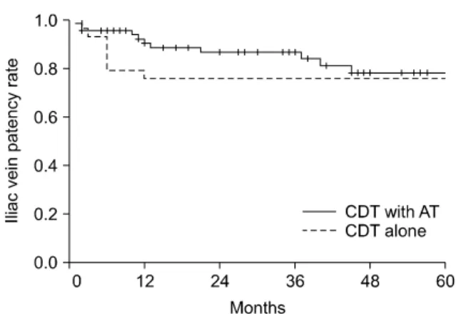
Fig. 1. The patency rate of iliac vein by thrombus dissolution (P = 0.546). CDT, catheter directed thrombolysis; AT, aspiration thrombectomy.
(P < 0.001) and dose of urokinase used during the proce- dure significantly decreased in the CDT with AT group compared to CDT-alone group (P < 0.001).
Complete thrombus removal (≥99% elimination of thrombus) based on angiographic evidence was achieved in 48 of 71 patients (67.6%) in the CDT with AT group. In contrast, complete thrombus removal occurred in 15 of 29 patients (51.7%) in the CDT alone (P = 0.136) (Table 3).
Complete symptom resolution as judged by the elimi- nation of edema on the affected extremity within 24 hours after intervention, occurred in 61 cases (85.9%) of CDT with AT group and in 24 cases (82.8%) of CDT-alone group. There was no significant difference in symptom improvement (P = 0.456) (Table 3). PTA with stent insertion was performed in 61 patients with severe stenosis or complications. Stenting in the CDT with AT group and CDT-alone group was necessary in 45 (63.4%) and 17 cases (58.6%), respectively. There was no technical failure in stent insertion. The patency rate of iliac vein in the groups with stent (28.95%) was better than in the groups without stent (11.29%) (P = 0.026). PTS was defined as signs and symptoms including pain, heaviness, tingling sensation and swelling. There was no significant difference in the frequency of PTS between the groups with stent (10.53%) and the groups without stent (16.13%) (P = 0.433).
There were three patients with minor wound hema- toma in all groups. No surgical interventions were re- quired for these hemorrhagic complications. There was no complication related to AT or thrombolytic catheter itself.
PE was not noted in all groups at arterial phase of 3D ve- nous CT. There was no major morbidity or mortality.
Follow-up was possible on all patients for an average of
37.62 ± 25.21 months (range, 1 to 82 months). No patient died as a direct result of the DVT. Follow-up information regarding lower extremity venous valvular function was collected in 72 patients by venous reflux test (20 in CDT-alone group, 52 in CDT with AT group). In venous re- flux test, reflux was noted 3 in CDT-alone group (15%) and 7 in CDT with AT group (13.7%). Forty-six patients under- went prophylactic temporary IVC filter for the prevention of floating thrombus. Tulip (Cook Ireland Ltd.) was used in 35 cases and OptEase (Cordis) was used in 11 cases.
Temporary IVC filter was removed within 2 weeks except patients having a free floating thrombus at the IVC after CDT with/without AT. There was no removal failure. The entrapped thrombi were founded 6 patients in only CDT with AT group. Thrombus recurrence was founded 7 pa- tients in CDT-alone group (24.1%) and 11 patients in CDT with AT group (15.5%). The PTS was appeared 9 cases (12.68%) in CDT with AT group and 5 cases (17.24%) in CDT-alone group. There was no significant difference in PTS between the two groups (P = 0.550).
Over all patency rates of iliac vein by Kaplan-Meier method were 88.4%, 83.5%, 81.9%, 78.6% at 12, 24, 36, 48 months, respectively. Patency rates of CDT-alone group were 75.9%, 75.9%, 75.9%, 75.9% and patency rates of CDT with AT group were 90.4%, 86.6%, 83.9%, 78.2% at 12, 24, 36, 48 months, respectively (Fig. 1). There was no differ- ence in long-term patency rate between the two groups (P
= 0.546).
DISCUSSION
CDT has been accepted to be more effective and safer than systemic infusion but its many disadvantages have limited its widespread acceptance. Bleeding complica- tions have been reported to occur in 0–25% of patients in a single-center series, and in 11% of patients in a multicenter venous registry study [10-12,20]. By adding AT during CDT procedure, shorter treatment time and more reduced thrombolytic agents contribute the decrease of bleeding complication. We found that combining CDT with AT sig- nificantly reduced the mean treatment duration compared with CDT alone (P < 0.001). And, we identified a sig- nificant reduction in mean amount of drug associated with therapy in the group treated with CDT with AT com- pared with CDT alone (P < 0.001). Vedantham et al. [20]
investigated the effect of adjunctive mechanical thrombec- tomy with pharmacologic catheter-directed lower ex- tremity venous thrombolysis. In a retrospective study of 20 patients in CDT with AT group, they demonstrated a procedural success rate of 82% and reduced doses of thrombolytic agents and infusion time compared with a previously published series of patients treated with CDT alone. The fact that the required treatment time and doses of thrombolytic agents decreased in CDT with AT group compared to CDT-alone group coincided with our data. In addition, CDT with AT was associated with a reduction in the cost of interventional material and the number of ve- nographic assessments in the interventional suites [21]. AT is also associated with shorter intensive care unit and hos- pital stays as well as reduced hospital costs without differ- ence of thrombus removal rate between CDT with AT and CDT alone [20]. We did not check the hospital costs.
Because the procedure time and the amount of thrombo- lytic agents were decreased, medical costs might be reduced.
There were no statistically significant differences in clinical outcomes judged by complete thrombus removal and elimination of symptom between the two groups. But, the frequency of complete thrombus removal was higher in CDT with AT group (67.6%) than CDT-only group (51.7%). The frequency of symptom resolution was also higher in CDT with AT group (85.9%) than CDT-only
group (82.8%).
With mechanical clot dissolution, clot migration or em- bolization is of clinical concern. The incidence of sympto- matic PE and fatal PE during CDT was reported as 1% and 0% to 0.2%, respectively [12,22]. Kinney et al. [23] reported that CDT with AT had a higher rate of PE complications when used for hemodialysis graft declotting but not when used for DVT treatment. Cases of PE were not noted in each group in our series, but entrapped thrombus during procedure was noted in 6 of 37 cases in the CDT with AT group and 0 of 9 cases in the CDT-alone group among 46 patients previously inserted with prophylactic IVC filter.
Noguchi et al. [24] reported that temporary IVC filters are useful and safe for the management of an acute proximal DVT, especially in patients who require aggressive throm- bus removal. When we viewed the above results, prophy- lactic IVC filter insertion should be considered for the pre- vention of PE by floating thrombus in AT treatment.
In conclusion, our study suggested that the use of AT of- fers more effective thrombus removal in less time and doses of thrombolytic agents required. CDT with AT is safe and effective for the treatment of an acute iliofemoral DVT. In AT treatment, prophylactic IVC filter insertion should be considered for the prevention of PE by floating thrombus.
CONFLICTS OF INTEREST
No potential conflict of interest relevant to this article was reported.
REFERENCES
1. White RH. The epidemiology of venous thromboem- bolism. Circulation 2003;107(23 Suppl 1):I4-8.
2. Cushman M, Tsai AW, White RH, Heckbert SR, Rosamond WD, Enright P, et al. Deep vein thrombosis and pulmonary embolism in two cohorts: the longitudinal investigation of thromboembolism etiology. Am J Med 2004;117:19-25.
3. Jang MJ, Bang SM, Oh D. Incidence of venous throm- boembolism in Korea: from the Health Insurance Review and Assessment Service database. J Thromb Haemost 2011;
9:85-91.
4. Cohen AT, Agnelli G, Anderson FA, Arcelus JI, Bergqvist
D, Brecht JG, et al. Venous thromboembolism (VTE) in Europe. The number of VTE events and associated morbid- ity and mortality. Thromb Haemost 2007;98:756-64.
5. Nyamekye I, Merker L. Management of proximal deep vein thrombosis. Phlebology 2012;27 Suppl 2:61-72.
6. Sharafuddin MJ, Sun S, Hoballah JJ, Youness FM, Sharp WJ, Roh BS. Endovascular management of venous throm- botic and occlusive diseases of the lower extremities. J Vasc Interv Radiol 2003;14:405-23.
7. Meissner MH, Gloviczki P, Comerota AJ, Dalsing MC, Eklof BG, Gillespie DL, et al. Early thrombus removal strat- egies for acute deep venous thrombosis: clinical practice guidelines of the Society for Vascular Surgery and the American Venous Forum. J Vasc Surg 2012;55:1449-62.
8. Okrent D, Messersmith R, Buckman J. Transcatheter fi- brinolytic therapy and angioplasty for left iliofemoral ve- nous thrombosis. J Vasc Interv Radiol 1991;2:195-7.
9. Palombo D, Porta C, Brustia P, Peinetti F, Udini M, Antico A, et al. Loco-regional thrombolysis in deep venous thrombosis. Phlebologie 1993;46:293-302.
10. Semba CP, Dake MD. Iliofemoral deep venous thrombosis:
aggressive therapy with catheter-directed thrombolysis.
Radiology 1994;191:487-94.
11. Bjarnason H, Kruse JR, Asinger DA, Nazarian GK, Dietz CA Jr, Caldwell MD, et al. Iliofemoral deep venous throm- bosis: safety and efficacy outcome during 5 years of cathe- ter-directed thrombolytic therapy. J Vasc Interv Radiol 1997;8:405-18.
12. Mewissen MW, Seabrook GR, Meissner MH, Cynamon J, Labropoulos N, Haughton SH. Catheter-directed throm- bolysis for lower extremity deep venous thrombosis: re- port of a national multicenter registry. Radiology 1999;
211:39-49.
13. Cho JS, Martelli E, Mozes G, Miller VM, Gloviczki P. Effects of thrombolysis and venous thrombectomy on valvular competence, thrombogenicity, venous wall morphology, and function. J Vasc Surg 1998;28:787-99.
14. Enden T, Haig Y, Klow NE, Slagsvold CE, Sandvik L, Ghanima W, et al. Long-term outcome after additional catheter-directed thrombolysis versus standard treatment for acute iliofemoral deep vein thrombosis (the CaVenT
study): a randomised controlled trial. Lancet 2012;379:31-8.
15. Murphy KD. Mechanical thrombectomy for DVT. Tech Vasc Interv Radiol 2004;7:79-85.
16. Kearon C, Kahn SR, Agnelli G, Goldhaber S, Raskob GE, Comerota AJ, et al. Antithrombotic therapy for venous thromboembolic disease: American College of Chest Physicians Evidence-Based Clinical Practice Guidelines (8th Edition). Chest 2008;133(6 Suppl):454S-545S.
17. Sniderman KW, Bodner L, Saddekni S, Srur M, Sos TA.
Percutaneous embolectomy by transcatheter aspiration.
Work in progress. Radiology 1984;150:357-61.
18. Kwon SH, Oh JH, Seo TS, Ahn HJ, Park HC. Percutaneous aspiration thrombectomy for the treatment of acute lower extremity deep vein thrombosis: is thrombolysis needed?
Clin Radiol 2009;64:484-90.
19. O'Sullivan GJ. The role of interventional radiology in the management of deep venous thrombosis: advanced therapy. Cardiovasc Intervent Radiol 2011;34:445-61.
20. Vedantham S, Vesely TM, Parti N, Darcy M, Hovsepian DM, Picus D. Lower extremity venous thrombolysis with adjunctive mechanical thrombectomy. J Vasc Interv Radiol 2002;13:1001-8.
21. Kim HS, Patra A, Paxton BE, Khan J, Streiff MB. Adjunctive percutaneous mechanical thrombectomy for low- er-extremity deep vein thrombosis: clinical and economic outcomes. J Vasc Interv Radiol 2006;17:1099-104.
22. Enden T, Klow NE, Sandvik L, Slagsvold CE, Ghanima W, Hafsahl G, et al. Catheter-directed thrombolysis vs. anti- coagulant therapy alone in deep vein thrombosis: results of an open randomized, controlled trial reporting on short- term patency. J Thromb Haemost 2009;7:1268-75.
23. Kinney TB, Valji K, Rose SC, Yeung DD, Oglevie SB, Roberts AC, et al. Pulmonary embolism from pulse-spray pharmacomechanical thrombolysis of clotted hemodial- ysis grafts: urokinase versus heparinized saline. J Vasc Interv Radiol 2000;11:1143-52.
24. Noguchi M, Eishi K, Sakamoto I, Nakamura S, Yamachika S, Hazama S, et al. Thrombus removal with a temporary vena caval filter in patients with acute proximal deep vein thrombosis. Heart Vessels 2003;18:197-201.