ISSN 0378-6471 (Print)⋅ISSN 2092-9374 (Online)
http://dx.doi.org/10.3341/jkos.2016.57.7.1144
Case Report
안와에 발생한 원발성 지방층염양 말초 T세포 림프종 1예
A Case of Primary Orbital Peripheral T-cell Lymphoma with Panniculitis-like Features
강민구⋅성윤미⋅백지선⋅양석우
Min Ku Kang, MD, Youn Mi Sung, MD, Ji Sun Paik, MD, PhD, Suk Woo Yang, MD, PhD
가톨릭대학교 의과대학 서울성모병원 안과 및 시과학교실
Department of Ophthalmology and Visual Science, Seoul St. Mary’s Hospital, College of Medicine, The Catholic University of Korea, Seoul, Korea
Purpose: To report a case of complete remission of primary orbital peripheral T-cell lymphoma with panniculitis-like features af- ter chemotherapy.
Case summary: A 57-year-old healthy female presented with periorbital swelling and symptoms of diplopia. The patient was first treated with high-dose systemic corticosteroids, however, symptoms persisted. Therefore, anterior orbitotomy with excisional bi- opsy was performed for diagnostic purposes. On microscopic examination, the excised mass showed localized dense lympho- cyte infiltrates, and cytologic atypia was observed under a high-power field. On immunehistochemical examination, tumor cells were positive for CD3 and CD8 but negative for CD4, CD20 and CD56. Based on histopathological results, primary orbital pe- ripheral T-cell lymphoma with panniculitis-like features was diagnosed. Additionally, molecular pathological testing was positive for Epstein-Barr virus. Subsequently, the patients underwent chemotherapy and complete remission was obtained.
Conclusions: Peripheral T-cell lymphoma often manifests as systemic symptoms, including lymph node enlargement and B symptom. The primary form of the disease in an orbit is very rare, and has a poor prognosis with a high mortality rate because the disease quickly progresses. Herein, the authors report a rare case of a healthy patient without any past medical history who ach- ieved complete remission of a fast-growing primary orbital T-cell lymphoma with no preceding systemic symptoms.
J Korean Ophthalmol Soc 2016;57(7):1144-1149
Keywords: Orbital tumor, Panniculitis-like features, Peripheral T-cell lymphoma
■Received: 2015. 11. 19. ■ Revised: 2016. 1. 14.
■Accepted: 2016. 2. 26.
■Address reprint requests to Suk Woo Yang, MD, PhD Department of Ophthalmology, The Catholic University of Korea Seoul St. Mary’s Hospital, #222 Banpo-daero, Seocho-gu, Seoul 06591, Korea
Tel: 82-2-3779-1848, Fax: 82-2-761-6869 E-mail: [email protected]
* This study was presented as an e-poster at the 114th Annual Meeting of the Korean Ophthalmological Society 2015.
ⓒ2016 The Korean Ophthalmological Society
This is an Open Access article distributed under the terms of the Creative Commons Attribution Non-Commercial License (http://creativecommons.org/licenses/by-nc/3.0/) which permits unrestricted non-commercial use, distribution, and reproduction in any medium, provided the original work is properly cited.
림프종은 림프구계열 세포들의 악성신생물로 주로 림프 절, 비장, 그리고 비조혈성 조직들을 침범한다.1 안와에 발 생하는 림프세포증식성질환들은 다양한 임상소견을 가지므 로 안와에 발생하는 다른 염증성 질환들과 감별하는 것이
중요하다.2 안와 림프종의 발현율은 결절외 비호지킨 림프 종(extra-nodal non-Hodgkin lymphoma)의 8-10%를 차지하 며, 전체 림프종의 1% 정도를 차지한다.3,4 말초 T세포 림프 종은 비호지킨 림프종의 일종으로 서양에서는 비호지킨 림 프종의 약 12%를 차지하는 것으로 알려져 있다. 환자들은 보통 발열, 체중 감소, 야간 발한의 B증상을 동반하며, 전신 적인 림프절종대가 흔한 증상이다. 림프절 외 침범도 드물 지 않으며 피부와 소화기관이 가장 대표적인 침범 기관이 다.5 말초 T세포 림프종의 치료로는 전신 항암제를 사용하 며, 치료에 따른 반응이 좋지 않고 재발이 흔하며 5년 생존 율도 20-30%에 지나지 않아 비호지킨 림프종 중 예후가 가 장 불량하다.6
A B C
Figure 2. Serial orbital computed tomography. Axial (A) and coronal (B, C) computed tomography images show a large infiltrative
mass with homogenous and high-density lesion in the right preseptal and retrobulbar region.A
B
C
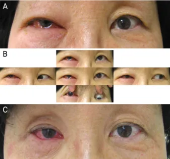
Figure 1. Clinical photographs. (A) The right eye, erythema-
tous, eyelid swelling and (B) extraocular movement limitation at initial presentation. (C) After chemotherapy, eyelid swelling was improved.현재까지 국내에서 안와에 원발성으로 발생한 말초 T세 포 림프종 1예가 보고된 적이 있으나 상기 환자의 경우 항 암치료 거부 후 병의 진행으로 진단 후 한 달 내로 사망하 였고, 조직학적으로 기타 상세불명의 말초 T세포 림프종으 로 진단된 증례였다.2 이에 저자들은 기왕력이나 전신 증상 이 없는 57세 여자 환자의 안와에 원발성으로 발생한 지방 층염양 말초 T세포 림프종의 진단 및 치료 후 완전관해에 도달한 경우를 경험하였기에 문헌 고찰과 함께 보고하는 바이다.
증례보고
특별한 과거력이 없는 57세 여자 환자가 2개월 전부터 복시증상을 동반한 우측 안와 주위의 부종을 주소로 내원 하였다(Fig. 1). 내원 당시 나안시력은 우안 0.4, 좌안 0.5였 고 안압은 비접촉성 안압계로 우안 24 mmHg, 좌안 11 mmHg로 측정되었으며, 안구돌출계 검사상 우안 15.5 mm, 좌안 14.5 mm였다. 우안의 안구운동장애 소견 및 세극등현 미경 검사상 우안 결막 부종 소견이 관찰되었으나 안과적 소견 외에 림프절종대, B증상과 같은 전신증상은 나타나지 않았다. 안와 전산화단층촬영에서 우측 안구 뒤쪽으로 고 음영의 병변이 관찰되어 안와가성종양 의심하에 전신고용 량 스테로이드요법을 3일(총 12회) 시행하였다. 치료 후 안 구운동장애 및 안와 주위 부종이 호전되었으나 2주 뒤부터 우안 충혈 증상 및 안와 주위 부종 등 재발 소견을 보여 조 영제 증강 안와 자기공명영상 시행, 우측 원추 내 및 원추 외 공간에 조영 증강되는 병변이 관찰되어 염증성 안와가 성종양 또는 안와림프종 의심하에 국소마취하 눈확절개술 을 통한 절개생검을 시행하였다(Fig. 2, 3).
절제된 종괴에서는 조직학적 검사상 국소적으로 치밀한 림프구의 침윤 및 고배율상에서 세포학적 비정형 및 림프구
가 지방 소엽을 둘러싸는 듯한 형태를 보였으며, 면역조직 화학검사상 CD3, CD8에 양성이었고 CD4, CD20, CD56 음 성이며 Ki-67 증가된 것으로 관찰되어 지방층염양 말초 T 세포 림프종으로 확진하였고(Fig. 4), 추가적으로 시행한 분 자병리학적 검사상 Epstein-Barr virus (EBV) 양성이었다.
진단 이후 타 장기 전이 여부를 감별하기 위하여 흉부, 복부, 골반 전산화단층촬영 및 전신 양전자방출 단층촬영(positron emission tomography-computed tomography, PET-CT)과 신기 능, 간기능검사를 포함한 혈액 검사 및 골수 생검을 시행하 였다. 골수 생검에서 침범 소견이 없었고 복부 전산화단층촬 영 및 양전자방출 단층촬영(PET-CT)상 췌장에 침범 소견을 보여 Ann Arbor stage3, international prognostic index (IPI) 2 였으며, 이를 토대로 전신화학항암치료 cyclophosphamide,
C D
A B
Figure 3. Serial orbital magnetic
resonance imaging. T1-weighted (fat suppressed) axial (A) pre-con- trast, (B) post-contrast and (C, D) coronal magnetic resonance imaging showed a contrast- enhancing lesion occupying the right conal and ex- traconal space, with mild expansion.doxorubicin, vincristine, prednisolone (CHOP) regimen으로 4 차례 시행하였다. 전신화학항암치료 완료 후 4개월 추척관찰 하면서 시행한 재평가에서 완전관해에 도달하였으며 6개월 째 경과 관찰 중이다(Fig. 5).
고 찰
안와 및 안부속기에 발생되는 림프종은 최근 들어 빈도 가 증가하는 추세이며, 이는 환경적인 요인과 생활양식의 변화와 관련이 있을 뿐 아니라 지난 수십 년간 염증성 가성 종양, 림프증식성질환, 그리고 가성 림프종 등으로 정확한 구분 및 분류가 되지 못했던 질환들이 최근 들어 면역조직 화학검사와 분자유전학적 분석 등의 조직병리학적 진단방 법의 발전으로 인하여 그 정확한 진단이 가능해진 것과도 무관하지 않다고 할 수 있다.7
다수의 보고에 따르면 대부분의 안와 림프세포증식성질 환들은 (1) 말초 결절외 B세포 비호지킨 악성림프종(peri- pheral extra-nodal B-cell non-Hodgkin’s malignant lympho- ma), (2) 비정형림프증식(atypical lymphoid hyperplasia), (3) 양성림프증식증(benign reactive lymphoid hyperplasia)으로
분류할 수 있다.8 눈 부속 기관의 림프종은 대부분 B세포 기원의 림프종이며, 결절외 변연부 B세포 림프종(extrano- dal marginal zone B-cell lymphoma of mucosa-associated lymphoid tissue type)이 가장 흔하며 T세포 림프종은 드물 다. Ferry et al9은 31년 동안 내원했던 353명의 눈 부속 기 관의 림프종 환자들을 대상으로 한 연구에서 단지 2명만이 T세포 림프종으로 진단 받았다고 보고하였으며, Hatef et al10도 T세포 기원의 림프종은 드물다고 보고하였다.
말초 T세포 림프종은 T세포 비호지킨 림프종의 한 종류 로, 안과적으로 말초 T세포 림프종은 결막, 안와, 눈꺼풀 등 에 침범하는 것으로 알려져 있다.11 이 중 원발성 안와 말초 T세포 림프종은 매우 드문 질환으로 몇몇 예가 보고된 적 이 있으며,8,11 국내에서도 통증눈근육마비를 동반한 원발성 기타 상세불명의 안와 말초 T세포 림프종이 1예 보고된 적 이 있으나 환자의 치료 거부로 진단 후 한 달 내에 사망하 였으며,2 저자들의 증례와 같이 지방층염양 말초 T세포 림 프종이 안와에 원발성으로 발생된 예는 보고된 바 없었다. 말초 T세포 림프종은 B세포 림프종에 비해 예후가 불량 하며, 조직학적으로 지방층염양 T 세포 림프종의 경우 소 엽상 지방층염과 유사한 소견을 보이며 림프구가 지방 소
Figure 4. Microscopic examinat-
ion. (A) dense lobular panniculitis composed of small to medium- sized lymphoid cells with some atypical nuclei (H&E, ×100), (B) atypical lymphoid cells rim- ming individual lipocytes (H&E,×400). On immunohistochemi- cal staining, (C) atypical lym- phocytes show diffuse strong im- munoreactivity for CD3 along the cytoplasmic membrane (×200), negative for both (D) CD56 and (E) CD20 (×200), which is con- sistent with peripheral T-cell lymphoma and panniculitis-like features.
엽을 둘러싸는 듯한 형태를 보이는 것이 특징이고, 면역조 직화학검사상 CD3+, CD8+, CD56-인 경우가 전형적이 다. 또한 예후는 경미한 것부터 강력한 항암치료에도 반응 하지 않는 치명적인 것까지 다양한 것으로 알려져 있다.
Epstein-Barr 바이러스(EBV)의 경우 버킷림프종, 호지킨림 프종, 그리고 비강형 자연살해/T세포 림프종과 연관되어 있 는 것으로 알려져 있다.12 대부분의 말초 T세포 림프종의 경우 EBV 음성인 경우가 많으나,13 EBV가 유행하는 지역 에서는 말초 T세포 림프종 환자의 62% 이상에서 발견되었 다.14 말초 T세포 림프종에서 EBV genome이 CD30의 발현
과 관련성이 있을 것으로 추정되며, 여러 연구들을 통해 EBV는 더 빠르게 진행하는 경과와 연관이 있는 것으로 알 려져 있다.14-17
임상적으로는 국제예후지표(International prognostic in- dex, IPI)가 가장 효과적인 예후의 예측 모델이며, 60세 이 상의 연령, Stage III 또는 IV, 혈청 유산탈수소효소(lactase dehydrogenase, LDH)의 상승, 수행 상태(performance sta- tus)의 저하, 1개 이상의 결절 외 침범이 있는 경우 예후가 더 불량하다.18 한국에서도 IPI가 말초 T세포 림프종에서 환 자의 생존율을 예측할 수 있는 가장 중요한 모델로 보고된
A
B C
D E
CD56 CD20
CD3
Figure 5. Chemotherapy results
after 4 months. Axial (A) and co- ronal (B) contrast-enhanced mag- netic resonance images show de- creased size and a mildly enhanced lesion in the right conal and ex- traconal space as well as regressed thickening and enhancement of the right eyelids.바 있다.19
말초 T세포 림프종을 진단 받은 경우 Anthracycline을 포 함한 항암 치료가 표준 치료이며 약 60%에서 치료에 반응 하나 5년 무병생존율(failure free survival rate)이 약 20%로 재발이 잦아 5년 생존율은 약 20-30%로 예후가 매우 불량 하다.6 또한 자가 또는 동종 골수 이식, 자가 줄기세포 이식 이 시도되고 있으나 질병이 드물고 다른 아형의 말초 T세포 림프종과 함께 연구가 진행되어 개별적인 아형에 대한 치료 효과가 확실히 알려지지 않아 항암화학요법이 표준 치료로 시행되고 있다. 항암 화학요법에 반응하지 않거나 재발하는 경우의 치료는 아직 정립되지 않았으나, 고용량의 화학항암 요법과 자가 줄기세포 이식이 시도되고 있다.20,21 안와에 발 생한 지방층염양 말초 T세포 림프종이 특이 과거력 없이 건 강한 사람에서 다른 전신 증상 없이 생겨 진단 후 전신화학 항암치료를 통해 완전관해에 도달한 경우는 보고되지 않은 바, 이와 같은 증례를 경함하였기에 보고하는 바이다.
REFERENCES
1) Amit S, Purwar N, Agarwal A, Kanchan S. Primary orbital non-Hod- gkin's lymphoma. BMJ Case Rep 2012;2012. pii: bcr2012006847.
2) Lee DS, Woo KI, Chang HR. T-cell lymphoma presenting as pain- ful ophthalmoplegia. Korean J Ophthalmol 2006;20:192-4.
3) Fitzpatrick PJ, Macko S. Lymphoreticular tumors of the orbit. Int J Radiat Oncol Biol Phys 1984;10:333-40.
4) Freeman C, Berg JW, Cutler SJ. Occurrence and prognosis of ex- tranodal lymphomas. Cancer 1972;29:252-60.
5) Sabattini E, Bacci F, Sagramoso C, Pileri SA. WHO classification of tumours of haematopoietic and lymphoid tissues in 2008: an overview. Pathologica 2010;102:83-7.
6) Vose J, Armitage J, Weisenburger D; International T-Cell Lympho- ma Project. International peripheral T-cell and natural killer/T-cell lymphoma study: pathology findings and clinical outcomes. J Clin Oncol 2008;26:4124-30.
7) Cho EY, Han JJ, Ree HJ, et al. Clinicopathologic analysis of ocular
adnexal lymphomas: extranodal marginal zone b-cell lymphoma constitutes the vast majority of ocular lymphomas among Koreans and affects younger patients. Am J Hematol 2003;73:87-96.
8) Janatpour KA, Choo PH, Lloyd WC 3rd. Primary orbital peripheral T-cell lymphoma: histologic, immunophenotypic, and genotypic features. Arch Ophthalmol 2007;125:1289-92.
9) Ferry JA, Fung CY, Zukerberg L, et al. Lymphoma of the ocular ad- nexa: a study of 353 cases. Am J Surg Pathol 2007;31:170-84.
10) Hatef E, Roberts D, McLaughlin P, et al. Prevalence and nature of systemic involvement and stage at initial examination in patients with orbital and ocular adnexal lymphoma. Arch Ophthalmol 2007;125:1663-7.
11) Shields CL, Shields JA, Eagle RC. Clinicopathologic reports, case reports, and small case series: rapidly progressive T-cell lympho- ma of the conjunctiva. Arch Ophthalmol 2002;120:508-9.
12) Okano M, Osato T, Koizumi S, et al. Epstein-Barr virus infection and oncogenesis in primary immunodeficiency. AIDS Res 1986;2 Suppl 1:S115-9.
13) Stein H, Foss HD, Dürkop H, et al. CD30(+) anaplastic large cell lymphoma: a review of its histopathologic, genetic, and clinical features. Blood 2000;96:3681-95.
14) Zhou XG, Hamilton-Dutoit SJ, Yan QH, Pallesen G. High fre- quency of Epstein-Barr virus in Chinese peripheral T-cell lymphoma. Histopathology 1994;24:115-22.
15) Dupuis J, Emile JF, Mounier N, et al. Prognostic significance of Epstein-Barr virus in nodal peripheral T-cell lymphoma, unspeci- fied: a Groupe d'Etude des Lymphomes de l'Adulte (GELA) study.
Blood 2006;108:4163-9.
16) Cheng AL, Su IJ, Tien HF, et al. Characteristic clinicopathologic features of adult B-cell lymphoblastic lymphoma with special em- phasis on differential diagnosis with an atypical form probably of blastic lymphocytic lymphoma of intermediate differentiation origin. Cancer 1994;73:706-10.
17) Su IJ, Hsieh HC, Lin KH, et al. Aggressive peripheral T-cell lym- phomas containing Epstein-Barr viral DNA: a clinicopathologic and molecular analysis. Blood 1991;77:799-808.
18) Gallamini A, Stelitano C, Calvi R, et al. Peripheral T-cell lympho- ma unspecified (PTCL-U): a new prognostic model from a retro- spective multicentric clinical study. Blood 2004;103:2474-9.
19) Kim K, Kim WS, Jung CW, et al. Clinical features of peripheral T-cell lymphomas in 78 patients diagnosed according to the
A B
= 국문초록 =
안와에 발생한 원발성 지방층염양 말초 T세포 림프종 1예
목적: 안와에 원발성으로 발생한 지방층염양 말초 T세포 림프종 진단 및 화학항암요법 후 완전관해에 도달한 증례 1예를 경험하였기 에 이를 보고하고자 한다.
증례요약: 57세 여자가 복시증상을 동반한 우측 안와주위의 부종을 주소로 내원하였다. 안와 가성종양 의심하에 전신 고용량 스테로 이드요법을 시행하였으나 호전이 없어 눈확절개술을 통해 절개생검을 시행하였다. 절개된 종괴는 조직학적 검사상 국소적으로 치밀 한 림프구의 침윤 및 세포학적 비정형을 보였고, 면역조직화학검사상 CD3, CD8 양성이었으며, CD4, CD20, CD56 음성소견을 보여 지방층염양 말초 T세포 림프종으로 확진되었다. 또한 분자병리학적 검사상 Epstein-Barr 바이러스 양성이었다. 이후 화학항암치료를 시행하였으며 완전관해에 도달하였다.
결론: 말초 T세포 림프종에서는 림프절종대, B 증상과 같은 전신증상이 흔하게 나타나며, 안와에 원발성으로 발생하는 경우는 매우 드물고 병의 진행이 빠르며 치사율이 높아 예후가 매우 불량하다. 저자들은 특이 과거력 없이 건강한 사람에게 전신증상 없이 발생한 안와 원발성 말초 T세포 림프종 진단 및 치료 후 완전관해에 도달한 드문 경우를 경험하였기에 보고하고자 한다.
<대한안과학회지 2016;57(7):1144-1149>
Revised European-American lymphoma (REAL) classification.
Eur J Cancer 2002;38:75-81.
20) Jagasia M, Morgan D, Goodman S, et al. Histology impacts the outcome of peripheral T-cell lymphomas after high dose chemo- therapy and stem cell transplant. Leuk Lymphoma 2004;45:2261-7.
21) Rodriguez J, Caballero MD, Gutierrez A, et al. High dose chemo- therapy and autologous stem cell transplantation in patients with peripheral T-cell lymphoma not achieving complete response after induction chemotherapy. The GEL-TAMO experience. Haemato- logica 2003;88:1372-7.