11
Original Article http://dx.doi.org/10.7461/jcen.2012.14.1.11
Cognitive Function of Korean Neurosurgical Patients:
Cross-sectional Study Using the Korean Version of the Mini-mental Status Examination
Jiha Kim, MD,1 Chi Heon Kim, MD, PhD,2,3,4 Hyun-Seung Kang, MD, PhD,2,3,4
Chul-Kee Park, MD, PhD,2,3,4 Chun Kee Chung, MD, PhD2,3,4
Department of Neurosurgery, Kangwon National University Hospital,1 Department of Neurosurgery, Seoul National University Hospital,2 Neuroscience Research Institute, Seoul National University Medical Research Center,3 Clinical
Research Institute, Seoul National University Hospital, Seoul, Korea4 ABSTRACT
Objective: As interest in life quality and expectancy increases, cognitive dysfunction is becoming an important topic.
Although there are many foreign articles on this topic, they require cultural interpretation to be applicable to Koreans.
The purpose of this study was to assess cognitive function in Korean neurosurgical patients. Methods: We recruited 214 adult Korean patients with various cerebral pathologies and treatments from an outpatient clinic. The male-to-fe- male ratio was 88:126, and their ages ranged from 28 to 81 years (mean: 57.9 years). The Korean version of the mini-mental status examination (K-MMSE) was adopted as an instrument for measuring cognitive function, and a score
≤23 was defined as cognitive dysfunction. K-MMSE scores were analyzed considering the patients’ gender, age, time elapsed since treatments, pathology and treatment modality. A serial analysis was performed for 59 patients who com- pleted the K-MMSE more than once. Results: The mean K-MMSE score of 214 patients was 22.3, and 133 patients (62.1%) had a score ≤ 23. Cognitive dysfunction was common regardless of age, gender, pathology or treatment modality. Serial analysis revealed K-MMSE score improvement in 45 of 59 patients (76.3%). The mean time interval between two tests was 11.9 months, and the mean K-MMSE score improvement was 2.7, which was statistically sig- nificant (P = 0.000). However, many still had cognitive impairment. Conclusion: Most Korean neurosurgical patients showed cognitive dysfunction despite improvement after several months. Patients with trauma or ischemic disease were more vulnerable. More attention should be paid to neuropsychological complications such as cognitive dysfunction.
(J Cerebrovasc Endovasc Neurosurg. 2012 Mar;14(1):11~21) KEY WORDS Cognitive dysfunction·Craniotomy·Korea·MMSE·Neurosurgery
Received : 5 December 2011 Revised : 4 January 2012 Accepted : 1 February 2012
Corresponding Author : Chun Kee Chung, MD, PHD
Address : Department of Neurosurgery, Seoul National University Hospital, 28 Yeongeon-dong, Jongno-gu Seoul, 110-744, South Korea
Tel : (001) 82-2-2072-2352 Fax : (001) 82-2-744-8459 E-mail : [email protected]
INTRODUCTION
Public interest in quality of life is increasing with the improvement of general health and economic conditions.
Many studies are now being conducted that focus on both mental and physical health. An increasing elderly population is also enhancing the number of studies on cognitive impairment.7) As Korean life expectancy in-
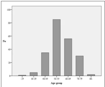
Fig. 1. Bar graphs showing the age distribution of 214 patients.
creases, interest in cognitive impairment, including de- mentia, continues to increase.7)9)10)
Although there are many published articles about cog- nitive function, most articles focus on non-surgical dis- eases or population-based cognitively normal groups.7)9)10) Moreover, most published postoperative reports are on foreign patients who have different cultural backgrounds.
1)13-15)18)20)22)23) Compared with foreign countries, a rela- tively small number of studies have been conducted in Korea. Although many diseases and neurosurgical proce- dures have the potential to cause cognitive impairment, only limited information is available in the Korean neu- rosurgical field.
This paucity of information is likely to stem from the lack of previous normative data and a verified measur- ing instrument.7) There are many difficulties in applying knowledge from foreign articles directly to Korean patients. Cognitive functional evaluation results can be influenced by many individual socio-cultural factors, such as age, gender, race and education level; unlike other clinical or laboratory tests, they require cultural interpretation and compensation.3)5)7)17) In addition, there are deviations when certain measuring instruments are applied. Although many instruments have been de- signed to measure the cognitive function of various groups, most of them were written in English and adapted to Western socio-cultural conditions. Therefore, they are difficult to apply directly to Korean patients who have different socio-cultural background; these as- sessments need to be translated and adapted according to Korean cultural backgrounds.7)13)
Here, we present the cognitive function assessment results of 214 Korean neurosurgical patients who vis- ited a neurosurgical outpatient clinic. We used the Korean version of the mini-mental status examination (K-MMSE) questionnaire as a measuring instrument.
Our study includes patients with various pathologies who underwent surgical or interventional procedures.
The aim of this cross-sectional study was to present a general perspective on the cognitive functional state of Korean neurosurgical patients, rather than detailed in-
formation about specific diseases or procedures.
MATERIALS AND METHODS
We conducted this cross-sectional study in patients who visited the outpatient clinic of the Department of Neurosurgery in our hospital between January 2009 and December 2010. All of them underwent either brain sur- gery or endovascular intervention. The male-to-female ratio was 88:126, and their ages ranged from 28 to 81 years (mean: 57.9 years) (Fig. 1). They presented with a range of cerebral pathologies including aneurysm, cer- ebral ischemic disease, vascular anomaly, spontaneous hemorrhage, tumor, epilepsy and trauma. They under- went various treatment modalities, such as aneurysm clipping, endovascular coiling, vascular anastomosis, tu- mor surgery and epilepsy surgery. The time interval from treatment to study ranged from 1 to 194 months (mean: 30.9 months) (Table 1). We excluded pediatric patients under 15 years of age and those who refused to fill out our questionnaires or had poor compliance.
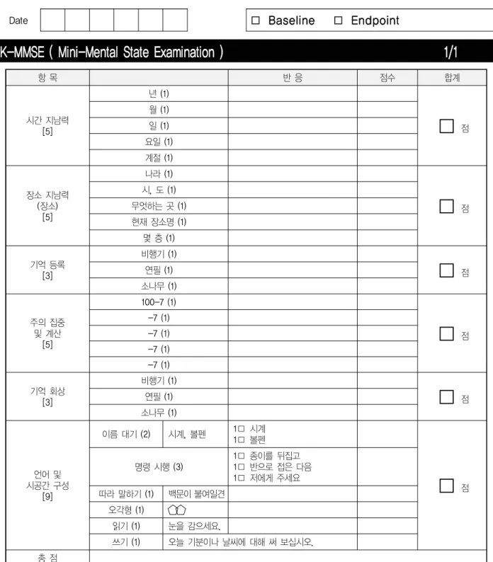
In our study, the K-MMSE questionnaire was adopted as an instrument to measure cognitive function (Fig. 2).
All patients spoke Korean as their first language and did not experience any linguistic difficulties in filling out our questionnaire. Considering many other published our questionnaire. Considering many other published articles
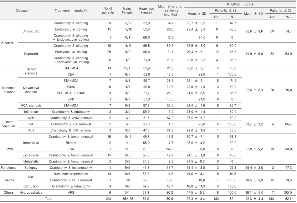
Disease Treatment modality No of patients
Male/
Female
Mean age (years)
Mean time after treatments
(months)
K-MMSE score Mean ± SD Patients ≤ 23
Mean ± SD Patients ≤ 23
No % No %
Aneurysm
Unruptured
Craniotomy & clipping 31 8/23 62.3 14.7 22.7 ± 3.6 21 67.7
22.6 ± 3.6 29 61.7
Endovascular coiling 15 3/12 62.4 20.5 22.4 ± 3.9 8 53.3
Craniotomy & clipping
+ Endovascular coiling 1 0/1 68.0 12.0 24.0 0 0
Ruptured
Craniotomy & clipping 15 4/11 59.8 80.7 22.9 ± 3.0 9 60.0
21.9 ± 5.0 32 64.0
Endovascular coiling 29 8/21 56.6 21.7 21.4 ± 6.1 19 65.5
Craniotomy & clipping
+ Endovascular coiling 6 1/5 61.0 61.7 22.0 ± 2.3 4 66.7
Ischemic disease
Carotid stenosis
STA-MCA 13 12/1 62.4 27.8 22.2 ± 3.1 10 76.9
22.6 ± 2.3 28 70.0
CEA 1 0/1 62.0 18.0 22.0 1 100.0
Moyamoya disease
STA-MCA 7 4/3 50.7 28.6 22.1 ± 2.1 5 71.4
EDAS 6 1/5 52.0 30.7 22.8 ± 1.3 3 50.0
STA-MCA + EDAS 3 3/0 51.7 25.0 24.0 ± 3.5 2 66.7
EVD 1 0/1 57.0 15.0 24.0 0 0
MCA stenosis STA-MCA 7 5/2 57.3 23.6 22.4 ± 1.6 6 85.7
Infarction Craniotomy & lobectomy 2 2/0 69.0 6.0 23.0 ± 1.4 1 50.0
Other Vascular
AVM Craniotomy & AVM removal 2 1/1 51.5 57.0 26.0 ± 5.7 1 50.0
23.7 ± 3.2 4 66.7
CA Craniotomy & CA removal 2 1/1 50.0 4.5 22.0 2 100.0
ICH Craniotomy & ICH removal 2 2/0 47.5 27.0 23.0 ± 1.4 1 50.0
Tumor
Intra-axial
Craniotomy & tumor removal 16 5/11 49.7 43.9 20.7 ± 7.1 11 68.8
22.8 ± 5.2 18 50.0
Biopsy 2 1/1 68.0 7.5 23.0 ± 4.2 1 50.0
TSA 1 0/1 61.0 187.0 26.0 0 0
Extra-axial Craniotomy & tumor removal 15 2/13 55.5 42.3 24.1 ± 1.9 6 40.0
Metastatic Craniotomy & tumor removal 2 2/0 54.5 9.5 27.5 ± 0.7 0 0
Functional Epilepsy Craniotomy & lesionectomy 11 6/5 46.3 32.7 25.4 ± 3.0 3 27.3 25.4 ± 3.0 3 27.3
Trauma SDH Burr-hole trephination 13 8/5 68.2 17.5 21.6 ± 4.1 8 61.5
20.5 ± 5.8 12 70.6
Craniotomy & SDH removal 1 1/0 68.0 19.0 19.0 1 100.0
Contusion Craniotomy & lobectomy 3 3/0 52.3 40.7 16.0 ± 11.3 3 100.0
Others Hydrocephalus VPS 6 5/1 59.8 20.2 17.5 ± 5.2 6 100.0 18.1 ± 5.0 7 100.0
Total 214 88/126 57.9 30.9 22.3 ± 4.4 133 62.1 22.3 ± 4.4 133 62.1
K-MMSE = Korean mini-mental status examination; SD = standard deviation; STA-MCA = Superficial temporal artery-middle cerebral artery anastomosis; CEA = Carotid endarterectomy; EDAS = Encephalo-dura-arterio-synangiosis; EVD = Extra-ventricular drainage; MCA = Middle cerebral artery; AVM = Arteriovenous malformation; CA = Cavernous angioma; ICH = Intracerebral hematoma; TSA = Trans-sphenoidal approach; SDH = Subdural hematoma; VPS = Ventriculo-peritoneal shunt; ACM = Arnold-Chiari malformation; FMD = Foramen magnum decompression
Table 1. Summary of disease and treatment modalities in the patient cohort
Volume 14 · NUMBER 1 · MARCH 2012
13
JIHA KIM ET AL
Date
□ Baseline □ Endpoint
K-MMSE ( Mini-Mental State Examination ) 1/1
항 목 반 응 점수 합계
시간 지남력 [5]
년 (1)
□
점월 (1) 일 (1) 요일 (1) 계절 (1)
장소 지남력 (장소)
[5]
나라 (1)
□
점시, 도 (1) 무엇하는 곳 (1) 현재 장소명 (1)
몇 층 (1)
기억 등록 [3]
비행기 (1)
□
점연필 (1) 소나무 (1)
주의 집중 및 계산
[5]
100-7 (1)
□
점-7 (1) -7 (1) -7 (1) -7 (1)
기억 회상 [3]
비행기 (1)
□
점연필 (1) 소나무 (1)
언어 및 시공간 구성
[9]
이름 대기 (2) 시계, 볼펜 1□ 시계 1□ 볼펜
□
점명령 시행 (3)
1□ 종이를 뒤집고 1□ 반으로 접은 다음 1□ 저에게 주세요 따라 말하기 (1) 백문이 불여일견
오각형 (1)
읽기 (1) 눈을 감으세요.
쓰기 (1) 오늘 기분이나 날씨에 대해 써 보십시오.
총 점
Fig. 2. The questionnaire for the Korean mini-mental status examination (K-MMSE).
and our patient distribution, a K-MMSE score ≤ 23 was defined as the cut-off for decreased cognitive function.
The K-MMSE scores were analyzed considering the patients’ gender, age, time after treatments, pathologies and treatment modalities. We retrospectively reviewed
their medical records and radiological data, such as magnetic resonance imaging, computed tomography or angiography.
First, we classified each patient according to his or her pathologies and treatment modalities (Table 1). Then, we compared the K-MMSE score of 214 patients using a
JIHA KIM ET AL
Volume 14 · NUMBER 1 · MARCH 2012 15
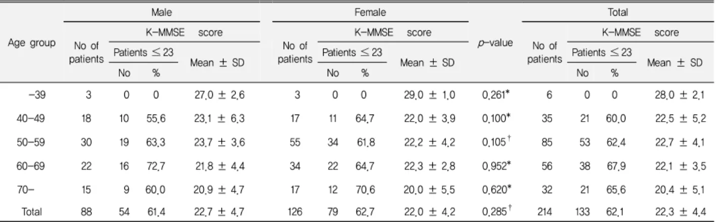
Age group
Male Female
p-value
Total
No of patients
K-MMSE score
No of patients
K-MMSE score
No of patients
K-MMSE score Patients ≤23
Mean ± SD Patients ≤23
Mean ± SD Patients ≤23
Mean ± SD
No % No % No %
-39 3 0 0 27.0 ± 2.6 3 0 0 29.0 ± 1.0 0.261* 6 0 0 28.0 ± 2.1
40-49 18 10 55.6 23.1 ± 6.3 17 11 64.7 22.0 ± 3.9 0.100* 35 21 60.0 22.5 ± 5.2 50-59 30 19 63.3 23.7 ± 3.6 55 34 61.8 22.2 ± 4.2 0.105† 85 53 62.4 22.7 ± 4.1 60-69 22 16 72.7 21.8 ± 4.4 34 22 64.7 22.3 ± 2.8 0.952* 56 38 67.9 22.1 ± 3.5 70- 15 9 60.0 20.9 ± 4.7 17 12 70.6 20.0 ± 5.5 0.620* 32 21 65.6 20.4 ± 5.1 Total 88 54 61.4 22.7 ± 4.7 126 79 62.7 22.0 ± 4.2 0.285† 214 133 62.1 22.3 ± 4.4 K-MMSE score difference between genders in each age group, p-value using Mann-Whitney test (*) or t-test (†)
Table 2. Patient summary according to gender and age group
fixed cut-off value of 23. For patients who completed our questionnaire more than once, only the first score was used for this analysis.
Next, we analyzed the score after stratifying by gender and age. We also compared significant differences be- tween genders in every age group using the Mann- Whitney test or the t-test.
Third, we analyzed the data collected from 90 aneur- ysm patients to determine the effect of pathologies or treatment modalities on cognitive function. We classified them into four groups according to their pathologies (unruptured or ruptured) and treatment modalities (endovascular coiling or clipping). Then, we compared the K-MMSE scores of each group using the Mann- Whitney tests. Seven patients who had both treatments were excluded in this analysis.
Next, we analyzed cognitive function changes by time interval. There were 59 patients (27.6%) who completed the K-MMSE more than once at different times. From 42 patients who answered twice, all scores were used.
From 17 patients who answered 3 times or more, only the first and last scores were used. There were 13 and 4 patients who answered 3 and 4 times, respectively. We compared their last K-MMSE score with their first one using Wilcoxon signed-ranks test or paired t-test.
In this study, every comparison or analysis of K-MMSE scores was conducted using a fixed cut-off value of 23, and a p-value < 0.05 was considered stat-
istically significant. All data analyses were performed with commercially available statistical software (SPSS version 17.0, SPSS Inc., Chicago, IL, USA).
RESULTS
General description
The mean K-MMSE score of all 214 patients was 22.3, and 133 patients (62.1%) had a K-MMSE score ≤ 23.
The decreased K-MMSE score was obvious in most pathologies, including unruptured aneurysm (61.7%), ruptured aneurysm (64.0%), ischemic disease (70.0%) and trauma (70.6%), although there were some differ- ences according to their treatment modalities. The pro- portion of patients with a K-MMSE score ≤ 23 was the highest in trauma and ischemic disease and the lowest in epilepsy and tumor patients (27.3% and 50.0%, re- spectively) (Table 1).
Effect of age and gender
A decreased K-MMSE score was prevalent in almost all age groups, regardless of gender. In age groups older than 39 years, over 60.0% of patients had a K-MMSE score ≤ 23. Inter-gender differences were not statistically significant in all age groups. Although the overall mean K-MMSE score was higher in males than in females (22.7 and 22.0, respectively), this difference was not statistically significant (p = 0.285, using t-test) (Table 2).
Disease Treatment modality No of patients
Male/
Female
Mean age (years)
Mean time after treatments
(months)
K-MMSE score Mean
interval*
(months)
Mean ± SD
p value First Last
Unruptured aneurysm
Craniotomy & clipping 13 3/10 64.8 12.4
11.6 21.9 ± 4.1 25.6 ± 5.6 0.001† Endovascular coiling 6 1/5 65.0 14.8
Ruptured aneurysm
Craniotomy & clipping 5 0/5 51.2 59.4
11.5 20.2 ± 5.0 24.3 ± 6.0 0.013† Endovascular coiling 9 3/6 66.2 27.6
Craniotomy & clipping
+ Endovascular coiling 2 0/2 66.5 87.0
Moyamoya disease
STA-MCA 3 2/1 54.3 22.0
11.2 22.4 ± 2.7 21.9 ± 7.0 0.700†
EDAS 1 0/1 52.0 7.0
STA-MCA+ EDAS 2 2/0 53.5 24.0
Carotid stenosis STA-MCA 3 3/0 69.0 40.0
MCA stenosis STA-MCA 1 1/0 68.0 18.0
CA Craniotomy & CA removal 1 1/0 50.0 6.0
ICH Craniotomy & ICH removal 2 2/0 47.5 27.0
SDH Burr-hole trephination 3 2/1 72.7 22.3
15.5 22.5 ± 2.4 25.5 ± 3.0 0.109† Craniotomy & lobectomy 1 1/0 68.0 19.0
Intra-axial tumor Craniotomy & tumor removal 3 1/2 51.0 75.3 12.0 21.7 ± 3.1 23.3 ± 5.5 0.276† Epilepsy Craniotomy & lesionectomy 2 0/2 49.5 24.5 13.0 22.0 ± 1.4 29.0 ± 1.4 0.180†
Hydrocephalus VPS 2 2/0 53.0 40.0 15.0 21.0 ± 2.8 20.0 ± 4.2 0.655†
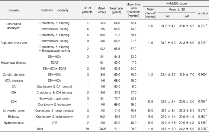
Total 59 24/35 61.1 29.3 11.9 21.6 ± 3.9 24.2 ± 5.9 0.000‡
*Mean interval = mean time interval from first to last K-MMSE score; p-value = difference between first and last K-MMSE score using Wilcoxon signed-ranks test (†) or paired t-test(‡)
Table 4. Summary of the patients who completed the K-MMSE questionnaire more than once Aneurysm Treatment
modality
No of patients
Male/
Female
Mean age (years)
Mean time after treatments
(months)
K-MMSE score
Groups No of patients (%)
Mean ± SD
≤20 21-23 24 25-2
6 27-30
Unruptured Clipping 31 8/23 62.3 14.7 2 ( 6.5) 19 (61.3) 7 (22.6) 0 3 ( 9.7) 22.7 ± 3.6 A Coiling 15 3/12 62.4 20.5 4 (26.7) 4 (26.7) 5 (33.3) 0 2 (13.3) 22.4 ± 3.9 B Ruptured Clipping 15 4/11 59.8 80.7 3 (20.0) 6 (40.0) 4 (26.7) 0 2 (13.3) 22.9 ± 3.0 C Coiling 29 8/21 56.6 21.7 9 (31.0) 10 (34.5) 3 (10.3) 0 7 (24.1) 21.4 ± 6.1 D Clipping = Craniotomy and clipping; Coiling = Endovascular coiling
Table 3. Summary of aneurysm patients according to treatment modality
Effect of pathology and treatment modality
We classified 90 aneurysm patients into four groups;
group A (unruptured aneurysm, clipping), group B (unruptured aneurysm, endovascular coiling), group C (ruptured aneurysm, clipping) and group D (ruptured aneurysm, endovascular coiling) (Table 3). The pro- portion of patients in these groups with a K-MMSE score ≤ 23 was 67.7%, 53.3%, 60.0% and 65.5%,
respectively. However, there was no statistically sig- nificant difference when patients were divided by treat- ment modality (group A vs. B and C vs. D, p = 0.914 and 0.478, respectively). There was also no difference in K-MMSE score between patients with unruptured vs.
ruptured aneurysms (group A vs. C and group B vs. D, p = 0.802 and 0.550, respectively).
JIHA KIM ET AL
Volume 14 · NUMBER 1 · MARCH 2012 17 Effect of time
From 59 patients (27.6%) who completed the K-MMSE more than once, the first and last scores were compared.
Their mean time after treatment was 29.3 months, and the mean time interval between test administrations was 11.9 months. The number of patients with a K-MMSE score ≤ 23 was 41 and 18 (69.5% and 30.5%) for the first and the last test, respectively. The mean K-MMSE score difference was 2.7, which was statistically sig- nificant (p = 0.000, using paired t-test) (Table 4). In all, 45 of 59 patients (76.3%) had an improved K-MMSE score. These improvements were notable in unruptured and ruptured aneurysm groups (89.5% and 75.0%, re- spectively). However, patients with vascular disease, such as Moyamoya disease or carotid stenosis had de- creased K-MMSE scores, although these changes were not statistically significant (p = 0.700, using Wilcoxon signed-ranks test) (Table 4). The linear correlation be- tween the time interval and the K-MMSE score change was not definite in the unruptured or ruptured aneurysm groups (R2= 0.078 and 0.002, respectively).
DISCUSSION
Study design
The purpose of this cross-sectional study was to meas- ure the cognitive function of general neurosurgical patients. To measure the effect of disease or treatment modality on cognitive function, a case-control or com- parative study can be good designs to compare two sets of data obtained from cases and controls or from the same individual before and after a procedure. However, these study designs are not always powerful. For exam- ple, in slowly progressing diseases such as tumor, we do not always know its outbreak to obtain initial data to use as a baseline. In emergency conditions, such as cerebral hemorrhage or trauma, we are also unable to know when we should obtain initial baseline data. Therefore, we thought the cross-sectional study design was a good choice to overcome these difficulties.
Measuring instrument
The MMSE was originally developed to evaluate eld- erly psychiatric patients.6)10) Although it is the most widely used screening instrument for dementia or cogni- tive dysfunction, it has some limitations.7)9)10) First, it cannot be fully administered to disabled individuals with motor impairment because it contains performance tests that assess praxis and visuospatial function.10) Therefore, we conducted our study in an outpatient clinic setting to exclude patients who were either in an acute post- operative period or critical condition. We also excluded those who never had brain surgery or interventions to maintain homogeneity of our case population; most of them had either very mild symptoms or very critical problems that were inoperable, and we thought they might cause a bias and skew the results. Second, it is difficult to distinguish postoperative cognitive dysfunc- tion from postoperative delirium using the MMSE.19)21) By conducting an outpatient clinic based-study, we ex- cluded postoperative delirium, which tends to be a tran- sient and fluctuating disturbance of consciousness that occurs shortly after surgery, whereas postoperative cog- nitive dysfunction is a more persistent problem of cogni- tive performance that can be assessed by neuro- psychological tests.16) Our patients’ mean time from treatment to study was 30.9 months, and over 60% (130 of 214) of patients participated more than 1 year after treatment. Third, the MMSE needs to be translated and adapted to the Korean cultural background.7) Because simply translating the tests into foreign languages would not provide comparative results, we needed to adopt a verified measuring instrument.7)13)
The MMSE was modified and translated into Korean by Kang et al., and the resulting K-MMSE has been widely used in clinical evaluations and research involv- ing Korean dementia patients.7)9) We defined a K-MMSE score ≤ 23 as the cut-off value for abnormal cognitive function for two reasons. First, it was used as a cut-off value in many other articles.4)9)11)14)21) With the same cut-off value, we can share established knowledge with
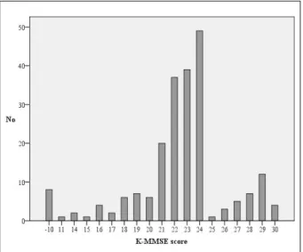
Fig. 3. Bar graphs showing the distribution of the Korean mini-mental status examination (K-MMSE) score of 214 patients.
other studies and compare them with ours. Second, we thought 23 is reasonable cut-off value, considering the mean K-MMSE score measured by Han et al. in 977 normal elderly Koreans.7) Although there are some varia- tions according to age, gender and education level, the mean K-MMSE score of each group ranged from 23.3 to 27.7.7) Setting the cut-off value was very critical be- cause more than 20% of patients (49 out of 214) were clustered around a score of 24 in our series, and this number was equivalent to more than half of normal pa- tients (Fig. 3). If the cut-off value was 24 instead of 23, the percentage of normal patients would decrease from 37.9% to 15.0%.
High incidence of cognitive dysfunction
More than 62% of patients (133 out of 214) had K-MMSE scores ≤ 23, and this decrease was common in almost all pathologies and treatment modalities in our study (Table 1). After stratification according to gender and age, most groups still had an average K-MMSE score
≤ 23. There was no statistically significant difference be- tween genders (Table 2). In other articles, cognitive dys- function has been documented in up to 50% of patients who have experienced subarachnoid hemorrhage.11)12) Several studies gave incidence estimates for post-stroke
dementia that range between 6 and 32%.2) Compared with other type of surgery, patients who undergo brain surgery seem to be more vulnerable to cognitive dysfunction. Abildstrom et al.1) reported 10.4% of pa- tients who underwent major abdominal, non-cardiac thoracic, or orthopedic surgery with general anesthesia had cognitive dysfunction 1-2 year postoperatively.
Moller et al.14) also demonstrated that 9.9% of patients had cognitive dysfunction 3 months postoperatively.
Although it is difficult to directly compare our results with other articles because of different study populations and various measurement instruments, our proportion seems much higher than that reported for non-brain surgery. We believe this difference stems from effects on the brain itself, either by pathologies or by treatments. Some studies on general versus regional or spinal anesthesia have not found differences in post- operative cognitive dysfunction.4)16) We think post- operative cognitive dysfunction is affected more by the type of surgery than anesthesia. Cognitive impairment was documented in up to 50% of patients of cardiac sur- gery patients.8)15)23) This is because cardiac surgery is a lot similar to brain surgery in that cardiopulmonary by- pass is thought to have cerebral effect.14)16)23)
Differences according to pathologies or treatment modalities
To figure out if the K-MMSE score was influenced by different pathologies or treatment modalities, we ana- lyzed the data collected from aneurysm patients (Table 3). Because both unruptured and ruptured aneurysms share the same treatment modalities, we could analyze the effect of pathologies when the effect of treatment modalities was controlled (group A vs. C and B vs. D).
At the same time, we could analyze the effect of treat- ment modalities when the effect of pathology type was controlled (group A vs. B and C vs. D). Because rup- tured aneurysms develop from unruptured aneurysms, we expected to estimate the additive effect of subarachnoid hemorrhage. However, we failed to find significant dif-
JIHA KIM ET AL
Volume 14 · NUMBER 1 · MARCH 2012 19 ferences in each analysis. There was no difference be-
tween pathologies or treatment modalities. Considering the articles that report no cognitive function change after craniotomy for unruptured aneurysm,20)22) it is reasonable that there was no difference between clipping and endo- vascular coiling. However, our result was different from that of Otawara et al.18) who demonstrated lower cogni- tive function in the ruptured aneurysm group compared with the unruptured group. We think our limited patient number might have masked differences between groups.
Difference according to time interval
We compared data from 59 patients who completed the K-MMSE more than once (Table 4). Although their pathologies and treatment modalities varied, 76.3% of (45 of 59) patients showed an improved K-MMSE score when they took the test again (mean: 11.9 months after the first test). However, it is unclear whether this change was due to cognitive function recovery or a practice ef- fect due to repeated trials. Only one version of the ques- tionnaire was available in our study, although other stud- ies have utilized different versions administered in a ran- dom order to minimize practice effects.1) Because there was an interval of 11.9 months between the two tests, we think that practice effect were not a significant contributor. Similar cognitive improvements were also documented in several non-brain surgeries studies.14)15) Moller et al.14) indicated cognitive dysfunction in 25.8%
and 9.9% of patients 1 week and 3 months after non-brain surgery, respectively. Newman et al.15) docu- mented cognitive dysfunction in 36% and 24% of pa- tients 6 weeks and 6 months after cardiac surgery, respectively. Abildstrom et al.1) reported that post- operative cognitive dysfunction is reversible in the ma- jority of elderly patients after general surgery, such as major abdominal, non-cardiac thoracic, or orthopedic surgery with general anesthesia. Our results are different from those in non-brain surgery studies in two ways.
First, our cognitive dysfunction rate was much higher, even after cognitive improvements. Although most pa-
tients showed an improved K-MMSE score and the number of the patients with cognitive dysfunction de- creased from 41 to 18 (69.5% to 30.5%), many patients in our series still had cognitive dysfunction. The K-MMSE score did not show any linear correlation with the time interval between testing or with the time after treatment (p > 0.05). Therefore, we did not expect this improvement would continue. Second, our mean time from treatments to test was much longer than that ob- served in non-brain surgery. According to Moller et al.,14) dynamic cognitive improvement occurred within 3 months postoperatively. However, our study subjects showed similar improvement after a mean of 29.3 months postoperatively.
Limitations of study
Our study has some limitations. First, we could not compensate all of the factors that might possibly bias K-MMSE scores; cognitive function is not only affected by pathologies or treatment modalities, it may also be influenced by many complicated socio-cultural factors, such as age, gender, education level, social class, marital status or income.3)5)7)17) This incomplete stratification and compensation stems from the uneven and relatively small number of patients, which is a drawback of re- cruiting various pathologies. Second, mortality and se- vere morbidity cases unable to visit the outpatient clinic were excluded from our study. We also excluded pa- tients who refused to fill out our questionnaire. It is like- ly that there is something intrinsically different between people who volunteer for our study and those who do not.13) This is particularly true of neuropsychological studies, where personality traits might influence willing- ness to participate and test results.13) Thus, our result might have limitation in representing all kinds of neuro- surgical patients.
Despite these limitations, however, our study contains unique information about the cognitive function of gen- eral neurosurgical patients, which can be used as a nor- mative data for Koreans.
CONCLUSION
Over 62% of patients who underwent a brain operation or endovascular intervention suffer from cognitive dysfunction. In our study, cognitive dysfunction was more prevalent in patients with trauma or ischemic dis- ease than in those with epilepsy or tumor. Although many patients showed improvement, it took time and the dysfunction was not completely resolved. More attention should be paid to neuropsychological complications, such as cognitive dysfunctions.
REFERENCES
1. Abildstrom H, Rasmussen LS, Rentowl P, Hanning CD, Rasmussen H, Kristensen PA, et al. Cognitive dysfunction 1-2 years after non-cardiac surgery in the elderly. ISPOCD group. International Study of Post-Operative Cognitive Dysfunction. Acta Anaesthesiol Scand. 2000 Nov;44(10):
1246-51.
2. Bowler JV. Editorial comment-dementia after stroke.
Stroke. 2004 Jun;35(6):1268-9.
3. Brayne C, Calloway P. The association of education and so- cioeconomic status with the Mini Mental State Examination and the clinical diagnosis of dementia in elderly people.
Age Ageing. 1990 Mar;19(2):91-6.
4. Chung FF, Chung A, Meier RH, Lautenschlaeger E, Seyone C. Comparison of perioperative mental function after gen- eral anaesthesia and spinal anaesthesia with intravenous sedation. Can J Anaesth. 1989 Jul;36(4):382-7.
5. Fillenbaum GG, Hughes DC, Heyman A, George LK, Blazer DG. Relationship of health and demographic char- acteristics to Mini-Mental State examination score among community residents. Psychol Med. 1988 Aug;18(3):
719-26.
6. Folstein MF, Folstein SE, McHugh PR. “Mini-mental state”. A practical method for grading the cognitive state of patients for the clinician. J Psychiatr Res. 1975 Nov;12(3):
189-98.
7. Han C, Jo SA, Jo I, Kim E, Park MH, Kang Y. An adaptation of the Korean mini-mental state examination (K-MMSE) in elderly Koreans: demographic influence and population- based norms (the AGE study). Arch Gerontol Geriatr. 2008 Nov-Dec;7(3):302-10.
8. Jensen BO, Hughes P, Rasmussen LS, Pedersen PU, Steinbruchel DA. Cognitive outcomes in elderly high-risk patients after off-pump versus conventional coronary artery bypass grafting: a randomized trial. Circulation. 2006 Jun;
113(24):2790-5.
9. Kang Y, Na DL, Hahn S.[A validity study on the korean mini-mental state examination (K-MMSE) in dementia patients.] J Korean Neurol Assoc. 1997 Apr; 15(2):300- 308. Korean.
10. Kim TH, Jhoo JH, Park JH, Kim JL, Ryu SH, Moon SW, et al. Korean version of mini mental status examination for dementia screening and its' short form. Psychiatry Investig.
2010 Jun;7(2):102-8.
11. King JT Jr, DiLuna ML, Cicchetti DV, Tsevat J, Roberts MS. Cognitive functioning in patients with cerebral aneur- ysms measured with the mini mental state examination and the telephone interview for cognitive status. Neurosurgery.
2006 Oct;59(4):803-10; discussion 810-1.
12. Kreiter KT, Copeland D, Bernardini GL, Bates JE, Peery S, Claassen J, et al. Predictors of cognitive dysfunction after subarachnoid hemorrhage. Stroke. 2002 Jan;33(1):200-8.
13. Lewis MB, Howdle PD. Cognitive dysfunction and health-related quality of life in long-term liver transplant survivors. Liver Transpl. 2003 Nov;9(11):1145-8.
14. Moller JT, Cluitmans P, Rasmussen LS, Houx P, Rasmussen H, Canet J, et al. Long-term postoperative cog- nitive dysfunction in the elderly ISPOCD1 study. ISPOCD investigators. International Study of Post-Operative Cogni- tive Dysfunction. Lancet. 1998 Mar;351(9106):57-61.
15. Newman MF, Kirchner JL, Phillips-Bute B, Gaver V, Grocott H, Jones RH, et al. Longitudinal assessment of neu- rocognitive function after coronary-artery bypass surgery.
N Engl J Med. 2001 Feb;344(6):395-402.
16. Newman S, Stygall J, Hirani S, Shaefi S, Maze M.
Postoperative cognitive dysfunction after noncardiac sur- gery: a systematic review. Anesthesiology. 2007 Mar;
106(3):72-90.
17. O'Connor DW, Pollitt PA, Treasure FP, Brook CP, Reiss BB. The influence of education, social class and sex on Mini-Mental State scores. Psychol Med. 1989 Aug;19(3):
771-6.
18. Otawara Y, Ogasawara K, Kubo Y, Kashimura H, Ogawa A, Yamadate K. Comparison of postoperative cognitive function in patients undergoing surgery for ruptured and
JIHA KIM ET AL
Volume 14 · NUMBER 1 · MARCH 2012 21 unruptured intracranial aneurysm. Surg Neurol. 2009 Dec;
72(6):592-5; discussion 595.
19. Park IS, Kim NS, Lim HJ, Jang SH.[Comparison of the Frequency of Postoperative Delirium in Elderly between General Anesthesia and Regional Anesthesia.] Korean J Anesthesiol. 1998 Mar;34(3):623-9. Korean.
20. Pereira-Filho AA, Pereira AG, Faria MB, Lima LC, Portuguez MW, Kraemer JL. Microsurgical clipping in for- ty patients with unruptured anterior cerebral circulation aneurysms: an investigation into cognitive outcome. Arq Neuropsiquiatr. 2010 Oct;68(5):770-4.
21. Sohn BK, Sung YB, Park EJ, Lee DW.[The Incidence and
Related Factors of Delirium in Elderly Patients with Hip Fracture after Surgery.] J Korean Geriatr Soc. 2010 Sep;
14(3):162-70. Korean.
22. Tuffiash E, Tamargo RJ, Hillis AE. Craniotomy for treat- ment of unruptured aneurysms is not associated with long-term cognitive dysfunction. Stroke. 2003 Sep;34(9):
2195-9.
23. van Dijk D, Spoor M, Hijman R, Nathoe HM, Borst C, Jansen EW, et al. Cognitive and cardiac outcomes 5 years after off-pump vs on-pump coronary artery bypass graft surgery. JAMA. 2007 Feb;297(7):701-8.