INTRODUCTION
Hyponatremia associated with cerebral disease was first described in 1950 by Peters et al. (1). They described a clini- cal entity in which central nervous system disease was asso- ciated with hyponatremia and paradoxically increased the renal sodium excretion. The patients also had a high urine osmolarity, high urine output, and a depleted extracellular vol- ume. As has been defined by Schwartz et al. in 1957 (2), the syndrome of inappropriate secretion of antidiuretic hormone (SIADH) became the usual explanation for any hyponatremia associated with neurological and neurosurgical disorders. Re- cently, however, the cerebral salt wasting (CSW) syndrome instead of SIADH has been reported frequently in patients with acute brain disease (3-5). CSW syndrome is a disease that leads to hyponatremia resulting from the renal loss of sodium in the presence of disorders of the central nervous system (6) and is different in its pathogenesis from SIADH showing dilutional hyponatremia due to an increase of antidi- uretic hormone (ADH). The treatment of CSW syndrome is to supply sufficient water and sodium through a normal saline
solution (7).
We here report two cases of postoperative CSW syndrome after a cranial vault remodeling surgery.
CASE REPORT Case 1
A 21-month-old boy had an operation for syndactyly of both hands at 6 months after the birth, and then underwent cranial vault remodeling due to craniosynostosis with a chief complaint of trigonocephaly in the Department of Plastic Surgery. The patient developed postoperative polyuria and hypotension, and his general condition was exacerbated after two postoperative days. The patient urinated 50-60 mL/hr (1,510 mL/24 hr) on the first postoperative day, and revealed 3-4 cmH2O of central venous pressure, 136 mL of blood loss through a suction drain, and 90/60 mmHg of blood pressure.
After the third postoperative day, polyuria was developed with a change of urination to over 80 mL/hr (1,965 mL/24
Soon-Ju Lee, Eun-Ju Huh, Jun-Hee Byeon*
Departments of Pediatrics and Plastic Surgery*, College of Medicine, The Catholic University of Korea, Seoul, Korea
Address for correspondence Jun-Hee Byeon, M.D.
Department of Plastic Surgery, College of Medicine, The Catholic University of Korea, St. Mary’s Hospital, 62 Youido-dong, Yongdungpo-gu, Seoul 150-713, Korea
Tel : +82.2-3779-2047, Fax : +82.2-780-9167 E-mail : [email protected]
627 J Korean Med Sci 2004; 19: 627-30
ISSN 1011-8934
Copyright � The Korean Academy of Medical Sciences
Two Cases of Cerebral Salt Wasting Syndrome Developing after Cranial Vault Remodeling in Craniosynostosis Children
Hyponatremia has been recognized as an important postoperative metabolic com- plication after central nervous system (CNS) operations in children. If not appropri- ately treated, the postoperative hyponatremia can cause several types of CNS and circulatory disorders such as cerebral edema, increased intracranial pressure. The postoperative hyponatremia after CNS surgery has been considered as one of the underlying causes of the syndrome of inappropriate secretion of antidiuretic hormone (SIADH). In some cases, however, the cerebral salt wasting (CSW) syndrome has been detected. CSW syndrome is far less well-known than SIADH and also differ- ent from SIADH in diagnosis and treatment. It causes an increase in urine output and urine sodium after a trauma of CNS and dehydration symptoms. The appropriate treatment of CSW syndrome is opposite the usual treatment of hyponatremia caused by SIADH. The latter is treated with fluid restriction because of the increased level of free water and its dilutional effect causing hyponatremia, whereas the former is treated with fluid and sodium resuscitation because of the unusual loss of high uri- nary sodium. Early diagnosis and treatment of CSW syndrome after CNS surgery are, therefore, essential. We made a diagnosis of CSW syndrome in two craniosyn- ostosis children manifesting postoperative hyponatremia and supplied them an appro- priate amount of water and sodium via intravenous route. The hyponatremia or natri- curesis of the children improved and neurologic and circulatory sequelae could be prevented.
Key Words : Inappropriate ADH Syndrome; Cerebral Salt Wasting Syndrome; Hyponatremia; Natriuresis;
Surgical Procedures, Operative; Craniosynostoses; Postoperative Complication
Received : 21 May 2003 Accepted : 5 September 2003
628 S.-J. Lee, E.-J. Huh, J.-H. Byeon
hr), and the blood pressure and central venous pressure (CVP) decreased to 70/30 mmHg and to 1-3 cm H2O of CVP, respec- tively, which was associated with blood loss (52 mL) through the surgical drain. From the sixth postoperative day, the am- ount of the urine reduced to 70 mL/hr (1,730 mL/24 hr) and the blood pressure was maintained within the normal range at 100/50 mmHg because there was no more blood loss. Since then, the amount of urine was gradually decreased and finally stabilized at 30-40 mL/hr from the twelfth postoperative day.
At admission to the hospital, the patient was 12.6 kg (50- 75 percentile) in weight and 85 cm (50-75 percentile) in height. The temperature was measured at 36.5℃, the pulse was at 100/min, and the respiration was at 25/min. The blood pressure was 100/60 mmHg. The patient had a clear conscious- ness, a little pale conjunctivae, and anicteric sclera. No cervical lymph node was palpated, and the thorax was symmetrically expanded without showing sunken thorax. We could not find any abdominal abnormalities but detected operational scars in both hands. There was not neurological abnormality.
Preoperatively microcytic anemia was observed with hemo- globin 9.7 g/dL, hematocrit 29.5%, and MCV 61.9 fL. Serum electrolyte levels were sodium (Na+) 138 mEq/L, potassium (K+) 4.5 mEq/L, and chloride (Cl-) 107 mEq/L, and urine specific gravity was 1.015. Postoperative data of electrolytes and osmolarity of serum and urine are shown in Table 1. Pre- operative levels of ADH and atrial natriuretic peptide (ANP) were 4 pg/mL and 19 pg/mL (<40 pg/mL), respectively. The ANP and human brain natriuretic peptide (HBNP) were 89 pg/mL and 68.7 pg/mL (<100 pg/mL) on the first postopera- tive day, respectively. On the fifth and ninth postoperative day ADH was 1.29 pg/mL and 3.68 pg/mL, ANP 65 pg/mL and 51 pg/mL, and HBNP 13.8 pg/mL and 31.9 pg/mL, respec- tively.
The infant was diagnosed as having CSW syndrome that developed after cranial vault remodeling, and was supplied with water and sodium through intravenous normal saline administration from the early stage. The amount of urine was decreased from the sixth postoperative day, and the sodium
level was stabilized. The infant currently shows normal in growth and development, weight gain, serum electrolytes, and urine osmolarity.
Case 2
A 4.3-yr-old boy came to the Department of Plastic Sur- gery with chief complaints of Down syndrome, syndactyly of the third and fourth toes on both feet, and trigonocephaly.
Plastic surgeons performed cranial vault remodeling for cran- iosynostosis and managed the patient cooperatively with pedi- atricians for the postoperative polyuria and exacerbation of the general condition. The findings on the first and second postoperative day were as follows; daily urine amounts of 798 mL and 1,790 mL during 24 hr, CVP 5-8 cmH2O and 2-6 cmH2O, blood loss of 99 mL and 64 mL, and blood pressure of 100/60 mmHg and 96/60 mmHg, respectively. On the third postoperative day, the values were changed to 2,375 mL of 24-hr urine amount, 3-6 cmH2O of CVP, 9.5 mL of blood loss, and 90/50 mmHg of blood pressure. The 24-hr urine amount, CVP, and blood pressure were 2,360 mL, 1-3 cmH2O, and 90/50 mmHg, respectively, on the fourth postoperative day and were 1,750 mL, 5-7 cmH2O, and 100/60 mmHg on the fifth postoperative day. The findings on the eighth post- operative day were as follows; 24-hr urine amount of 463 mL, CVP of 6-7 cmH2O, blood loss of 45 mL, and blood pressure of 110-60 mmHg. Since then, the 24-hr urine amount was maintained at less than 700 mL, and the blood pressure was also stabilized at 100-110/50-65 mmHg. On the thirteenth postoperative day, there was no more blood loss, and the suc- tion drain was removed.
Growth percentiles at admission were 15 kg (0-25 per- centile) in weight, 96 cm (3-10 percentile) in height, 45 cm (0-3 percentile) in head circumference. Vital signs were 37.7℃ of body temperature, 92 beats/min of pulse, 22 times/min of respirations, and 110/60 mmHg of blood pressure. While the patient had clear consciousness and good nutritional con- dition, the intelligence quotient and the physical quotient
Postoperative day Preoperative
1 3 5 9
Serum
Na+(mE/L) 138 133 137 133 140
K+(mE/L) 4.5 3.4 3.9 4.9 4.5
Cl-(mE/L) 107 103 101 106 108
Osmolarity 269 283 - 279
(mOsm/kg) Urine
Specific gravity 1.015 1.020 1.015 1.020 1.025
Na+(mE/L) - - 212 190 188
Osmolarity 390 246 568 970
(mOsm/kg)
Table 1.Changes of electrolyte values and osmolarity in the first case after operation
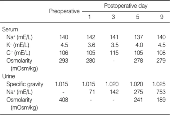
Serum
Na+(mE/L) 140 142 141 137 140
K+(mE/L) 4.5 3.6 3.5 4.0 4.5
Cl-(mE/L) 106 105 115 105 108
Osmolarity 293 280 - 278 279
(mOsm/kg) Urine
Specific gravity 1.015 1.015 1.020 1.020 1.025
Na+(mE/L) - 71 142 275 753
Osmolarity 408 - - 241 189
(mOsm/kg)
Table 2.Changes of electrolytes values and osmolarity of the sec- ond case after operation
Postoperative day Preoperative
1 3 5 9
Cerebral Salt Wasting Syndrome after Cranial Vault Remodeling 629
were comparatively retarded corresponding to about 15 months and 24 months, respectively. No abnormal rash or pigmented lesions were found on skin examination. The conjunctivae were a little pale but the sclera was anicteric. There were no pharyngeal injections or tonsillar hypertrophy. The thorax and the abdomen did not show any specific abnormalities. In extremities, syndactyly was shown on the third and the fourth toes of both feet. There was no neurological abnormality.
Preoperative laboratory findings included normocytic ane- mia with hemoglobin 9.5 mg/dL and MCV 83.5 fL. Bio- chemical test was normal, and electrolyte levels showed Na+ 140 mEq/L, K+4.5 mEq/L, Cl-106 mEq/L, urine-specific gravity of 1.015, PmOsm of 293 mOsm/kg, and UmOsm of 408 mOsm/kg. The changes of electrolytes and osmolarity of serum and urine after surgery are shown in Table 2. Pre- operative values of ADH, ANP, and HBNP were 3.25 pg/mL, 14 pg/mL, and HBNP 4.1 pg/mL, respectively. On the day of operation, the values were ADH 2.3 pg/mL, ANP 92 pg/
mL, and HBNP 63 pg/mL. On the fifth postoperative day, they were ADH 1.38 pg/mL, ANP 36 pg/mL, and HBNP 6.4 pg/mL. The patient was diagnosed as having CSW syn- drome when the postoperative 24-hr urine amount was over 1,500 mL, and did not show hyponatremia after the water and sodium supply through normal saline. Since the eighth postoperative day, the patient was in good general condition with the urine output gradually decreasing and normal blood pressure. Then, the patient was discharged from the hospital on the seventeenth postoperative day without circulatory or neurologic sequelae.
DISCUSSION
When hyponatremia develops in patients with CNS lesions such as brain damage, brain hemorrhage, brain tumor, intra- cranial infections, and stroke, an appropriate treatment based on an accurate diagnosis is the most important issue. If a treat- ment inhibiting water supply is put on a child with CSW syndrome, crucial hypotension and cerebral ischemia can be induced. Conversely, excess water and sodium supply into a patient with SIADH can cause osmotic demyelinolysis (7).
CSW syndrome is defined as a state of hyponatremia and extracellular water loss due to sodium loss into kidneys in the existence of a CNS lesion, while SIADH is a state of dilution- al hyponatremia due to increased ADH secretion (6). CSW is a diagnosis of exclusion on clinical basis. Two essential fea- tures of CSW are a cerebral lesion and renal sodium and chlo- ride wasting. We must be cautious in making diagnosis of the disease if the patient has an expanded extracelluar fluid (ECF) volumes and a condition causing a deficiency of a phys- iologic stimulator of renal sodium reabsorption (8). Our cases showed extracted ECF volumes and natriuresis without any other etiologic factors except brain surgery. CSW syndrome can be induced by the increased sodium excretion through
the urine due to the secretion of ANP and HBNP (9); how- ever, in the existence of a brain lesion, the mechanism to increase the secretion of natriuretic peptide can hardly be known. ANP and BNP are secreted from the walls of atri- um and ventricle, and the secretion is normally controlled by the heart volume and the pressure increase (6). The secre- tion of ANP and BNP promotes the sodium excretion but inhibits the vasodilation and the renin-aldosterone axis (10).
The treatment of CSW syndrome is to maintain a positive sodium balance by supplying sufficient water and sodium (11). The water and sodium supply was generally performed through an intravenous injection of 0.9% normal saline solu- tion or 3% salt solution. At this time, the correction hypona- tremia of needs to be done to the levels to prevent the occur- rence of central pontine myelinolysis. The rate of injection of saline solution should be at 1-2 mmol/L per hour and must be monitored not to exceed 25 mEq/L per day. Occasionally, injection of a normal saline solution with loop diuretics like furosemide needs to be considered (7).
The two cases of this report developed hypotension and natriuresis after a cranial vault remodeling surgery and man- ifested polyuria; however, any neurologic abnormalities such as reduced levels of consciousness were not observed in the cases. On the other hand, the first case showed an increase of the 24-hr urine amount up to 2,000 mL, a decrease of the central venous pressure down to 1-3 cmH2O, a continuous decrease of the blood pressure on the third postoperative day;
and thereupon, the infusion volume of 0.9% normal saline was increased. The second case showed an increase of the 24- hr urine amount up to 2,375 mL at the third postoperative day, but owing to the maintenance of the injection of 0.9%
normal saline, the decrease of blood pressure was not obvious as in the first case. Also, the two cases demonstrated normal ranges of ADH both pre- and the postoperatively. ANP and HBNP were temporarily increased postoperatively, which can be observed in CSW syndrome. Accordingly, considering that the patients had undergone cranial operations that could be accompanied by CSW syndrone, we closely observed the central venous pressure and urine output immediately after the operations, and measured the urine and blood sodium values. And then, we could make a diagnosis of CSW syn- drome in the two cases. The experience of the drop in the blood pressure due to polyuria in the first case made it pos- sible to prevent the drop of blood pressure in the second case by increasing the fluid volume from the early stage to prevent the water loss. The patients currently do not show any neu- rologic sequelae. Based on the experience, when hyponatrem- ia after CNS operation such as cranial surgery, the causal fac- tor should be verified first via checking the urine sodium excre- tion amount, serum electrolyte levels, ADH, and ANP, while monitoring the changes of postoperative urine amount and central venous pressure, and then, an appropriate treatment should follow.
In summary, we report our experience of two cases of chil-
630 S.-J. Lee, E.-J. Huh, J.-H. Byeon
dren with CSW syndrome after cranial surgery. The patients manifested natriuresis, polyuria, and hypotension, and showed improvement with the supply of water and sodium with 0.9%
normal saline. Therefore, CSW syndrome should always be considered when a child patient with a CNS lesion manifests hyponatremia and polyuria.
REFERENCES
1. Peters JP, Welt LG, Sims EA, Orloff J, Needham J. A salt wasting syndrome associated cerebral disease. Trans Assoc Am Physicians 1950; 63: 57-64.
2. Schwartz WB, Bennett W, Curelop S, Bartter FC. A syndrome of renal sodium loss and hyponatremia probably resulting from inappropriate secretion of antidiuretic hormone. Am J Med 1957; 23: 529-42.
3. Sengupta K, Ali U, Andankar P. Cerebral salt wasting: Case reports.
Indian Pediatrics 2002; 39: 488-91.
4. Coenraad MJ, Meinders AE, Taal JC, Bolk JH. Hyponatremia in in- tracranial disorders. Neth J Med 2001; 58: 123-7.
5. Levine JP, Stelnicki E, Weiner HL, Bradley JP, McCarthy JG. Hypona-
tremia in the postoperative craniofacial pediatric patient population:
A connection to cerebral salt wasting syndrome and management of the disorder. Plast Reconstruct Surg 2001; 108: 1501-8.
6. Betjes MG. Hyponatremia in acute brain disease: the cerebral salt wasting syndrome. Eur J intern Med 2002; 13: 9-14.
7. Roca-Ribas F, Ninno JE, Gasperin A, Lucas M, Llubia C. Cerebral salt wasting syndrome as a postoperative complication after surgical resection of acoustic neuroma. Otol neurotol 2002; 23: 992-5.
8. Singh S, Bohn D, Carlotti AP, Cusimano M, Rutka JT, Halperin ML.
Cerebral salt wasting: Truths, fallacies, theories, and challenges. Crit Care Med 2002; 30: 2575-9.
9. Berendes E, Walter M, Cullen P, Prien T, Van Aken H, Horsthemke J, Schulte M, von Wild K, Scherer R. Secretion of brain natriuretic peptide in patients with aneurysmal subarachnoid haemorrhage. Lancet 1997; 349: 245-9.
10. Espiner EA. Physiology of natriuretic peptides. J Intern Med 1994;
235: 527-41.
11. Sivakumar V, Rajshekhar V, Chandy MJ. Management of neurosur- gical patients with hyponatremia and natriuresis. Neurosurgery 1994;
34: 269-74.