INTRODUCTION
The incidence of pulmonary disease caused by nontuber- culous mycobacteria (NTM) in human immunodeficiency virus (HIV)-negative patients is increasing worldwide (1-5).
Lung disease due to NTM occurs commonly in structural lung disease, such as chronic obstructive lung disease, bronchiec- tasis, and prior tuberculosis. Of the various species of NTM, Mycobacterium avium-intracellulare complex (MAC), M. kansasii, and M. abscessus are the most common causes of NTM lung disease (1, 2).
Unlike M. kansasii lung disease, which is easily treated with conventional anti-tuberculosis drugs, including isoni- azid, rifampin, and ethambutol, both MAC lung disease and M. abscessus lung disease are usually resistant to conventional anti-tuberculosis agents. Therefore, these patients have long been considered candidates for surgical treatment when the disease is localized (6-9), and this has been regarded as essen- tial for the treatment of NTM lung disease. The major ther- apeutic advance in the treatment of NTM lung disease was the introduction of the newer macrolides, clarithromycin and azithromycin, which have substantial in vitro and clini- cal activity against MAC and M. abscessus. However, many
studies have found that pulmonary NTM disease due to MAC or M. abscessus is still difficult to eradicate despite long-term multiple antibiotic therapy, especially in patients with cavi- tary or bronchiectatic lesions (10-13).
There have been a few reports on the role of surgery in the management of NTM lung disease since the introduction of the newer macrolides. However, they included a substantial proportion of patients who did not receive the recommend- ed standardized combination antibiotic therapy (14-18).
Therefore, this study retrospectively examined the outcomes in patients who underwent pulmonary resections for NTM lung disease in recent years while taking the recommended standardized combination antibiotic therapy.
MATERIALS AND METHODS Patients
We retrospectively reviewed the surgical and medical records of all patients who had underwent pulmonary resec- tion for NTM lung disease at the Samsung Medical Center (a 1,250-bed referral hospital in Seoul, Korea) between Jan-
397
Won-Jung Koh, Yee Hyung Kim, O Jung Kwon, Yong Soo Choi*, Kwhanmien Kim*, Young Mog Shim*, and Jhingook Kim*
Division of Pulmonary and Critical Care Medicine, Department of Medicine, and Thoracic Surgery*, Samsung Medical Center, Sungkyunkwan University School of Medicine, Seoul, Korea
WJ Koh and YH Kim equally contributed to this work.
Address for correspondence Jhingook Kim, M.D.
Department of Thoracic Surgery, Samsung Medical Center, Sungkyunkwan University School of Medicine, Seoul 135-710, Korea
Tel : +82.2-3410-3483, Fax : +82.2-3410-0089 E-mail : [email protected]
*This work was supported by the Samsung Biomedical Research Institute grant (# SBRI C-A6-402-1).
DOI: 10.3346/jkms.2008.23.3.397
Surgical Treatment of Pulmonary Diseases Due to Nontuberculous Mycobacteria
Although the treatment of pulmonary diseases due to nontuberculous mycobacte- ria (NTM) requires the long-term use of antibiotics in combination, the treatment success rates are unsatisfactory. We evaluated the clinical characteristics and sur- gical outcomes of 23 patients with NTM lung diseases who had underwent pul- monary resection. The median age of the patients was 45 yr. Of the 23 patients, 10 had Mycobacterium avium-intracellulare complex infection, 12 had M. absces- sus infection, and one had M. xenopi infection. The indications for surgery were antibiotic therapy failure (n=11), remnant cavitary lesion with high probability of relapse (n=8), and massive hemoptysis (n=4). The most common procedure was lobectomy (48%). Postoperative complications occurred in eight patients (35%), including postoperative pneumonia (n=3) and late bronchopleural fistula (n=2).
Negative sputum culture conversion was achieved and maintained in all except two mortalities. Although it is associated with a relatively high complication rate, patients with NTM lung disease whose disease is localized to one lung and who can tolerate resectional surgery might be considered for surgery, if there has been poor response to drug therapy or if the patients develop significant disease-related complications such as hemoptysis.
Key Words : Mycobacteria, Atypical; Mycobacterium avium Complex; Lung Diseases; Treatment; Surgery
Received : 30 May 2007 Accepted : 8 October 2007
uary 2002 and January 2007. During this period, 23 patients underwent pulmonary resection for NTM lung disease. Of the 23 patients, 10 patients were identified as having MAC infection (7 with M. intracellulare and 3 with M. avium), 12 patients had M. abscessus infection, and one patient had M.
xenopi infection. All the patients met the diagnostic criteria for NTM lung disease, according to the American Thoracic Society (1).
The institutional review board gave permission to retro- spectively review and publish the patient records.
Standardized antibiotic regimens
The patients with NTM lung disease were managed accord- ing to the guidelines recommended by the American Tho- racic Society in 1997 (1).
For treating MAC lung disease, we typically prescribe initial daily dosing with clarithromycin (500 mg twice a day), rifampin (600 mg for body weight >50 kg, 450 mg for body weight ≤50 kg), and ethambutol (25 mg/kg per day for 2 months followed by 15 mg/kg per day). Strepto- mycin is also used for several months in all patients (1, 4).
The patients with M. abscessus lung disease are treated with clarithromycin (500 mg twice a day), ciprofloxacin (500 mg twice a day), and doxycycline (200 mg twice a day) in com- bination with parenteral antibiotics, including amikacin (15 mg/kg per day) and cefoxitin (200 mg/kg per day), for the first 4 weeks of hospitalization (1, 4).
Indications for pulmonary resection
Chest computed tomography (CT) was performed preop- eratively in all patients to evaluate their lesions, including cavities and bronchiectasis, and underlying pulmonary con- dition.
Patients were selected for surgery based on consensus by medical and surgical specialists. In general, indications for pulmonary resection included failure of medical therapy, remnant cavitary lesion with a high possibility of relapse, and the development of complications such as massive hemoptysis. Surgical candidates had to have sufficient pul- monary function to tolerate resection and a localized lesion with a high bacterial burden, such as a cavity. For patients with bilateral lesions, the area with the higher bacterial bur- den was resected, and the remaining lesion with the lower bacterial burden in the ipsilateral or contralateral lung was controlled with medical therapy.
The standard preoperative work-up included chest radio- graphy, chest CT, pulmonary function tests with a quantita- tive lung perfusion scan, electrocardiogram, echocardiogra- phy, and bronchoscopy.
Surgery was performed under general anesthesia using a double-lumen endobronchial tube. The majority of pul- monary resections (16/23, 70%) were performed on the right
side. Postoperatively, the patients were scheduled to have intensive antibiotic regimens that were generally same as the preoperative ones. After the sputum acid-fast bacilli (AFB) culture became negative with either medical treatment or surgery, standardized antibiotic therapy was continued for at least 12 months.
Treatment outcomes
Sputum conversion was defined as three consecutive neg- ative sputum cultures tested at monthly intervals. If the patient could not expectorate during the treatment period, the sputum was considered to have converted to negative.
The conversion date was recorded as the date of the first neg- ative sputum culture in a patient with negative sputum con- version.
RESULTS Patient characteristics
During the study period, pulmonary resections for NTM lung disease were performed in 23 patients (7 men and 16 women) ranging in age between 24 and 66 yr (median, 45;
interquartile range [IQR], 37 to 57). The main characteristics of the patient population are shown in Table 1. No patients suffered from immunodeficiency disorders such as HIV infec- tion. All the patients underwent lung resection to treat NTM
The data are presented as the median (IQR) or number (%).
AFB, acid-fast bacilli; FVC, forced vital capacity; FEV1, forced expirato- ry volume in one second.
Variable Value
Age (yr) 45 (37-57)
Gender (male/female) 7 (30%)/16 (70%)
Body mass index (kg/m2) 18.7 (17.6-21.3) Etiology
M. avium-intracellulare complex 10 (44%)
M. abscessus 12 (52%)
M. xenopi 1 (4%)
Duration of preoperative antibiotic therapy 7.5 (5-17) (months)
Preoperative sputum status
Positive AFB smear 15 (65%)
Positive AFB culture 17 (74%)
Radiographic findings
Cavity-predominant 16 (70%)
Bronchiectasis-predominant 7 (30%)
Pulmonary function tests
FVC (L) 2.56 (1.99-2.74)
Percent FVC 77 (64-89)
FEV1(L) 1.95 (1.48-2.30)
Percent FEV1 78 (61-90)
Table 1. Clinical characteristics of the patients
lung disease or the complications of NTM lung disease and not for diagnostic purposes.
The preoperative sputum smear and culture were positive in 15 (65%) and 17 (74%) patients, respectively. All patients had either a cavitary or bronchiectatic lesion on chest CT.
The lesions were predominantly cavities in 16 (70%) and predominantly bronchiectasis in seven (30%) patients.
Antibiotic treatment and indication for surgery
A multi-antibiotic regimen based on the standardized pro- tocol for NTM lung infection was initiated preoperatively in 20 patients. The duration of the preoperative antibiotic therapy in these patients was a median of 7.5 months (IQR, 5 to 17; Table 1). Three patients who developed massive hemoptysis did not receive preoperative antibiotic therapy for NTM lung disease.
The indications for surgery included failure of medical treatment in 11 patients (48%), remnant cavitary lesion with a high possibility of relapse in eight patients (35%), and mas- sive hemoptysis in four patients (17%).
In particular, surgery was performed in eight patients who showed some microbiologic improvements (preoperative negative sputum culture in six patients and positive culture in two patients), but had remnant cavitary lesion after antibi- otic therapy for a median of 10 months (IQR, 4 to 19). These patients were considered for indication of surgery because of the remnant cavitary lesion with a high probability of relapse.
Four patients had MAC lug disease, and four had M. absces- sus lung disease. Microscopic findings showed parenchymal destruction with cavity (or multiple cavities) and granuloma- tous inflammation with caseation necrosis in these patients.
Surgical treatment
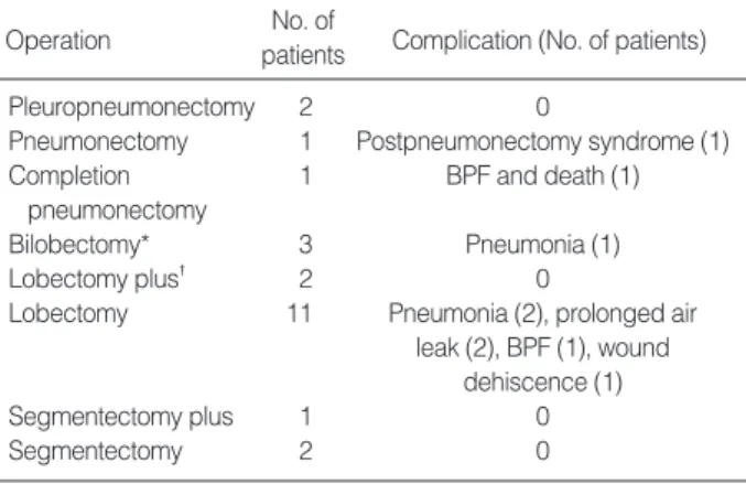
The pulmonary resections included lobectomy in 11 patients, pleuropneumonectomy or pneumonectomy including com- pletion pneumonectomy in four, bilobectomy in three, seg- mentectomy in three, and lobectomy plus segmentectomy in two patients (Table 2). Nineteen patients underwent open thoracotomy. One patient underwent segmentectomy through a minithoracotomy with video assistance, and three patients underwent video-assisted thoracic surgery without a minitho- racotomy (lobectomy in two patients and bilobectomy in one patient). The pathologic findings showed granuloma- tous inflammation with necrosis in all cases.
The median operating time was 180 min (IQR, 156 to 228 min). The median estimated intraoperative blood loss was 400 mL (IQR, 325 to 575 mL). In nine patients, the blood loss exceeded 500 mL (500 to 4,000 mL), and these patients were given blood transfusions. The median dura- tion of postoperative hospitalization was 9 days (IQR, 6 to 15 days).
Postoperative complications
There were no intraoperative deaths. There was one post- operative death during hospitalization; a 66-yr-old man with M. xenopi lung infection died of respiratory failure associated with the development of a late bronchopleural fistula 47 days after his completion pneumonectomy (19). The right upper lobe had been removed 44 yr earlier for tuberculosis in this patient. There was one late outpatient death; a 44-yr-old man with M. intracellulare lung infection who had been discharged home after an uneventful postoperative course with right upper lobectomy died in an accident 50 days after the oper- ation.
Postoperative complications occurred in eight patients (35%) and included postoperative pneumonia (n=3), late bronchopleural fistula (n=2), prolonged air leak (n=2), wound dehiscence (n=1), and postpneumonectomy syndrome (n=1) (Table 2).
Treatment outcomes
Postoperative antibiotic therapy was administered to 22 patients. In one patient with M. abscessus infection who had bronchiectasis in the right middle lobe and developed mas- sive hemoptysis, neither preoperative nor postoperative antibi- otic therapy for M. abscessus infection was prescribed, as there was no residual lesion on chest CT after pulmonary resection.
Negative sputum culture conversion was achieved within 1 or 2 months postoperatively and maintained in all survivors
*Right upper and middle lobectomy in one patient and right middle and lower lobectomy in two patients; �left upper lobectomy plus lower lobe superior segmentectomy in one patient and left lower lobectomy plus upper lobe lingular segmentectomy in one patient; lobectomy: right upper lobectomy in five patients, right middle lobectomy in two patients, right lower lobectomy in two patients, left upper lobectomy in one patient, and left lower lobectomy in one patient; segmentectomy plus: left upper lobe lingular segmentectomy plus lower lobe superior segmentectomy;
segmentectomy: right upper lobe posterior segmentectomy in one patient and right lower lobe posterior basal segmentectomy in one patient. BPF, bronchopleural fistula.
Operation No. of
Complication (No. of patients) patients
Pleuropneumonectomy 2 0
Pneumonectomy 1 Postpneumonectomy syndrome (1)
Completion 1 BPF and death (1)
pneumonectomy
Bilobectomy* 3 Pneumonia (1)
Lobectomy plus� 2 0
Lobectomy 11 Pneumonia (2), prolonged air leak (2), BPF (1), wound
dehiscence (1)
Segmentectomy plus 1 0
Segmentectomy 2 0
Table 2. Operative procedures and postoperative complications
with preoperative culture-positive sputum. Antibiotic treat- ment was completed in seven patients, who received post- operative antibiotics for a median duration of 12 months (IQR, 6 to 26 months) and were followed up for a median duration of 14 months (IQR, 1 to 22 months) after the end of treatment. In addition, 14 patients received postoperative antibiotic therapy for a median duration of 8 months (IQR, 5.5 to 11 months). Of these 21 patients, no patient has been known to have relapsed during the follow-up period.
DISCUSSION
NTM lung disease has become a significant health prob- lem. However, NTM lung disease remains difficult to treat with medication alone, although the introduction of newer macrolides has improved the outcome of its medical treat- ment. Therefore, pulmonary resection surgery has been advo- cated for selected patients who have localized disease and are able to tolerate resection to remove gross lesions that con- tain large numbers of bacilli (1, 2).
Regarding the treatment of NTM lung disease, the Amer- ican Thoracic Society has recommended that the initial ther- apy for patients with MAC lung disease consist of a mini- mum three-drug regimen of clarithromycin (or azithromycin), rifampin (or rifabutin), and ethambutol (1, 2). In addition, intermittent streptomycin for the first 2 to 3 months of ther- apy was recommended for extensive disease. Recently, one prospective, randomized trial revealed that a better microbi- ological response was observed in patients treated with the regimen including streptomycin (20). Nevertheless, the suc- cess rates of these medical treatments have peaked at 70-80%
(10, 20), and the relapse rate in patients with sputum con- version at the completion of medical treatment is high (21).
For the treatment of M. abscessus lung disease, combined intravenous antibiotic therapy including amikacin and cefox- itin for 2-4 weeks for clinical and microbiologic improve- ment was recommended in addition to the oral antibiotics, including clarithromycin (or azithromycin) (1). However, the antibiotic treatment of lung disease caused by M. absces- sus is usually unsuccessful because of high levels of in vitro resistance, the need for injectable antimicrobial drugs, poten- tially toxic drugs, and the long treatment duration. There- fore, surgery is usually recommended for patients with local- ized lung disease who can withstand lung resection after an initial period on antimicrobials to reduce the microbial bur- den (1, 2, 12, 13).
In our study, all the patients who had received preopera- tive antibiotic therapy were treated with the recommended standardized regimen. Moreover, streptomycin was routine- ly administered for several months to patients with MAC lung disease. In addition, the patients with M. abscessus lung disease also received standardized antibiotic therapy, which included 4 weeks of initial intravenous antibiotics while hos-
pitalized. Therefore, the patients included in this study must have received the most effective medication in terms of med- ical treatment, as compared with those in previous studies (14-18). Nevertheless, the medical treatment outcome was unsatisfactory in our patients.
Therefore, the patients whose disease was mainly localiz- ed to one lung and who could tolerate pulmonary resection were considered for surgery. In this study, we were able to assess the surgical outcomes of 23 patients with NTM lung disease. Although most of our patients (n=17, 74%) had preoperative culture-positive sputum for NTM, negative sputum culture conversion was achieved within 1 or 2 months postoperatively and was maintained in all survivors, exclud- ing the two patients who died postoperatively. In addition, no survivor relapsed during the postoperative follow-up peri- od. These results indicate that pulmonary resection can play an important role in achieving a better outcome in selected patients, especially patients in whom medical therapy failed or who had a remnant lesion with a high possibility of relapse.
However, there is no rule concerning the optimal timing of surgery. One retrospective study recommended that patients with localized NTM lung disease be considered for pulmonary resection as early as possible (14). In our study, the median duration of the preoperative antibiotic therapy was 7.5 months, which was shorter than in previous studies (14-18). This was possible because, in recent years at our institution, surgery has been actively considered in patients with localized NTM lung disease when medical treatment appeared ineffective, before the lesions had become inoperable. This treatment strategy may lead to more favorable outcomes. Early surgery with standardized antibiotic therapy may be necessary for patients who are likely to fail medical therapy yet have suf- ficient predicted postoperative pulmonary function.
Despite the favorable treatment outcomes, postoperative complications were relatively high (35%) with NTM lung disease in our study. This was similar to previous studies that reported a high incidence of postoperative morbidity (14- 18, 22, 23).
In conclusion, despite its relatively high surgical compli- cation rate, patients with NTM lung disease whose disease is localized to one lung and who can tolerate resectional surgery might be considered for surgery, if there has been a poor response to drug therapy or if the patients develop sig- nificant disease-related complications such as hemoptysis.
REFERENCES
1. Wallace RJ Jr, Cook JL, Glassroth J, Griffith DE, Olivier KN, Gordin F. Diagnosis and treatment of disease caused by nontuberculous mycobacteria. This official statement of the American Thoracic Society was approved by the Board of Directors, March 1997. Med- ical Section of the American Lung Association. Am J Respir Crit Care Med 1997; 156: S1-25.
2. Griffith DE, Aksamit T, Brown-Elliott BA, Catanzaro A, Daley C, Gordin F, Holland SM, Horsburgh R, Huitt G, Iademarco MF, Ise- man M, Olivier K, Ruoss S, von Reyn CF, Wallace RJ Jr, Winthrop K. An official ATS/IDSA statement: diagnosis, treatment, and pre- vention of nontuberculous mycobacterial diseases. Am J Respir Crit Care Med 2007; 175: 367-416.
3. Field SK, Cowie RL. Lung disease due to the more common nontu- berculous mycobacteria. Chest 2006; 129: 1653-72.
4. Koh WJ, Kwon OJ, Lee KS. Diagnosis and treatment of nontuber- culous mycobacterial pulmonary diseases: a Korean perspective. J Korean Med Sci 2005; 20: 913-25.
5. Yim JJ, Park YK, Lew WJ, Bai GH, Han SK, Shim YS. Mycobac- terium kansasii pulmonary diseases in Korea. J Korean Med Sci 2005; 20: 957-60.
6. Hattler BG Jr, Young WG Jr, Sealy WC, Gentry WH, Cox CB. Sur- gical management of pulmonary tuberculosis due to atypical mycobac- teria. J Thorac Cardiovasc Surg 1970; 59: 366-71.
7. Elkadi A, Salas R, Almond CH. Surgical treatment of atypical pul- monary tuberculosis. J Thorac Cardiovasc Surg 1976; 72: 435-40.
8. Corpe RF. Surgical management of pulmonary disease due to Mycobac- terium avium-intracellulare. Rev Infect Dis 1981; 3: 1064-7.
9. Moran JF, Alexander LG, Staub EW, Young WG Jr, Sealy WC.
Long-term results of pulmonary resection for atypical mycobacteri- al disease. Ann Thorac Surg 1983; 35: 597-604.
10. Field SK, Fisher D, Cowie RL. Mycobacterium avium complex pul- monary disease in patients without HIV infection. Chest 2004; 126:
566-81.
11. Lam PK, Griffith DE, Aksamit TR, Ruoss SJ, Garay SM, Daley CL, Catanzaro A. Factors related to response to intermittent treat- ment of Mycobacterium avium complex lung disease. Am J Respir Crit Care Med 2006; 173: 1283-9.
12. Griffith DE, Girard WM, Wallace RJ Jr. Clinical features of pul- monary disease caused by rapidly growing mycobacteria. An analy- sis of 154 patients. Am Rev Respir Dis 1993; 147: 1271-8.
13. Daley CL, Griffith DE. Pulmonary disease caused by rapidly grow-
ing mycobacteria. Clin Chest Med 2002; 23: 623-32.
14. Shiraishi Y, Fukushima K, Komatsu H, Kurashima A. Early pul- monary resection for localized Mycobacterium avium complex dis- ease. Ann Thorac Surg 1998; 66: 183-6.
15. Nelson KG, Griffith DE, Brown BA, Wallace RJ Jr. Results of opera- tion in Mycobacterium avium-intracellulare lung disease. Ann Tho- rac Surg 1998; 66: 325-30.
16. Shiraishi Y, Nakajima Y, Takasuna K, Hanaoka T, Katsuragi N, Konno H. Surgery for Mycobacterium avium complex lung disease in the clarithromycin era. Eur J Cardiothorac Surg 2002; 21: 314-8.
17. Shiraishi Y, Nakajima Y, Katsuragi N, Kurai M, Takahashi N. Pneu- monectomy for nontuberculous mycobacterial infections. Ann Tho- rac Surg 2004; 78: 399-403.
18. Watanabe M, Hasegawa N, Ishizaka A, Asakura K, Izumi Y, Eguchi K, Kawamura M, Horinouchi H, Kobayashi K. Early pulmonary resection for Mycobacterium avium complex lung disease treated with macrolides and quinolones. Ann Thorac Surg 2006; 81: 2026-30.
19. Park HY, Koh WJ, Kwon OJ, Lee NY, Shim YM, Park YK, Bai GH, Mun HS, Kim BJ. Pulmonary disease caused by Mycobacteri- um xenopi: the first case in Korea. Yonsei Med J 2007; 48: 871-5.
20. Kobashi Y, Matsushima T, Oka M. A double-blind randomized study of aminoglycoside infusion with combined therapy for pulmonary Mycobacterium avium complex disease. Respir Med 2007; 101:
130-8.
21. Kobashi Y, Matsushima T. The microbiological and clinical effects of combined therapy according to guidelines on the treatment of pulmonary Mycobacterium avium complex disease in Japan - includ- ing a follow-up study. Respiration 2007; 74: 394-400.
22. Pomerantz M, Madsen L, Goble M, Iseman M. Surgical manage- ment of resistant mycobacterial tuberculosis and other mycobacte- rial pulmonary infections. Ann Thorac Surg 1991; 52: 1108-11;
discussion 12.
23. Sherwood JT, Mitchell JD, Pomerantz M. Completion pneumonec- tomy for chronic mycobacterial disease. J Thorac Cardiovasc Surg 2005; 129: 1258-65.