Extraskeletal Ewing’s
Sarcoma of the Thoracic Nerve Root: A Case Report
흉부 신경근에서 발생한 골외성 유잉육종: 증례 보고
Jung Won Kim, MD1 , Jihae Lee, MD1* , Jae Hyung Kim, MD1, Myeong Ja Jeong, MD1, Soung Hee Kim, MD1, Ji-Young Kim, MD1,
Soo Hyun Kim, MD1, Mi-Jin Kang, MD1, Tae-gyu Kim, MD1, Kyung Eun Bae, MD1, Jun Jae Shin, MD2, Hyun-Jung Kim, MD3, Jung Yeon Kim, MD3
Departments of 1Radiology, 2Neurosurgery, 3Pathology, Inje University College of Medicine, Sanggye Paik Hospital, Seoul, Korea
Extraskeletal Ewing’s sarcoma (EES) is a rare malignant soft tissue tumor which is morphologi- cally indistinguishable from skeletal ES. EES usually occurs in young adults and children and there has been only one case reported in a patient aged over 70 years old. We report a case of an EES arising from the first thoracic spinal nerve root in a 73-year-old female, which was mis- diagnosed as benign nerve sheath tumor in preoperative imaging evaluation.
Index terms Sarcoma, Ewing; Spinal Nerve Root; Thoracic Nerves; Soft Tissue Neoplasm
INTRODUCTION
Extraskeletal Ewing’s sarcoma (EES), especially involving spine accounts for only 5%
of all ES (1). It is known to be found mainly in the paravertebral and epidural space (2).
And ES usually occurs in children or young adults (1). Primary EES of spinal nerve has been reported only a few cases and there has been only one case of a senior patient in eighth decade (3).
Herein, we report a case of spinal epidural EES in a 73-year-old female.
CASE REPORT
A 73-year-old female visited to our hospital complaining pain in the posterior neck and left arm. The symptoms started 6 months ago and aggravated 2 days ago. Physical examination revealed hypoesthesia on her left ulnar nerve territory without decrease of muscle strength. She underwent plain radiography and CT of cervical spine. Plain ra-
Received September 29, 2017 Revised October 25, 2017 Accepted November 7, 2017
*Corresponding author Jihae Lee, MD
Department of Radiology, Inje University
College of Medicine, Sanggye Paik Hospital, 1342 Dongil-ro, Nowon-gu, Seoul 01757, Korea.
Tel 82-2-950-1182 Fax 82-2-950-1220 E-mail [email protected] This is an Open Access article distributed under the terms of the Creative Commons Attribu- tion Non-Commercial License (https://creativecommons.org/
licenses/by-nc/4.0) which permits unrestricted non-commercial use, distribution, and reproduc- tion in any medium, provided the original work is properly cited.
ORCID iDs Jihae Lee https://
orcid.org/0000-0002-4884-5876 Jung Won Kim
https://
orcid.org/0000-0003-3417-3260
https://doi.org/10.3348/jksr.2019.80.3.568 569
T2WI T1WI
FS-T1WI with Gd-CE FS-T2WI
diographs of the cervical spine showed no significant abnormality other than disc space nar- rowing in C5–C6 level (not shown).
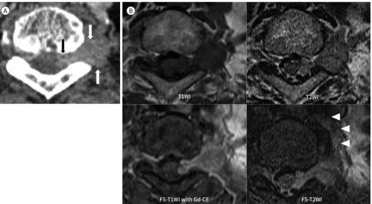
CT revealed a dumbbell-shaped mass in the left neural foramen of T1–T2. No calcification was seen in the mass. There was mild widening of left neural foramen (Fig. 1A). For further evaluation, MRI of the cervical and thoracic spine was performed. MR images showed that the medial part of the mass is in the intradural and extramedullary region and the lateral part of the mass extends to the paravertebral area through the left neural foramen. The le- sion shows intermediate signal intensity on T1-weight images and homogenously slightly high signal intensity on T2-weighted images (Fig. 1B). After administration of intravenous contrast material, the mass is enhanced homogeneously. Size of the lesion is measured ap- proximately 2.6 × 1.3 × 1.5 cm. The mass is branching outside the left neural foramen and ex- tending along the first branch of left T1 nerve (Fig. 1B).
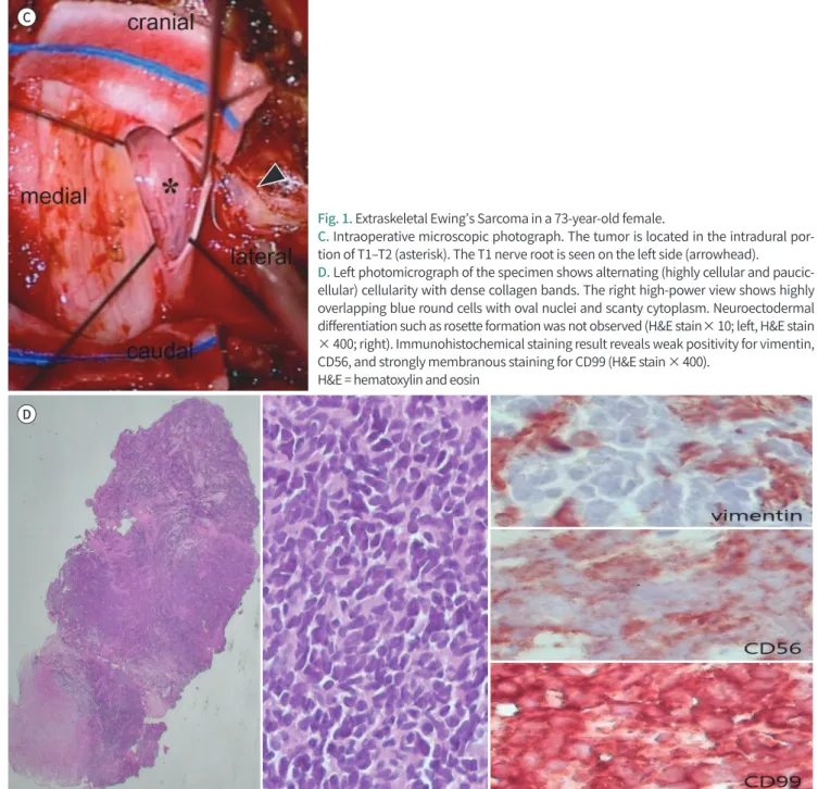
Based on the CT and MRI findings, the benign nerve sheath tumor was suggested. And then, the patient underwent surgery. In the operation, the tumor was revealed after laminec- tomy of T1 vertebra and also after incision of dura (Fig. 1C). The mass was strongly attached to the nerve root, partly intermingled with the nerve fibers. Subtotal resection of the tumor was done with moter evoked potential monitoring to minimize injury of the nerve.
Microscopic examination revealed that the tumor was composed of alternating highly cel-
Fig. 1. Extraskeletal Ewing’s Sarcoma in a 73-year-old female.
A. Thoracic spine CT with contrast enhancement shows a dumbell-shaped mass in the T1–T2 left neural foramen extending inside the central canal and outside the neural foramen, with mild homogeneous enhancement (arrows). Mild widening of the neural foramen is noted.
B. On MRI with contrast enhancement, the lesion shows intermediate signal intensity on the axial T1WI and slightly high signal intensity on the T2WI. Homogeneous enhancement of the lesion is seen on the FS-T1WI after administration of Gd-CE. The FS-T2WI shows the tumor ex- tending along the first branch of left T2 nerve root (arrowheads).
Gd-CE = gadolinium contrast-enhanced, FS = fat saturation, T1WI = T1-weighted image, T2WI = T2-weighted image
A B
lular and paucicellular components with dense collagen bands (× 10). The high-power field view showed highly overlapping blue round cells with oval nuclei and scanty cytoplasm, com- patible with blue round cell tumor. No evidence of neuroectodermal differentiation, such as rosette formation was identified (× 400) (Fig. 1D). The result of immunohistochemical stain- ing revealed weak positivity for vimentin and CD56, and strong membranous staining for CD99, compatible with ES (Fig. 1D). Further gene mutation study revealed EWSR1 gene translocation and the final pathological diagnosis was made as EES.
After confirmative diagnosis, the patient transferred to cancer clinic for adjuvant chemo- therapy and radiation therapy. After twelve months from the surgery, the tumor was reduced
Fig. 1. Extraskeletal Ewing’s Sarcoma in a 73-year-old female.
C. Intraoperative microscopic photograph. The tumor is located in the intradural por- tion of T1–T2 (asterisk). The T1 nerve root is seen on the left side (arrowhead).
D. Left photomicrograph of the specimen shows alternating (highly cellular and paucic- ellular) cellularity with dense collagen bands. The right high-power view shows highly overlapping blue round cells with oval nuclei and scanty cytoplasm. Neuroectodermal differentiation such as rosette formation was not observed (H&E stain × 10; left, H&E stain
× 400; right). Immunohistochemical staining result reveals weak positivity for vimentin, CD56, and strongly membranous staining for CD99 (H&E stain × 400).
H&E = hematoxylin and eosin C
D
https://doi.org/10.3348/jksr.2019.80.3.568 571 in size and the patient was in stable condition without local recurrence, only complaining mild tingling sensation on her elbow. However, recently, after post-operative sixteen months, local recurrence and leptomeningeal seeding developed and the patient is receiving chemo- therapy.
DISCUSSION
EES is a rare form of round cell malignant tumor with extraskeletal origin (1, 2, 4). It ac- counts for about 10% of ES in children and 5% of ES in adults (3). The primary site of EES in- volving extradural and intradural extramedullary space is extremely rare (1-10). Prefered an- atomic origin of EES is paravertebral and epidural space, so associated symptoms are variable such as local pain, gait disturbance, motor deficit, radicular pain, etc (1, 4, 5).
Imaging features of EES are nonspecific but, usually appears solid, iso signal intensity on T1-weighted image, intermediate to high signal intensity on T2-weighted image and moder- ate to strong enhancement after gadolinium administration (1-10). Some reported lesions show slightly inhomogeneous signal intensity or low signal intensity on T2-weighted images (1, 3-5) and heterogeneous enhancement (1, 3, 5, 6).
In our case, the mass was iso signal intensity in T1-weighted image and slightly high signal intensity in T2-weighted image with marked enhancement, similar to previous cases.
The radiologic differential diagnosis of EES involving epidural space includes benign nerve sheath tumor such as schwannoma, lymphoma and malignant nerve sheath tumor (2-4, 7).
Schwannoma has smooth margin and dumbell shaped with intense enhancement. On T2- weighted image, schwannoma appears heterogeneously high signal intenstiy with target sign (3, 4, 7). Lymphoma involving epidural space originates from body of vertebra or paraverte- bral lymph node then extends to epidural space. It is slightly high signal intensity to muscle on T1-weighted image and T2-weighted image involving multi-segment of spine with homoge- neous enhancement along epidural infiltration (9). Malignant nerve sheath tumor of spine appears expansile soft tissue tumor destructing adjacent vertebra with paraspinal and epidur- al extension. On MR, the mass appears low signal intensity on T1-weighted image and high signal intensity on T2-weighted image. It is mostly associated with neurofibromatosis type 1 (10).
This case was initially presumed to be a benign nerve sheath tumor, however the final di- agnosis was an EES. There was no definite aggressive feature such as ill-defined margin, ad- jacent bony destruction, heterogeneous signal intensity or attenuation of the mass, or large size of the mass. However, in the literature, there are some reported cases of EES mimicking benign nerve sheath tumors (4, 6, 7) and it was difficult to differentiate EES from benign nerve sheath tumor only with radiologic imaging, without pathologic confirmation in those cases.
In our case, the lesion extended to the 1st branch of thoracic nerve root and this feature may indicate relatively more aggressive kinds of tumor such as lymphoma or malignant peripher- al nerve sheath tumor rather than benign nerve sheath tumor, even though ES is very rare in elder patients. However, this finding may be seen in neurofibromas.
Therefore, even there is well-marginated dumbbell-shaped extradural mass whick looks benign, if there is subtle ill-defined enhancing or high signal intensity lesion extended to pe- ripheral nerve, possibility of malignancy might be considered.
We reported a case of EES arising from thoracic nerve root in 73-year-old woman. Because of its rarity and varying morphology on imaging modality, it is difficult to diagnosis from ra- diologic finding before pathologic confirmation. We suggest that in case of nerve root tumor showing atypical radiologic findings of nerve sheath tumors, EES also could be considered as differential diagnoses although these tumors are very rare in elder patients.
Conflicts of Interest
The authors have no potential conflicts of interest to disclose.
REFERENCES
1. Gong HS, Huang QS, Liu GJ, Chen FH, Zhao HB. Cervical primary Ewing’s sarcoma in intradural and extra- medullary location and skip metastasis to cauda equina. Turk Neurosurg 2015;25:943-947
2. Lozupone E, Martucci M, Rigante L, Gaudino S, Di Lella GM, Colosimo C. Magnetic resonance image find- ings of primary intradural Ewing sarcoma of the cauda equina: case report and review of the literature.
Spine J 2014;14:e7-e11
3. Mardekian SK, Gandhe A, Miettinen M, Pack S, Curtis MT, Abdullaev Z. Two cases of spinal, extraosseous, in- tradural Ewing’s sarcoma/peripheral neuroectodermal tumor: radiologic, pathologic, and molecular analy- sis. J Clin Imaging Sci 2014;4:6
4. Zhao M, Zhang B, Liang F, Zhang J. Primary spinal intradural extraskeletal Ewing sarcoma mimicking a gi- ant nerve sheath tumor: case report and review of the literature. Int J Clin Exp Pathol 2014;7:9081-9085 5. Saeedinia S, Nouri M, Alimohammadi M, Moradi H, Amirjamshidi A. Primary spinal extradural Ewing’s sar-
coma (primitive neuroectodermal tumor): report of a case and meta-analysis of the reported cases in the literature. Surg Neurol Int 2012;3:55
6. Haresh KP, Chinikkatti SK, Prabhakar R, Rishi A, Rath GK, Sharma DN, et al. A rare case of intradural extra- medullary Ewing’s sarcoma with skip metastasis in the spine. Spinal Cord 2008;46:582-584
7. Zhu Q, Zhang J, Xiao J. Primary dumbbell-shaped Ewing’s sarcoma of the cervical vertebra in adults: four case reports and literature review. Oncol Lett 2012;3:721-725
8. Shin JH, Lee HK, Rhim SC, Cho KJ, Choi CG, Suh DC. Spinal epidural extraskeletal Ewing sarcoma: MR find- ings in two cases. AJNR Am J Neuroradiol 2001;22:795-798
9. Liou DC, Chou JM. MRI of primary spinal epidural lymphoma: case report. Chin J Radiol 2007;32:147-151 10. Senapati SB, Mishra SS, Dhir MK, Das S. Malignant peripheral nerve sheath tumor in spine: two case re-
ports. South Asian J Cancer 2013;2:141
https://doi.org/10.3348/jksr.2019.80.3.568 573
흉부 신경근에서 발생한 골외성 유잉육종: 증례 보고
김정원1 · 이지혜1* · 김재형1 · 정명자1 · 김성희1 · 김지영1 · 김수현1 강미진1 · 김태규1 · 배경은1 · 신준재2 · 김현정3 · 김정연3
골외성 유잉육종은 드문 악성 종양으로 육안적으로는 골성 유잉육종과 감별할 수 없다. 골외 성 유잉육종은 대부분 젊은 성인이나 20세 이하의 소아청소년에서 발생하며 70세 이상에서 발생한 경우는 현재까지 1예가 보고되었다. 우리는 양성 신경초종양으로 오인되었던 73세 여 성의 첫 번째 흉부 신경근에서 기원한 골외성 유잉육종의 증례를 보고하고자 한다.
인제대학교 의과대학 상계백병원 1영상의학과, 2신경외과, 3병리과