Pregnancy and Delivery in a Generalized Dystonia Patient Treated with Internal Globus Pallidal Deep Brain Stimulation:
a Case Report
Internal globus pallidus (GPi) deep brain stimulation (DBS) has been widely accepted as an effective treatment modality of medically refractory dystonia. However, there have been few studies regarding the safety issue of pregnancy and childbirth related with DBS. This report describes a female patient who was pregnant and delivered a baby after GPi DBS surgery. A 33-year-old female patient with acquired generalized dystonia underwent bilateral GPi DBS implantation. She obtained considerable improvement in both movement and disability after DBS implantation. Four years later, she was pregnant and the
obstetricians consulted us about the safety of the delivery. At 38-weeks into pregnancy, a scheduled caesarian section was carried out under general anesthesia. After induction using thiopental and succinylcholine, intubation was done quickly, followed by DBS turn off. For hemostasis, only bipolar electrocautery was used. Before awakening from the anesthesia, DBS was turned on as the same parameters previously adjusted. After delivery, she could feed her baby by herself, because the dystonia of left upper extremity and hand was improved. Until now, she has been showing continual improvement and being good at housework, carrying for children, with no trouble in daily life. This observation indicates that the patients who underwent DBS could safely be pregnant and deliver a baby.
Keywords: Deep Brain Stimulation; Dystonia; Globus Pallidus; Child Birth and Care Hye Ran Park,1 Jae meen Lee,2
Hyeyoung Park,3 Chae Won Shin,3 Han-Joon Kim,3 Hee Pyoung Park,4 Dong Gyu Kim,2,5 Beom Seok Jeon,3,6 and Sun Ha Paek2,5
1Department of Neurosurgery, Soonchunhyang University Seoul Hospital, Seoul, Korea;
2Department of Neurosurgery, Seoul National University Hospital, Seoul, Korea;
3Department of Neurology, Seoul National University Hospital, Seoul, Korea;
4Department of Anesthesiology and Pain Medicine, Seoul National University Hospital, Seoul, Korea;
5Department of Neurosurgery, Seoul National University College of Medicine, Seoul, Korea;
6Department of Neurology, Seoul National University College of Medicine, Seoul, Korea Received: 24 August 2015
Accepted: 2 January 2016 Address for Correspondence:
Sun Ha Paek, MD
Department of Neurosurgery, Seoul National University Hospital, 103, Daehak-ro, Jongno-gu, Seoul 03080, Korea
E-mail: [email protected]
https://doi.org/10.3346/jkms.2017.32.1.155 • J Korean Med Sci 2017; 32: 155-159
INTRODUCTION
Dystonia, one of the most prevalent forms of movement disor
der, is defined as sustained or intermittent muscle contractions usually producing twisting and repetitive movement or abnor
mal posture (1,2). Oral medications and botulinum toxin injec
tions have been the mainstays of treatment for a time, but are not sufficiently effective in many patients. Internal globus palli
dus (GPi) deep brain stimulation (DBS) has been widely accept
ed as an effective treatment modality of medically refractory dystonia (3,4). However, few studies have been reported regard
ing the safety issue of pregnancy and childbirth and its long term outcome of the GPi DBS during the pregnancies. This re
port describes a female patient with generalized dystonia im
planted with GPi DBS who delivered a baby during 84month followup period after DBS surgery.
CASE DESCRIPTION History
A 33yearold female patient, a mother of one child, developed
a left upper limb dystonia after being asphyxiated under blan
kets at the age of 4 weeks. The dystonia in both lower limbs pro
gressively developed into a generalized form, accompanied by a left facial spasm. She had undergone orthopedic surgery be
cause of right ankle eversion and lateral deviation of the right great toe 2 years ago. The most painful symptom to her was a dystonia of left upper limb accompanied by tremor occurred in both resting and exercise. The symptoms were not improved by medical treatment including nortriptyline, levodopa, clonaze
pam, baclofen, and biperiden. There was no family history of movement disorders. The patient was referred to our institution for evaluation of the DBS (November 14, 2006).
Examination
She had alert consciousness and normal intellectual ability. Mo
tor power grades were V in all extremities, and sensory was also intact. After orthopedic surgery, the right ankle was nearly cor
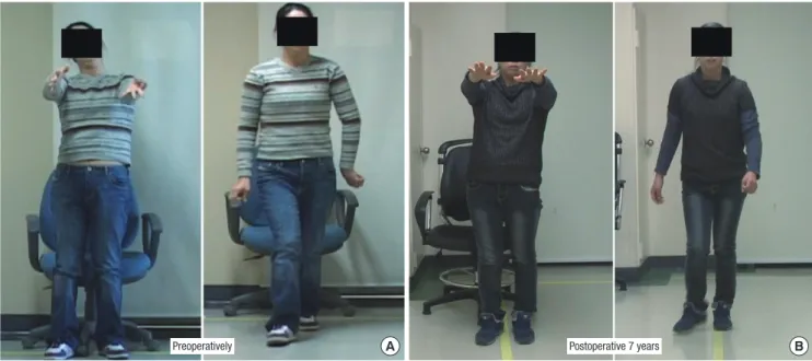
rected but the right great toe was still deviated laterally. Her gait was mildly ataxic. The left arm was adducted in the shoulder and extended in the elbow. The left wrist was hyperextended, with continuing rhythmic clenching and opening of the fist (Fig.
1A). The dystonia severity was measured using BurkeFahn
Marsden Dystonia Rating Scale (BFMDRS); her preoperative movement score was 33 points, and functional disability score was 10 points. Minimental state examination (MMSE) was 29 points, and Beck’s depression inventory (BDI) scale was 3 points.
DYT1 gene was not detected. On magnetic resonance imaging (MRI), there were no definite focal lesions in the brain paren
chyma.
Operation
In November 2006, she underwent bilateral GPi implantation.
A Leksell stereotactic frame was secured to the patient’s head after application of a local anesthetic and she was transferred to the MR suite. The posteroventral portion of the GPi was target
ed by means of axial, sagittal, and coronal MRI images (5). The pallidal target was 12.4 mm anterior to the midcommisural point, 20 mm lateral to the midline, and 3 mm below the intercommi
sural line in both side. The procedure was performed under gen
eral anesthesia, with the assistance of microelectrode recording (MER). A set of four microelectrodes (Differential microTarget
ing® Electrodes; FHC, Chemnitz, Germany; 1.5 MΩ impedance) were sequentially inserted toward the anatomical target within the GPi, which was vertically on the axial slice at the level of an
terior commissure and horizontally at the junction between the two posterior quarters of the GPi (6). Permanent DBS electrodes (DBS 3387; Medtronic, Minneapolis, MN, USA) placements were determined without interpreting vessels, ventricles, and sulci.
The electrode of the left side was inserted earlier than that of the right side, to minimize the error of the dominant side by brain shifting after cerebrospinal leakage. The electrodes were fixed to the burr hole and connected to pulse generators (IPG, Sole
tra 7426; Medtronic) implanted on both subclavicular pouch.
There were no adverse events. One day after surgery, stimulation was begun using an N’vision programmer (Medtronic). The ini
tial setting was as follows: monopolar stimulation by using Con
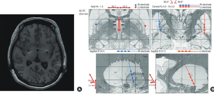
tact 1 as the negative and the internal pulse generator case as the positive pole with an amplitude of 3.72 V, pulse width 60 μsec, and frequency 130 Hz. Six months later, we performed a repeat computed tomography (CT) scan and fused it to the preopera
tive MRI to confirm the locations of the leads (Fig. 2).
Postoperative course
The movement and disability scores and the stimulation para
meters were recorded, as described in Tables 1 and 2. After DBS implantation, considerable improvement was noted. Three mon
ths later, she showed improvement in dystonic dyskinesia, but facial dyskinesia on left eyelid was still noted. Subjectively, she handled the cleaning supplies more easily. Her movement score was decreased to 29 points and disability score to 5 points, but Karnofsky performance scale (KPS) score was still checked as 80 points at 6month followup. The stimulation parameters were adjusted based on the patient’s symptom. The leftsided dystonic posture and walking had kept improving (Fig. 1B). She found it easier to clean, but still had difficulty in washing. At 12
month followup, the movement score decreased to 28 points (improvement rates, 15%) and disability score to 5 points (im
provement rates, 50%) were checked, and she had sustained such improved state. She could do housework including clean
ing and doing the laundry without difficulty. Four years later, the obstetricians consulted us about her 38 weeks pregnancy state. The maternal serum triple test and amniocentesis were performed because of her old age, and revealed low risk of con
Fig. 1. Movie frames obtained from preoperative and postoperative video. (A) Movie frames obtained from a preoperative video showing the patient lifting both arms and walk- ing. (B) Movie frame obtained from a postoperative video showing the same patient 7 years after deep brain stimulation (DBS) surgery.
A B
Preoperatively Postoperative 7 years
Table 1. The movement and disability scores during the follow periods
Movements Pre 3 mon 6 mon 12 mon 24 mon 36 mon 60 mon 84 mon
Off On Off On Off On Off On Off On Off On Off On
Movement scale
Eyes (n = 8) 4 5 4 4 4 6 4 6 4 6 4 3 1 4 1.5
Mouth (n = 8) 2 6 4 4 4 4 3 4 2 4 2 4.5 4.5 6 6
Speech/swallowing (n = 16) 2 2 2 2 2 2 2 2 2 4 2 6 4 3 2
Neck (n = 8) 4 2 2 0 0 0 0 0 0 4 4 4 6 3 3
Rt arm (n = 16) 4 1 1 4 0 3 3 3 3 4 2 2 4 4 4
Lt arm (n = 16) 12 9 9 12 12 12 12 10 8 9 6 4 6 2 2
Trunk (n = 16) 1 1 1 0 0 0 0 0 0 0 0 4 4 4 4
Rt leg (n = 16) 2 2 2 6 6 2 2 2 2 2 2 6 2 6 4
Lt leg (n = 16) 2 3 3 2 1 2 2 1 1 1 1 4 1 6 4
Movement sum (n = 120) 33 31 28 34 29 31 28 28 22 34 23 37.5 32.5 38 30.5
Disability scale
Speech (n = 4) 2 - 1 - 1 - 1 - 1 - 1 - 1 - 1
Writing (n = 4) 1 - 1 - 1 - 1 - 1 - 1 - 1 - 1
Feeding (n = 4) 1 - 1 - 0 - 0 - 2 - 1 - 0 - 0
Eating (n = 4) 0 - 0 - 0 - 0 - 0 - 0 - 0 - 0
Hygiene (n = 4) 1 - 0 - 0 - 0 - 0 - 0 - 0 - 0
Dressing (n = 4) 2 - 2 - 1 - 1 - 0 - 1 - 1 - 0
Walking (n = 6) 3 - 2 - 2 - 2 - 1 - 1 - 1 - 0
Disability sum (n = 30) 10 - 7 - 5 - 5 - 5 - 5 - 4 - 2
Rt = right, Lt = left.
Table 2. The stimulation parameters during the follow up periods
Stimulation Postoperatively 3 mon 6 mon 12 mon 24 mon 36 mon 60 mon 84 mon
Left
Amplitude, V 3.72 3.72 3.74 3.74 3.72 3.72 3.69 3.72
Pulse width, μsec 60 60 60 60 60 60 60 60
Frequency, Hz 130 130 130 130 130 130 130 130
Right
Amplitude, V 3.74 3.72 3.74 3.72 3.72 3.72 3.69 3.72
Pulse width, μsec 60 60 60 120 120 120 60 120
Frequency, Hz 130 130 130 130 130 130 130 130
Fig. 2. Postoperative imaging showing the location of the electrodes. (A) Postoperative magnetic resonance imaging (MRI) scans demonstrating the bilateral deep brain stimula- tion electrodes in the posteroventral internal globus pallidus (GPi) Axial fluid-attenuated inversion recovery (Axial FLAIR). (B) Postoperative assessment of implanted electrodes by image fusion of a postoperative computed tomography (CT) scan with the corresponding preoperative inversion-recovery image. The bilateral electrodes located in the exter- nal globus pallidus (GPe).
AC-PC 20.6 mm
Axial Hv -1.5
Sagittal SI 24.5
79.8° 70.9°
2.1 mm 4.3 mm
Sagittal SI 27.5
Rt electrode Lt electrode
Coronal Fa 4.0 - Fa 3.0
85.0° 89.8°
Rt electrode Lt electrode
20.7 23.3 Rt electrode
Lt electrode
A B
genital anomaly. A scheduled caesarian section was carried out under general anesthesia. After induction using thiopental and succinylcholine, intubation was done quickly, followed by DBS turn off. The surgery lasted for one hour, and the blood pressure was maintained within the normal range during the surgery.
For hemostasis, only bipolar electrocautery was used. Before awakening from the anesthesia, DBS was turned on as the same parameters previously adjusted. After delivery, she could feed her baby by herself, because the dystonia of left upper extremity and hand was improved. Six years after DBS surgery, her gener
al condition was more improved, and she started to play ping
pong and billiards. Until now, she has been showing continual improvement and being good at housework, carrying for chil
dren, with no trouble in daily life based on her KPS score of 90 points and MMSE score of 30 points. She even obtained a driv
er’s license in April 2014 and now drives a car by herself.
DISCUSSION
This report is one of the rare reports on GPi DBS for dystonia with longterm follow up over 7 years. Dystonia can be classi
fied according to the involved body distribution: focal, segmen
tal, multifocal, generalized, and hemidystonia, or according to the etiology: inherited dystonia of proven genetic origin, acquir
ed dystonia with a known specific cause (e.g., perinatal brain injury, infection, drugs, toxicity, vascular, neoplastic, or brain injury), and idiopathic dystonia of unknown cause. Previous reports have shown promising results of GPi DBS especially in the patients diagnosed as primary generalized dystonia (PGD) with DYT1 positive, focal, and tardive dystonia (710).
The patient was diagnosed as a patient with acquired dysto
nia that has been known to show less response to GPi DBS (11
13). However, Vercueil et al. (14) reported a few cases with sec
ondary dystonia showing successful response. Speelman et al.
(15) also reported that GPi DBS was useful in some secondary dystonia patients, but many patients diagnosed as tardive dys
tonia and HallervordenSpatz disease (HSD) were included into their secondary dystonia group. Since March 2005, the au
thor experienced 12 cases of acquired dystonia including this patient. The improvement rate of the patients diagnosed as ac
quired dystonia was about 29% at 12month follow up accord
ing to our unpublished study. Although the improvement rate was not high as much as those of PGD with or without DYT1 positive, the patients diagnosed as acquired dystonia also gained benefit from GPi DBS. The reason for favorable outcome in this patient might be an absence of the structural abnormality on preoperative MRI.
MER facilitates the selection of the final target in DBS in our experience, although some authors reported no benefit from MER (16). All procedures of GPi DBS for dystonia were perform
ed under general anesthesia because of the patients’ abnormal
posture and muscle contractions. General anesthesia did not interfere with the MER signals from the GPi. The typical burst
ing pattern could be identified, whereas amplitude was decre
ased and bursting pattern was emphasized more than in awak
en surgery.
One notable finding was that this patient revealed improved outcome despite the bilateral electrodes located in the external globus pallidus (GPe), as shown in Fig. 2B. A possible explana
tion is that GPi might receive near impact from GPe stimulation.
Another assumption is that the gammaaminobutyric acider
gic (GABAergic) pathway from striatum to GPi could be stimu
lated.
Few authors reported several cases about pregnancy and de
livery in patients who underwent DBS surgery. Paluzzi et al. (17) reported three women who were pregnant and had babies by vaginal delivery after bilateral GPi DBS surgery. These women showed no worsening of dystonic symptoms during the preg
nancy and labor periods, and had no problems in breast feed
ing. There is no evidence of that the pregnancy and delivery should not be allowed to the patients who underwent DBS im
plantation. GwinnHardy et al. (18) tried to figure out the corre
lation between hormones and dystonia in 279 female patients, but they found no clearcut relationship between pregnancy, menopause, postmenopausal hormone replacement therapy, and worsening of dystonic symptoms.
DBS is also not a contraindication for general or regional an
esthesia. If the surgeon is careful not to use short wave (around 2,727 MHz), microwave (2.45 GHz) diathermy, or therapeutic ultrasound (1–5 MHz) diathermy, delivery can be performed safely. The bipolar electrocautery should be used at least 15 cm away from the IPG device, extension cable, and lead (17). As the indication for DBS increases, the patients’ distribution will be more diverse. A standardized protocol for DBS implanted pa
tients who undergo pregnancy, delivery, and surgery irrelevant to neurosurgery would be required. In conclusion, GPi DBS is a safe and effective therapeutic method for treatment of dystonia.
Favorable outcomes could be expected even for the patients with not only PGD, but also acquired dystonia. The patients who un
derwent DBS could safely be pregnant and deliver a baby.
DISCLOSURE
The authors have no potential conflicts of interest to disclose.
AUTHOR CONTRIBUTION
Study concept and design of article: Park HR, Jeon BS, Paek SH.
Data collection and analysis: Park HR, Lee JM, Park H, Shin CW, Kim HJ. Writing draft: Park HR, Park H, Kim DG, Jeon BS, Paek SH. Revision: Park HR, Lee JM, Park H, Shin CW. Approval of fi
nal manuscript and agreement of submission: all authors.
ORCID
Hye Ran Park http://orcid.org/0000000305064882 Jae meen Lee http://orcid.org/0000000257081644 Hyeyoung Park http://orcid.org/0000000335044612 Chae Won Shin http://orcid.org/000000024157492X HanJoon Kim http://orcid.org/0000000182199663 Hee Pyoung Park http://orcid.org/0000000247720780 Dong Gyu Kim http://orcid.org/0000000257406189 Beom Seok Jeon http://orcid.org/0000000324913544 Sun Ha Paek http://orcid.org/0000000330078653
REFERENCES
1. Fahn S. Concept and classification of dystonia. Adv Neurol 1988; 50: 1-8.
2. Albanese A, Bhatia K, Bressman SB, Delong MR, Fahn S, Fung VS, Hallett M, Jankovic J, Jinnah HA, Klein C, et al. Phenomenology and classification of dystonia: a consensus update. Mov Disord 2013; 28: 863-73.
3. Toda H, Hamani C, Lozano A. Deep brain stimulation in the treatment of dyskinesia and dystonia. Neurosurg Focus 2004; 17: E2.
4. Meoni S, Zurowski M, Lozano AM, Hodaie M, Poon YY, Fallis M, Voon V, Moro E. Long-term neuropsychiatric outcomes after pallidal stimulation in primary and secondary dystonia. Neurology 2015; 85: 433-40.
5. Krauss JK. Deep brain stimulation for cervical dystonia. J Neurol Neuro- surg Psychiatry 2003; 74: 1598.
6. Coubes P, Vayssiere N, El Fertit H, Hemm S, Cif L, Kienlen J, Bonafe A, Frere- beau P. Deep brain stimulation for dystonia. Surgical technique. Stereo- tact Funct Neurosurg 2002; 78: 183-91.
7. Kupsch A, Benecke R, Müller J, Trottenberg T, Schneider GH, Poewe W, Eisner W, Wolters A, Müller JU, Deuschl G, et al. Pallidal deep-brain stim- ulation in primary generalized or segmental dystonia. N Engl J Med 2006;
355: 1978-90.
8. Coubes P, Cif L, El Fertit H, Hemm S, Vayssiere N, Serrat S, Picot MC, Tuffery S, Claustres M, Echenne B, et al. Electrical stimulation of the globus palli-
dus internus in patients with primary generalized dystonia: long-term re- sults. J Neurosurg 2004; 101: 189-94.
9. Vidailhet M, Vercueil L, Houeto JL, Krystkowiak P, Benabid AL, Cornu P, Lagrange C, Tézenas du Montcel S, Dormont D, Grand S, et al. Bilateral deep-brain stimulation of the globus pallidus in primary generalized dys- tonia. N Engl J Med 2005; 352: 459-67.
10. Krauss JK, Loher TJ, Weigel R, Capelle HH, Weber S, Burgunder JM. Chron- ic stimulation of the globus pallidus internus for treatment of non-dYT1 generalized dystonia and choreoathetosis: 2-year follow up. J Neurosurg 2003; 98: 785-92.
11. Vidailhet M, Jutras MF, Grabli D, Roze E. Deep brain stimulation for dys- tonia. J Neurol Neurosurg Psychiatry 2013; 84: 1029-42.
12. Lee JY, Deogaonkar M, Rezai A. Deep brain stimulation of globus pallidus internus for dystonia. Parkinsonism Relat Disord 2007; 13: 261-5.
13. FitzGerald JJ, Rosendal F, de Pennington N, Joint C, Forrow B, Fletcher C, Green AL, Aziz TZ. Long-term outcome of deep brain stimulation in gen- eralised dystonia: a series of 60 cases. J Neurol Neurosurg Psychiatry 2014;
85: 1371-6.
14. Vercueil L, Pollak P, Fraix V, Caputo E, Moro E, Benazzouz A, Xie J, Koudsie A, Benabid AL. Deep brain stimulation in the treatment of severe dysto- nia. J Neurol 2001; 248: 695-700.
15. Speelman JD, Contarino MF, Schuurman PR, Tijssen MA, de Bie RM. Deep brain stimulation for dystonia: patient selection and outcomes. Eur J Neu- rol 2010; 17 Suppl 1: 102-6.
16. Bour LJ, Contarino MF, Foncke EM, de Bie RM, van den Munckhof P, Speel- man JD, Schuurman PR. Long-term experience with intraoperative mi- crorecording during DBS neurosurgery in STN and GPi. Acta Neurochir (Wien) 2010; 152: 2069-77.
17. Paluzzi A, Bain PG, Liu X, Yianni J, Kumarendran K, Aziz TZ. Pregnancy in dystonic women with in situ deep brain stimulators. Mov Disord 2006;
21: 695-8.
18. Gwinn-Hardy KA, Adler CH, Weaver AL, Fish NM, Newman SJ. Effect of hormone variations and other factors on symptom severity in women with dystonia. Mayo Clin Proc 2000; 75: 235-40.