Hydrogen Peroxide-induced Alterations in Na
+-phosphate Cotransport in Renal Epithelial Cells
Soon-Hee Jung
Department of Clinical Pathology, Jinju Health College, Jinju 660-757, Korea
This study was undertaken to examine the effect of oxidants on membrane transport function in renal epithelial cells. Hydrogen peroxide (H2O2) was used as a model oxidant and the membrane transport function was evaluated by measuring Na+-dependent phosphate (Na+-Pi) uptake in opossum kidney (OK) cells. H2O2
inhibited Na+-Pi uptake in a dose-dependent manner. The oxidant also caused loss of cell viability in a dose-dependent fashion. However, the extent of inhibition of the uptake was larger than that in cell viability.
H2O2 inhibited Na+-dependent uptake without any effect on Na+-independent uptake. H2O2-induced inhibition of Na+-Pi uptake was prevented completely by catalase, dimethylthiourea, and deferoxamine, suggesting involvement of hydroxyl radical generated by an iron-dependent mechanism. In contrast, antioxidants Trolox, N,N’-diphenyl-p-phenylenediamine, and butylated hydroxyanisole did not affect the H2O2 inhibition. Kinetic analysis indicated that H2O2 decreased Vmax of Na+-Pi uptake with no change in the Km value.
Phosphonoformic acid binding assay did not show any difference between control and H2O2-treated cells.
H2O2 also did not cause degradation of Na+-Pi transporter protein. Reduction in Na+-Pi uptake by H2O2 was associated with ATP depletion and direct inhibition of Na+-K+-ATPase activity. These results indicate that the effect of H2O2 on membrane transport function in OK cells is associated with reduction in functional Na+-pump activity. In addition, the inhibitory effect of H2O2 was not associated with lipid peroxidation.
Received 12 April 2009/Returned for modification 3 June 2009/Accepted 25 June 2009 Key Words : Na+-dependent phosphate, Membrane transport, H2O2, Renal epithelial cells.
I. Introduction
Reactive oxygen species (ROS) have been implicated in the pathogenesis of a number of renal diseases including ischemic and nephrotoxicant-induced acute renal failure (Baliga et al, 1999). The cell injury by ROS involved alterations in physical integrity of the cell membrane, such as rigidity and permeability, and functional integrity, such as membrane transport and enzymes functions (Gardes-
Corresponding author: Soon-Hee Jung, Department of Clinical Pathology, Jinju Health College, Jinju 660-757, Korea
TEL : 055-740-1847, 010-3584-1847 E-Mail : [email protected]
Albert et al, 1993). ROS cause DNA damage (Schraufstatter et al, 1986), rapid depression of intracellular ATP (Andreoli et al, 1993), a fast rise of intracellular Ca2+
(Hyslop et al, 1986), oxidation of susceptible amino acids in proteins (Aruoma et al, 1989), and gross perturbations to the cytoskeleton and plasma membrane (Andreoli et al, 1993). All these processes occur before loss of plasma membrane integrity, as measured by vital dye staining (Hyslop et al, 1986; Schraufstatter et al, 1986) or loss of preloaded 51Cr (Andreoli and Mallett, 1997). In renal epithelial cells, most of the studies on acute oxidative stress have focused on the physical integrity of the cell membrane (Schnellmann, 1988; Andreoli and McAteer,
1990; Walker and Shah, 1991; Sheridon et al, 1996) Many reports and propose that lipid peroxidation plays a critical role in oxidant-induced cell death (Salahudeen, 1995; Sheridan et al, 1996; Schnllmann, 1998).
In vivo studies have shown that ischemia and various nephrotoxicants produce significant changes in structure and function of the proximal tubule, a major site where exhibits many energy-dependent, specialized functions including reabsorption of solutes such as phosphate, glucose, amino acids. Although a recent study shows that H2O2 inhibits phosphate transport through a lipid peroxidation-independent mechanism in opossum kidney (OK) cells (Min et al, 2000), the mechanism by which H2O2 alters the transport function in renal epithelial cells remains to be identified. Therefore, the present study was carried out to examine the mechanism by which H2O2
alters membrane transport function by measuring Na+-Pi uptake in renal proximal tubular epithelial cells.
II. Materials and Methods
1. Chemicals
[32P]-phosphate and [14C]-phosphonoformic acid were obtained from Amersham international (Amersham, UK).
H2O2, deferoxamine (DFO), 3-aminobenzamide (AB), ca- talase, Tolox, dimethythiourea (DMTU), N,N’-diphenyl- p-phenylenediamine (DPPD), butylated hydroxyanisole (BHA), and malondialdehyde tetraethylacetal were pur- chased from Sigma-Aldrich Chemical (St. Louis, MO).
All other chemicals were of the highest commercial grade available.
2. Culture of OK cells
OK cells were obtained from ATCC and maintained by serial passages in 75-cm2 culture flasks (Costar, Cambridge, MA). The cells were grown in Dulbecco’s modified Eagle’s medium/Ham’s F12 (DMEM/F12, Sigma Chemical Co. St. Louis USA) containing 10% fetal
bovine serum at 37℃ in 95% air/5% CO2 incubator.
When the cultures reached confluence, subculture was prepared using a 0.02% EDTA-0.05% trypsin solution.
The cells were grown on 24-well tissue culture plates in DMEM/F12 medium containing 10% fetal bovine serum.
All experiments started 3~4 days after plating when a confluent monolayer culture was achieved.
3. Induction of oxidant injury
Cells were treated with H2O2 of the indicated con- centration in Hanks’ balanced salt solution (HBSS) containing the following; 115 mM NaCl, 5 mM KCl, 25 mM NaHCO3, 2 mM NaH2PO4, 1 mM MgSO4, 1 mM CaCl2, and 5 mM glucose (pH 7.4) for 120 min at 37℃.
Following oxidant stress, uptakes of solutes and ATP content were measured as described below.
4. Uptake studies
The uptake of solutes was determined in cell monola- yers grown on 24 well plates. After an exposure to oxidant stress, the reaction buffer was removed and washed twice with the uptake buffer containing the following; 137 mM NaCl, 5.4 mM KCl, 2.8 mM CaCl2, 1.2 mM MgSO4, and 10 mM Hepes (pH 7.4). The cells were incubated for 30 min at 37℃ in the uptake buffer containing 5 μM [32P]-phosphate. For kinetic studies, the cells were incubated for 20 min at 37℃ in the uptake buffer containing [32P]-phosphate of various concentrations (0.005-1 mM). For measurement of Na+-independent phosphate uptake, NaCl was replaced by 137 mM N-methyl-D-glucamine (NMG). At the end of the incubation period, the cells were washed three times with ice-cold uptake buffer and solubilized in 0.5 mL of 0.2 % Triton X-100. Aliquots of each sample were transferred to scintillation vials and the radioactivity was counted in a liquid scintillation counter (TRI-CARB 2100TR, Packard, USA). Protein was measured by the method of Bradford (Bradford, 1976).
5. [14C]PFA binding studies
The binding of [14C]PFA was measured in cell mono- layers grown on 24 well plates. After an exposure to oxidant stress, the reaction buffer was removed and washed twice with the uptake buffer described above. The cells were incubated for 30 min at 37℃ in the uptake buffer containing 1 mM [14C]PFA. At the end of the incubation period, the cells were washed three times with ice-cold buffer and solubilized in 0.5 mL of 0.2 % Triton X-100. Aliquots of each sample were transferred to scintillation vials and the radioactivity was counted in a liquid scintillation counter as described above
6. Electrophoresis and Immunoblotting
The intrinsic type II Na+-dependent phosphate trans- porter protein (NaPi-4) in OK cells was analyzed as described by Pfister et al. (1997). Cells were grown to confluency in Φ 10 cm Petri dishes. After treatment with H2O2, the cells were scraped off the dish and washed twice with TBS (0.9% NaCl and 10 mM Tris-HCl, pH 7.4). The scraped cells were homogenized in solution containing 10 mM NaCl, 1 mM EGTA, 1 mM EDTA, 20 mM Tris-HCl (pH 7.4), and 1% Triton X-100. The homogenate was centrifuged at 2000 rpm for 10 min at 4
℃. The supernatant was saved and centrifuged at 16,000 rpm for 40 min at 4℃. The pellet corresponding to a crude membrane preparation was resuspended in 100 μL of 50 mM mannitol and 10 mM Hepes-Tris (pH 7.2).
All samples were prepared by heating to 100℃ for 10 min in SDS gel-loading buffer. A 30μg of total protein was used for SDS-polyacrylamide gel electrophoresis (10%) and subsequent transfer to nitrocellulose membrane.
To confirm identical loading, nitrocellulose membrane was stained with Ponceau-S. Nonspecific binding was then blocked by incubating the nitrocellulose at room temperature for 1 hr in TBS containing 5% nonfat dry milk and 1% Triton X-100. Expression of the NaPi-4 protein was estimated using a affinity pure polyclonal antibody (Alpha Diagnostic, Inc.) raised against the
C-terminal 12 amino acids of the published NaPi-4 sequence (antibody dilution, 1/100). Incubation with the primary antibody took place overnight at room tem- perature. The nitrocellulose was washed four times with TBS containing 1% Triton X-100. The nitrocellulose was then incubated with a 1:10,000 dilution of an anti-rabbit IgG labeled with horseradish peroxidase (Amersham Life Science, Inc.) for 1 hr at room temperature. Blots were developed by ECL kit.
7. Measurement of cell viability
The cell viability was estimated by a trypan blue ex- clusion assay. Cells were grown to confluence in 24-well dishes, treated with H2O2, and then harvested using 0.025% trypsin. Cells were incubated with 4% trypan blue solution. Cells failing to exclude the dye were considered nonviable, and the data are expressed as percentage of nonviable cells.
8. Measurement of ATP content
ATP levels were measured on OK cells with a luci- ferin-luciferase assay. After an exposure to oxidant stress, the cells were solubilized with 500 μL of 0.5 % Triton X-100 and acidified with 100 μL of 0.6 M perchloric acid and placed on ice. Cell suspension was then diluted with 10 mM potassium phosphate buffer containing 4 mM MgSO4 (pH 7.4), and 100 μL of 20 mg/ml luciferin- luciferase was added to 10 μL of diluted sample. Light emission was recorded at 20 sec with a luminometer (MicroLumat LB96P, Berthold, Germany). Protein content was determined on a portion of the cell sample.
9. Measurement of Na+-K+-ATPase activity
The Na+-K+-ATPase activity was measured in the microsomal fraction prepared from OK cells. For the preparation of microsomal fraction, cells were grown to confluence in 100 mm dish, scraped from the dish in 10 mM mannitol and 2 mM Tris/HCl (pH 7.1) at 4℃, and briefly sonicated. Then, the cell lysate was centrifuged for
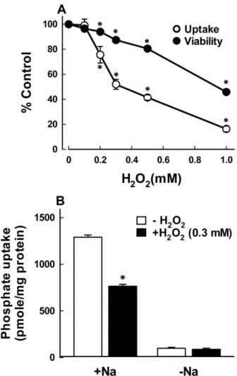
Fig. 1. (A) Concentration dependent effect of H2O2 on phosphate uptake and cell viability in OK cells. Cells were exposed to various concentrations of H2O2 for 120 min, and the uptake and cell viability were estimated.
The uptake was measured for 30 min and cell viability was evaluated by a trypan blue exclusion assay. Data are mean ± SE of four experiments. *P<0.05 compared with the absence of H2O2 (control). (B) Effect of H2O2 on Na+-dependent and –independent phosphate uptake in OK cells. Cells were exposed to 0.3 mM H2O2 for 120 min and the uptake was measured for 30 min in a buffer with or without Na+ (replaced by NMG). Data are mean SE of four experiments. *P<0.05 compared with control.
2 min at 2,000×g to remove unbroken cells and the supernatant was saved, centrifuged for 12 min at 15,000
×g. Pale-pink layer on top of pellet was removed and resuspended in 10 mM mannitol and 2 mM Tris/HCl (pH 7.1). Microsomal fraction was treated with ethanol for 60 min at 37℃ and Na+-K+-ATPase was measured.
The ATPase activity was determined by measuring inorganic phosphate (Pi) released by ATP hydrolysis during incubation of microsome with an appropriate medium containing 3 mM ATP (Sigma) as the substrate.
The total ATPase activity was determined in the presence of 100 mM Na+, 20 mM K+, 3 mM Mg, 2 mM EDTA, and 40 mM imidazole (pH 7.4). The Mg2+-ATPase activity was determined in the absence of K+ and in the presence of 1 mM ouabain. The difference between the total and the Mg2+-ATPase activities was taken as a measure of the Na+-K+-ATPase activity. At the end of a 10-min incubation, the reaction was terminated by the addition of ice-cold 6% perchloric acid. The mixture was then centrifuged at 3,500×g, and Pi in the supernatant fraction was determined by the method of Fiske and SubbaRow (Fiske and SubbaRow, 1925).
10. Statistical analysis
Data are expressed as mean ± SEM. Comparisons between two groups were made using the unpaired t test.
Multiple group comparisons were done using one-way analysis of variance followed by the Turkey post hoc test.
P < 0.05 was considered statistically significant.
III. Results
The concentration dependent effect of H2O2 on Na+-Pi uptake in OK cells was examined. Based on preliminary studies, the uptake was determined at 30 min following exposure of cells to various concentrations of H2O2 for 120 min (Fig. 1A). When cells were exposed to 0, 0.1, 0.2, 0.3, 0.5, and 1 mM, Na+-Pi uptake was reduced by
H2O2 in a dose-dependent manner, with an IC50 (the inhibitor concentration for 50% inhibition) of 0.33 mM.
In order to determine whether the inhibition of Na+-Pi uptake was attributed to irreversible cell injury, the effect of H2O2 on cell death as measured by trypan blue
Fig. 2. Effects of radical scavengers (A) and antioxidants (B) on H2O2-induced inhibition of Na+-Pi uptake in OK cells.
Cells were exposed to 0.3 mM H2O2 for 120 min in the presence of 500 units/mL catalase (CAT), 30 mM dimethythiourea (DEMTU), 2 mM deferoxamine (DFO), and 1 mM Trolox (Tro). The uptake was measured for 30 min. Data are mean SE of five experiments. *P<0.05 compared with control (C). #P<0.05 compared with H2O2
alone.
exclusion was examined. As shown in Fig. 1A, H2O2
caused cell death in a dose-dependent manner similar to the inhibition of uptake. However, the extent of cell death was much less than that of the uptake.
Phosphate uptake in the presence of Na+ was sig- nificantly reduced by 0.3 mM H2O2, whereas the uptake in the absence of Na+ was not substantially altered by H2O2 (Fig. 1B). These data suggest that H2O2 inhibits the active phosphate uptake driven by the gradient of Na+ without any effect on Na+-independent passive uptake.
The uptake in control cells in the absence of Na+ was 99.04 ± 10.62 pmole/mg/30 min, which was approximately 7.7% of the total uptake (1289.40 ± 24.79 pmole/mg/30 min). Na+-independent uptake was therefore not routinely measured.
H2O2 is converted into hydroxyl radical, a more potent oxidant, in the presence of iron via the Fenton/
Haber-Weiss reactions. To confirm the role of H2O2 and hydroxyl radical in mediating the inhibition of Na+-Pi uptake, effects of the H2O2 scavenger catalase and the hydroxyl radical scavenger DMTU were examined.
H2O2-induced alteration in Na+-Pi uptake was nearly completely prevented by these scavengers (Fig. 2A). A similar protection was also obtained with the iron chelator deferoxamine (Fig. 2B). These results suggest that hydroxyl radicals are responsible for the uptake inhibition. Since hydroxyl radicals are a potent initiator of lipid peroxidation, H2O2-induced inhibition of Na+-Pi uptake could be resulted from lipid peroxidation. To test this hypothesis the effect of Trolox on the inhibition of Na+-Pi uptake was examined. The antioxidant Trolox (0.5 mM) did not prevent H2O2-induced inhibition of Na+-Pi uptake, indicating that the effect of H2O2 on Na+-Pi uptake is not mediated by lipid peroxidation. Similarly, the other lipophilic antioxidants DPPD and BHA at 20 and 50 μM, respectively, did not protect against H2O2-induced inhi- bition of Na+-Pi uptake (data not shown). These results are consistent with those of previous studies (Min et al, 2000). The concentrations of antioxidants used in the
present study was similar to or higher than concentrations that have completely prevent oxidant-induced cell injury and lipid peroxidation (Chen and Stevens, 1991; Kim and Kim, 1996; Robb and Connor, 1998; Lin and Ho, 2000).
In an attempt to gain insight into the mechanisms by which H2O2 modulates Na+-Pi uptake, the effect of H2O2
on the kinetics of Na+-Pi uptake was examined. The time course of Na+-Pi uptake was linear up to 30 min incubation in cells with or without H2O2 treatment (data not shown). Based on these findings, the effect of H2O2
on the kinetics of Na+-Pi uptake was analyzed by
Fig. 3. Initial rate of phosphate uptake in control and H2O2- treated cells as a function of phosphate concentrations.
(A) Cells were exposed to 0.3 mM H2O2 for 120 min and the uptake was measured for 20 min in a buffer with or without Na+ (replaced by NMG). (B) Na+-dependent uptake was calculated by subtracting the uptake in the absence of Na+ from the total uptake. Data are mean ± SE of eight experiments. (C) Data of the Na+-dependent uptake presented according to Eadie-Hofstee transformation of Michaelis-Menten equation. In this plot, the intercept of the line with Y-axis represents Vmax and the slope indicates the Km for phosphate.
measuring the initial velocity (20 min) of Na+-Pi uptake as a function of phosphate concentration in the presence or absence of external Na+. The results are summarized in Fig. 3. Total phosphate uptake measured in the presence of external Na+ increased curvilinearly as the external phosphate concentration increased in control and H2O2- treated cells, whereas the uptake in the absence of Na+ increased linearly with increasing phosphate concentration in both groups. H2O2 inhibited the total uptake without a significant change in the Na+-independent uptake (Fig.
3A). The Na+-dependent uptake, computed by subtracting the uptake in the absence of Na+ from the total uptake in each group are illustrated in Fig. 3B. An Eadie-Hofstee transformation of the Na+-dependent uptake shows that the relationship between the initial rate of phosphate uptake (V) and V/[phosphate] was linear in both control and H2O2-treated cells (Fig. 3C). This indicates that in both cases the Na+-dependent phosphate uptake follows a simple Michaelis-Menten kinetics, i.e., V = Vmax × [S]/(Km + [S]), where Vmax is the maximal uptake, [S]
is the substrate concentration, and Km is the apparent Michaelis constant indicating the concentration of phosphate for ½Vmax. H2O2 caused a significant redu- ction in the Vmax for Na+-dependent phosphate uptake (5.41 ± 0.50 vs. 12.01 ± 0.03 pmole/mg/20 min in control cells), with no change in the apparent Km (0.050 ± 0.004 vs. 0.042 ± 0.009 mM in control cells).
PFA acts as a specific, competitive, and reversible inhibitor of Na+-Pi cotransport across the renal brush- border membrane and has been employed as a probe for studies of this transport system (Szczepanska et al, 1986;
Szczepanska et al, 1989). Therefore, we examined the effect of H2O2 on PFA binding in OK cells. The results depicted in Fig. 4A indicated that PFA binding was not different between control and H2O2-treated cells. These data suggest that H2O2-induced reduction in Vmax of Na+-Pi uptake is not attributed to a decrease in the number of Na+-Pi cotransporters.
Expression of the NaPi-4 protein in OK cells was also
Fig. 4. (A) Effect of H2O2 on phosphonoformic acid (PFA) binding in OK cells. Cells were exposed to 0.3 mM H2O2 for 120 min, and PFA binding was measured for 30 min. Data are mean ± SE of five experiments. (B) Effect of H2O2 on expression of the Na+-dependent phosphate transporter (NaPi-4) protein. Cells were exposed to 0.3 mM H2O2 for 120 min, and the expression of NaPi-4 protein was analyzed by immunoblotting using a affinity pure polyclonal antibody.
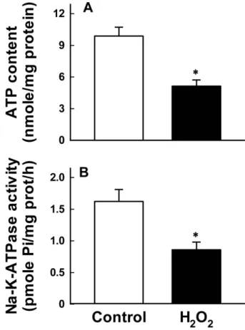
Fig. 5. Effect of H2O2 on ATP depletion in whole cells (A) and Na+-K+-ATPase activity in disrupted cells (B). Cells were exposed to 0.3 mM H2O2 for 120 min, and ATP content and the enzyme activity were measured. Data are mean SE of five experiments. *P<0.05 compared with control.
analyzed by immunoblotting. As shown in Fig. 4B, treatment with H2O2 did not caused any change in NaPi-4 protein. Together with data from kinetic analysis and PFA binding assay, these results suggest that H2O2-induced inhibition in Na+-Pi uptake is not a result of a decrease in the number of the transporters.
Since maintenance of intracellular Na+ gradient needed to drive Na+-dependent cotransport requires normal activity of the Na+-pump, H2O2 treatment may reduce Na+-Pi uptake through the inhibition of Na+-pump activity.
Reduction in Na+-pump activity could be resulted from ATP depletion and/or direct inhibition of Na+-K+-ATPase in whole cells. To test the possibility, we measured ATP content in whole cells and Na+-K+-ATPase activity in disrupted cells. Both ATP content and Na+-K+-ATPase activity were decreased approximately 50% of control in cells treated with H2O2 (Fig. 5).
IV. Discussion
The mechanism by which oxidants alter membrane transport functions in renal proximal tubular cells is not clearly defined. Previous studies have shown that oxidants
inhibit Na+-dependent solute transport by lipid peroxi- dation and direct damage of the transport protein (Jourd’
heuil et al, 1993) and disruption of normal ion gradients resulting from ATP depletion and inactivation of Na+-K+-ATPase (Andreoli et al, 1993).
In the present study, we demonstrate that membrane transport function such as Na+-Pi transport is inhibited in response to oxidant stress. The extent of the inhibition was much larger than that in cell death, suggesting that most of the uptake inhibition is not attributed to irreversible cell death. H2O2-induced inhibition of Na+-Pi uptake was completely prevented by the H2O2 scavenger catalase, the hydroxyl radical scavenger DMTU, and the iron chelator deferoxamine. These data indicate that the H2O2 inhibition is mediated by the intracellular generation of an iron-dependent hydroxyl radical.
Because hydroxyl radicals are a potent initiator of lipid peroxidation, the inhibition of Na+-Pi uptake induced by H2O2 could be resulted from lipid peroxidation. However, the H2O2 inhibition was not altered by antioxidants.
Therefore, it is likely that H2O2 inhibits Na+-Pi uptake through a lipid peroxidation-independent mechanism..
Effects of oxidants on kinetic analysis of membrane transport were studied in Na+-glucose uptake by brush- border membrane vesicles (Jourd'heuil et al, 1993) and organic anion uptake by renal proximal tubular cells (Takeda et al, 2000). They found that oxidants cause a significant reduction in Vmax of these transport systems without any change in Km. The effect of oxidants on the kinetics of Na+-Pi uptake in OK cells has not been explored until now. In the present study, the Vmax of Na+-Pi uptake was significantly reduced in cells treated with H2O2, whereas the Km value remained unchanged. In the kinetic analysis of carrier-mediated transport, the Vmax is determined by two factors: firstly, the capacity of the carrier system and, secondly, the proportion of adsorbed molecules which dissociate in a forward direction in unit time. The former depends mainly on the number of carrier sites per unit area of membrane. The
latter depends on (1) the probability of a substrate molecule to dissociate from a carrier site in a given time, and (2) the rate of turnover of carrier across the membrane. Since the Km for phosphate in the present study was not changed, it is unlikely to have altered carrier-substrate dissociation. Therefore, the decrease in Vmax could be attributed to reduction in the number of functional carrier units or the rate of turnover. Assuming that PFA binding provides an accurate estimate of the number of Na+-Pi transport (Szczepanska et al, 1987), the results of PFA binding studies (Fig. 4A) indicate that the decrease in Vmax may be mediated by a decrease in the turnover rate but by not the number of functional carrier units. This assumption was supported by an immuno- blotting assay (Fig. 4B).
The present study demonstrates that the principal mechanism of H2O2-induced modulation in Na+-Pi uptake is the decrease in Na+-pump activity resulting from ATP depletion and direct inhibition of Na+-K+-ATPase (Fig. 5), consistent with previous studies in LLC-PK1 cells (Andreoli et al, 1993). It is not certain, however, whether H2O2-induced ATP depletion was attributed to impairment of ATP synthesis resulting from mitochondrial damage and/or to activation of poly (ADP-ribose) polymerase (PARP). Since PARP catalyzes the transfer of ADP-ribose from NAD to protein with the concomitant release of nicotinamide, the activation of this enzyme results in depletion of NAD and a consequent reduction in ATP. In previous study, H2O2 produces activation of poly (ADP-ribose) polymerase (Min et al, 2000). Such a reduction in functional Na+-pump activity may contribute to the decrease in Vmax.
In conclusion, H2O2 reduced Na+-Pi uptake through the inhibition of function Na+-pump activity. Such effects are mediated by iron-dependent hydroxyl radical generation, but not attributed to lipid peroxidation or mostly irreversible cell death.
Acknowledgments
This research was supported by Jinju Health College Grant in 2008.
REFERENCE
1. Andreoli SP, Mallett CP. Disassociation of oxidant- induced ATP depletion and DNA damage from early cytotoxicity in LLC-PK1 cells. Am J Physiol 272:
F729-F735, 1997.
2. Andreoli SP, McAteer JA. Reactive oxygen molecule- mediated injury in endothelial and renal tubular epithelial cells in vitro. Kidney Int 38:785-794, 1990 3. Andreoli SP, McAteer JA, Seifert SA, Kempson SA.
Oxidant-induced alterations in glucose and phosphate transport in LLC-PK1 cells: mechanisms of injury.
Am J Physiol 265:F377-F384, 1993
4. Aruoma OI, Halliwell B, Gajewski E, Dizdaroglu M.
Damage to the bases in DNA induced by hydrogen peroxide and ferric ion chelates. J Biol Chem 264;
20509-20512, 1989
5. Baliga R, Ueda N, Walker PD, Shah SV. Oxidant mechanisms in toxic acute renal failure. Drug Metab Rev 31:971-997, 1999.
6. Bradford M. A rapid and sensitive method for the quantitation of microgram quantities of protein utilizing the principle of protein-dye binding. Anal Biochem 72:248-254, 1976.
7. Chen Q, Stevens JL. Inhibition of iodoacetamide and t-butylhydroperoxide toxicity in LLC-PK1 cells by antioxidants: a role for lipid peroxidation in alkylation induced cytotoxicity. Arch Biochem Biophys 284:422- 430, 1991.
8. Fiske CH, Subbarow Y. The colorimetric determination of phosphorus. J Biol Chem 66:375-400, 1925.
9. Gardes-Albert M, Jore D, Ferradini C. Membrane
lipid peroxidation: Pulse and radiolysis in oxyradical research. In ;Membrane Lipid Oxidation . p1-30, CRC Press, Boston, 1993.
10. Hyslop PA, Hinshaw DB, Schraufstatter IU, Sklar LA, Spragg RG, Cochrane CG. Intracellular calcium homeostasis during hydrogen peroxide injury to cultured P388D1 cells. J Cell Physiol 129:356-366, 1986.
11. Jourd'heuil D, Vaananen P, Meddings JB. Lipid peroxidation of the brush-border membrane: mem- brane physical properties and glucose transport. Am J Physiol 264:G1009-G1015, 1993.
12. Kim YK, Kim YH. Differential effect of Ca2+ on oxidant-induced lethal cell injury and alterations of membrane functional integrity in renal cortical slices.
Toxicol Appl Pharmacol 141:607-616, 1996.
13. Lin AM, Ho LT. Melatonin suppresses iron-induced neurodegeneration in rat brain. Free Radic Biol Med 28:904-911, 2000.
14. Min SK, Kim SY, Kim CH, Woo JS, Jung JS, Kim YK. Role of lipid peroxidation and poly (ADP-ribose) polymerase activation in oxidant-induced membrane transport dysfunction in opposum kidney (OK) cells.
Toxicol Appl Pharmacol 166:196-202, 2000.
15. Pfister MF, Lederer E, Forgo J, Ziegler U, Lotscher M, Quabius ES, Biber J, Murer H. Parathyroid hormone-dependent degradation of type II Na+/Pi cotransporters. J Biol Chem 272:20125-20130, 1997.
16. Robb SJ, Connor JR. An in vitro model for analysis of oxidative death in primary mouse astrocytes. Brain Res 788:125-132, 1998.
17. Salahudeen AK. Role of lipid peroxidation in H2O2-induced renal epithelial (LLC-PK1) cell injury.
Am J Physiol 268:F30-F38, 1995.
18. Schnellmann RG. Mechanisms of t-butyl hydropero- xide-induced toxicity to rabbit renal proximal tubules.
Am J Physiol 255:C28-C33, 1988.
19. Schraufstatter IU, Hyslop PA, Hinshaw DB, Spragg RG, Sklar LA, Cochrane CG. Hydrogen peroxide- induced injury of cells and its prevention by inhibitors
of poly(ADP-ribose) polymerase. Proc Natl Acad Sci USA 83:4908-4912, 1986
20. Sheridan AM, Fitzpatrick S, Wang C, White MD, Lieberthal W. Lipid peroxidation contributes to hy- drogen peroxide induced cytotoxicity in renal epithelial cells. Kidney Int 49:88-93, 1996.
21. Szczepanska KM, Yusufi AN, Dousa TP. Interactions of [14C]phosphonoformic acid with renal cortical brush-border membranes. Relationship to the Na+-pho- sphate co-transporter. J Biol Chem 262:8000-8010, 1987.
22. Szczepanska KM, Yusufi AN, Lin JT, Dousa TP.
Structural requirement of monophosphates for inhibition of Na+-Pi cotransport in renal brush border membrane. Biochem Pharmacol 38:4191-4197, 1986.
23. Szczepanska KM, Yusufi AN, VanScoy M, Webster SK, Dousa TP. Phosphonocarboxylic acids as specific inhibitors of Na+-dependent transport of phosphate across renal brush border membrane. J Biol Chem 261:6375-6383, 1989
24. Takeda M, Hosoyamada M, Cha SH, Sekine T, Endou H. Hydrogen peroxide downregulates human organic anion transporters in the basolateral mem- brane of the proximal tubule. Life Sci 68:679-687, 2000.
25. Walker PD, Shah SV. Hydrogen peroxide cytotoxicity in LLC-PK1 cells: a role for iron. Kidney Int 40:891- 898, 1991.