J. Exp. Biomed. Sci. 15 (2009) 105–112
Synthesis of 2-Thio-4-aminopyrimidine Derivatives as Anti-cancer Agent
Sang-Hyo Lee and Jinho Lee†
Department of Chemistry, College of Natural Sciences, Keimyung University, Daegu 704-701, Korea
The screening of the anti-cancer activity of the chemical library provided 2-thio-4-aminopyrimidine as the initial hit.
The confirmation of structure and biological effect of hit was performed by synthesis and biological evaluation. The optimization of hit was performed by derivatization of substituents while keeping the core structure. The evaluation of growth inhibitory activity was carried out using SRB assay against 6 human cancer cell lines and human fibroblast. The growth inhibitory activity of compounds showed substituent dependency and more than 5 compounds showed complete growth inhibition of cancer cell lines at 10 μM concentration. Chemical library screening followed by synthetic modification provided possibility that 2-thio-4-aminopyrimidine can be used as a new scaffold for the development of anti-cancer agent.
Key Words: Anti-cancer agent, 2-Thio-4-aminopyrimidine, Growth inhibitory effect, SRB assay
INTRODUCTION
Cancer, a disease of worldwide importance, causes more than 7.4 million deaths in 2004 and occupies the second rank after heart disease as a killer (WHO data, 2009).
Despite the major achievements in different new areas of drug discovery research, the successful treatment of the cancer still remains a significant challenge (Marshall, 2000).
The search of potent new anticancer agents has been carried out by two general methods: (i) screening the anti- cancer activities of new natural products; (ii) screening the anticancer activities of synthetic compounds either related or unrelated to known anticancer agents (Hua et al., 2006;
Valler, 2000) In the early 1990, screening was carried out mostly with purified target proteins. However, the portion of cell/phenotype-based screening gradually increased recently.
The cell-based screening has several advantages: (i) no need for purification of protein; (ii) screening can be carried out at conditions which is similar to physiological condition;
(iii) cellular toxicity and cell penetration problems can be
excluded. Also, it is expected to be more effective in devel- oping drug for both genetically mutated disease such as cancer and aiming multi-targets (Li et al., 2006; Weinstein et al., 2000).
As an initial step of searching new scaffold for anti- cancer agent, cell-based screening of compounds that were unrelated to known anticancer agents was performed. After screening the anticancer activities of a number of synthetic libraries, a class of 2-thio-4-aminopyrimidne was found to possess potent anticancer activities. 2-Thiobenzyl moiety was chosen for further evaluation and optimization.
MATERIALS AND METHODS
1H- and 13C-NMR spectra were obtained on an AVANEC 400 (Bruker, Switzerland) and are reported in part per million (δ) relative to TMS as an internal standard. Spin multiplets are given as s (singlet), d (doublet), t (triplet), q (quartet), dd (doublet of doublet), td (triplet of doublet), qd (quartet of doublet) and m (multiplet). TLC was performed on E. Merck silica gel 60 F254 plates (0.25 mm). Silica gel column chromatography was performed using Merck TA- 1287685. HPLC runs were carried out using Waters 2690 with an X Bridge C18 5 μm column. Unless otherwise noted, all starting materials were obtained from commercially available sources and they were used without further puri-
*Received: March 31, 2009 Accepted after revision: June 1, 2009
†Corresponding author: Jinho Lee, Department of Chemistry, Keimyung University, 1000 Sindang-Dong, Dalseo-Gu, Daegu 704-701, Korea Tel: 82-53-580-5183, Fax: 82-53-580-5183
e-mail: [email protected]
fication. Tetrahydrofuran (THF) was freshly distilled from sodium and benzophenone. All reactions were performed under nitrogen atmosphere.
Representative procedure: synthesis of 4-[4-(4-benzyl- piperidine-1-yl)-6-chloro-pyrimidine-2-yl-sulfanylmethyl]
-N-(2-morpholine-4-ylethyl)-benzamide (1)
1. 4-(4,6-Dihydroxy-pyrimidin-2-ylsulfanylmethyl)- benzoic acid methyl ester (1-b)
To 3 g (20.8 mmol) of 4,6-dihydroxy-2-mercaptopyrimi dine in 20 ml of water was added 2.1 g (25 mmol) of sodium bicarbonate. The mixture was stirred at room temperature for 30 min. to which was added 4.77 g (20.8 mmol) of methyl 4-(bromomethyl)benzoate in 20 ml ethanol and stirred for 23 hours. Removal of ethanol provided pre- cipitates. Collection of precipitate and drying by air blowing provided the desired product in 97% yield.
1H NMR (CDCl3, 400 MHz): δ (ppm) 8.02 (s, 1H), 7.82 (d, J=8 Hz, 1H), 7.74 (d, J=8 Hz, 1H), 7.46 (t, J=7.7 Hz, 1H), 5.17 (s, 1H), 4.44 (s, 2H), 3.84 (s, 3H)
2. 4-(4,6-Dichloro-pyrimidine-2-ylsulfanylmethyl)- benzoic acid methyl ester (1-c)
1.0 g (3.42 mmol) of 1-b and 0.88 ml (8.90 mmol) of 2-picoline were added to 6 ml of phosphorous oxychloride and the mixture was refluxed for 3 hours. The reaction mixture was poured to ice water and the product was extracted three times with ethyl acetate. The combined organic layers were dried over magnesium sulphate. After removal of solvent in vacuo, the purification with column chromatography using hexane/ ethyl acetate (1:1) mixture as eluent provided the desired product in 82% yield.
1H NMR (CDCl3, 400 MHz): δ (ppm) 7.99 (d, J=8.28 Hz, 2H), 7.51 (d, J=8.28 Hz, 2H), 7.03 (s, 1H), 4.38 (s, 2H), 3.90 (s, 3H)
3. 4-[4-(4-Benzyl-piperidine-1-yl)-6-chloro-pyrimidine- 2-ylsulfanyl methyl]-benzoic acid methyl ester (1-d)
To 200 mg (0.61 mmol) of 1-c in 20 ml ethanol was added 0.11 ml (0.61 mmol) of 4-benzylpiperidine and the mixture was refluxed for 4 hours. The solvent was removed
in vacuo. To the residue was added 10% aqueous citric acid, and the product was extracted three times with ethyl acetate.
The combined organic layers were dried over magnesium sulphate. After removal of solvent in vacuo, the purification with column chromatography using hexane/ ethyl acetate (5:1) mixture as eluent provided the desired product in 98% yield.
1H NMR (CDCl3, 400 MHz): δ (ppm) 7.96 (m, 2H), 7.48 (d, J=7.81 Hz, 2H), 7.31 (m, 2H), 7.22 (m, 1H), 7.13 (m, 2H), 6.16 (s, 1H), 4.33 (s, 2H), 3.78 (s, 3H), 2.81 (t, J=12.5 Hz, 2H), 2.53 (d, J=6.9 Hz, 2H), 1.81 (m, 1H), 1.70 (d, J=14.2 Hz, 2H), 1.18 (m, 2H)
4. 4-[4-(4-Benzyl-piperidine-1-yl)-6-chloro-pyrimide-2- ylsulfanylmethyl]-benzoic acid
To 290 mg (0.62 mmol) of 1-d in 6 ml of tetrahydrofuran/
methanol/water (3/1/1) mixture was added 148 mg (3.72 mmol) of sodium hydroxide and the reaction mixture was stirred for 29 hours. After solvents were removed in vacuo, 10% aqueous citric acid was added to the residue. After extraction of the product three times with ethyl acetate, the combined organic layers were pooled together and dried over magnesium sulphate. After filtration, the removal of solvent provided the desired product in 85% yield.
5. 4-[4-(4-benzyl-piperidine-1-yl)-6-chloro-pyrimidine- 2-ylsulfanyl methyl]-N-(2-morpholine-4-ylethyl)-benza- mide (1)
To 187 mg (0.41 mmol) of 1-d in 10 ml of N,N- dimethylformamide (DMF) were added 0.06 ml (0.41 mmol) of 4-(2-aminoethyl)-morpholin, 54 mg (0.45 mmol) of N-(3- dimethylaminopropyl)-N'-ethylcarbodiimide hydrochloride (EDC), and 89 mg (0.66 mmol) of 1-hydroxybenzotriazole monohydrate (HOBt). After the reaction mixture was stirred at room temperature for 3 hours, the solvent was removed in vacuo. To the residue was added saturated aqueous sodium carbonate and the product was extracted three times with ethyl acetate. The combined organic layers were dried over magnesium sulphate. After removal of solvent in vacuo, the purification with column chromatography using methanol/dichloromethane (7:93) mixture as eluent provided the desired product in 18% yield (95.8% purity).
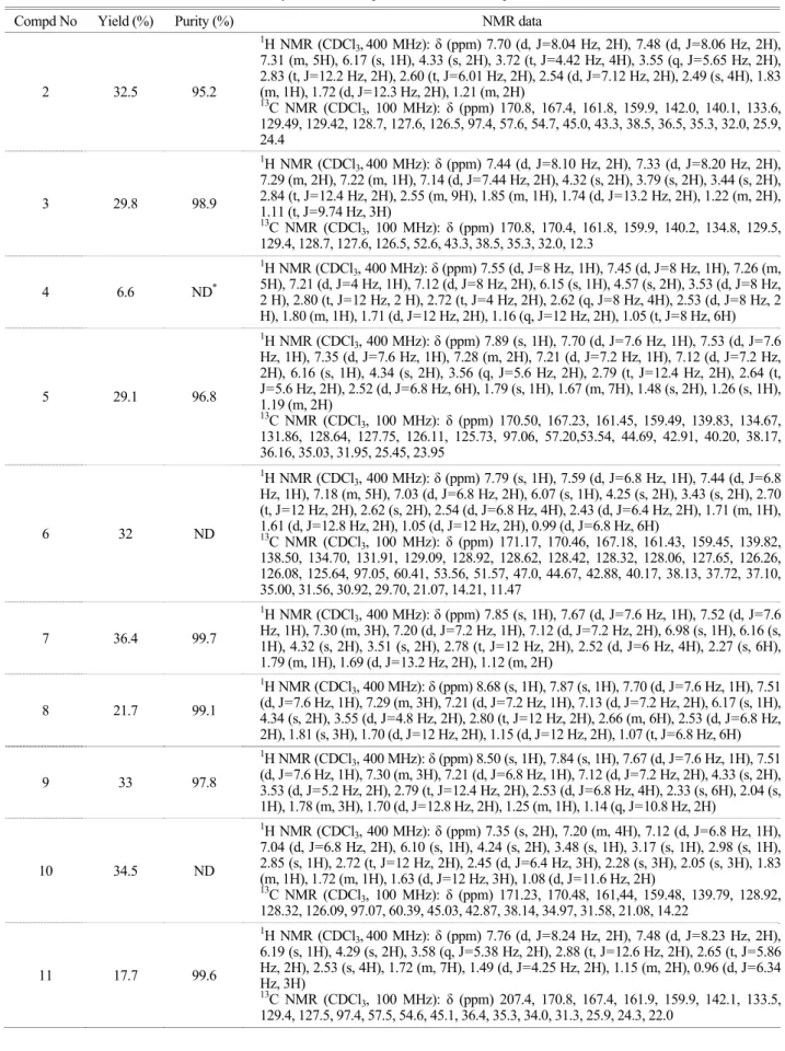
Table 1. Synthesis of compounds 2-14 and their spectra data
Compd No Yield (%) Purity (%) NMR data
2 32.5 95.2
1H NMR (CDCl3,400 MHz): δ (ppm) 7.70 (d, J=8.04 Hz, 2H), 7.48 (d, J=8.06 Hz, 2H), 7.31 (m, 5H), 6.17 (s, 1H), 4.33 (s, 2H), 3.72 (t, J=4.42 Hz, 4H), 3.55 (q, J=5.65 Hz, 2H), 2.83 (t, J=12.2 Hz, 2H), 2.60 (t, J=6.01 Hz, 2H), 2.54 (d, J=7.12 Hz, 2H), 2.49 (s, 4H), 1.83 (m, 1H), 1.72 (d, J=12.3 Hz, 2H), 1.21 (m, 2H)
13C NMR (CDCl3, 100 MHz): δ (ppm) 170.8, 167.4, 161.8, 159.9, 142.0, 140.1, 133.6, 129.49, 129.42, 128.7, 127.6, 126.5, 97.4, 57.6, 54.7, 45.0, 43.3, 38.5, 36.5, 35.3, 32.0, 25.9, 24.4
3 29.8 98.9
1H NMR (CDCl3,400 MHz): δ (ppm) 7.44 (d, J=8.10 Hz, 2H), 7.33 (d, J=8.20 Hz, 2H), 7.29 (m, 2H), 7.22 (m, 1H), 7.14 (d, J=7.44 Hz, 2H), 4.32 (s, 2H), 3.79 (s, 2H), 3.44 (s, 2H), 2.84 (t, J=12.4 Hz, 2H), 2.55 (m, 9H), 1.85 (m, 1H), 1.74 (d, J=13.2 Hz, 2H), 1.22 (m, 2H), 1.11 (t, J=9.74 Hz, 3H)
13C NMR (CDCl3, 100 MHz): δ (ppm) 170.8, 170.4, 161.8, 159.9, 140.2, 134.8, 129.5, 129.4, 128.7, 127.6, 126.5, 52.6, 43.3, 38.5, 35.3, 32.0, 12.3
4 6.6 ND*
1H NMR (CDCl3, 400 MHz): δ (ppm) 7.55 (d, J=8 Hz, 1H), 7.45 (d, J=8 Hz, 1H), 7.26 (m, 5H), 7.21 (d, J=4 Hz, 1H), 7.12 (d, J=8 Hz, 2H), 6.15 (s, 1H), 4.57 (s, 2H), 3.53 (d, J=8 Hz, 2 H), 2.80 (t, J=12 Hz, 2 H), 2.72 (t, J=4 Hz, 2H), 2.62 (q, J=8 Hz, 4H), 2.53 (d, J=8 Hz, 2 H), 1.80 (m, 1H), 1.71 (d, J=12 Hz, 2H), 1.16 (q, J=12 Hz, 2H), 1.05 (t, J=8 Hz, 6H)
5 29.1 96.8
1H NMR (CDCl3, 400 MHz): δ (ppm) 7.89 (s, 1H), 7.70 (d, J=7.6 Hz, 1H), 7.53 (d, J=7.6 Hz, 1H), 7.35 (d, J=7.6 Hz, 1H), 7.28 (m, 2H), 7.21 (d, J=7.2 Hz, 1H), 7.12 (d, J=7.2 Hz, 2H), 6.16 (s, 1H), 4.34 (s, 2H), 3.56 (q, J=5.6 Hz, 2H), 2.79 (t, J=12.4 Hz, 2H), 2.64 (t, J=5.6 Hz, 2H), 2.52 (d, J=6.8 Hz, 6H), 1.79 (s, 1H), 1.67 (m, 7H), 1.48 (s, 2H), 1.26 (s, 1H), 1.19 (m, 2H)
13C NMR (CDCl3, 100 MHz): δ (ppm) 170.50, 167.23, 161.45, 159.49, 139.83, 134.67, 131.86, 128.64, 127.75, 126.11, 125.73, 97.06, 57.20,53.54, 44.69, 42.91, 40.20, 38.17, 36.16, 35.03, 31.95, 25.45, 23.95
6 32 ND
1H NMR (CDCl3, 400 MHz): δ (ppm) 7.79 (s, 1H), 7.59 (d, J=6.8 Hz, 1H), 7.44 (d, J=6.8 Hz, 1H), 7.18 (m, 5H), 7.03 (d, J=6.8 Hz, 2H), 6.07 (s, 1H), 4.25 (s, 2H), 3.43 (s, 2H), 2.70 (t, J=12 Hz, 2H), 2.62 (s, 2H), 2.54 (d, J=6.8 Hz, 4H), 2.43 (d, J=6.4 Hz, 2H), 1.71 (m, 1H), 1.61 (d, J=12.8 Hz, 2H), 1.05 (d, J=12 Hz, 2H), 0.99 (d, J=6.8 Hz, 6H)
13C NMR (CDCl3, 100 MHz): δ (ppm) 171.17, 170.46, 167.18, 161.43, 159.45, 139.82, 138.50, 134.70, 131.91, 129.09, 128.92, 128.62, 128.42, 128.32, 128.06, 127.65, 126.26, 126.08, 125.64, 97.05, 60.41, 53.56, 51.57, 47.0, 44.67, 42.88, 40.17, 38.13, 37.72, 37.10, 35.00, 31.56, 30.92, 29.70, 21.07, 14.21, 11.47
7 36.4 99.7
1H NMR (CDCl3, 400 MHz): δ (ppm) 7.85 (s, 1H), 7.67 (d, J=7.6 Hz, 1H), 7.52 (d, J=7.6 Hz, 1H), 7.30 (m, 3H), 7.20 (d, J=7.2 Hz, 1H), 7.12 (d, J=7.2 Hz, 2H), 6.98 (s, 1H), 6.16 (s, 1H), 4.32 (s, 2H), 3.51 (s, 2H), 2.78 (t, J=12 Hz, 2H), 2.52 (d, J=6 Hz, 4H), 2.27 (s, 6H), 1.79 (m, 1H), 1.69 (d, J=13.2 Hz, 2H), 1.12 (m, 2H)
8 21.7 99.1
1H NMR (CDCl3, 400 MHz): δ (ppm) 8.68 (s, 1H), 7.87 (s, 1H), 7.70 (d, J=7.6 Hz, 1H), 7.51 (d, J=7.6 Hz, 1H), 7.29 (m, 3H), 7.21 (d, J=7.2 Hz, 1H), 7.13 (d, J=7.2 Hz, 2H), 6.17 (s, 1H), 4.34 (s, 2H), 3.55 (d, J=4.8 Hz, 2H), 2.80 (t, J=12 Hz, 2H), 2.66 (m, 6H), 2.53 (d, J=6.8 Hz, 2H), 1.81 (s, 3H), 1.70 (d, J=12 Hz, 2H), 1.15 (d, J=12 Hz, 2H), 1.07 (t, J=6.8 Hz, 6H)
9 33 97.8
1H NMR (CDCl3, 400 MHz): δ (ppm) 8.50 (s, 1H), 7.84 (s, 1H), 7.67 (d, J=7.6 Hz, 1H), 7.51 (d, J=7.6 Hz, 1H), 7.30 (m, 3H), 7.21 (d, J=6.8 Hz, 1H), 7.12 (d, J=7.2 Hz, 2H), 4.33 (s, 2H), 3.53 (d, J=5.2 Hz, 2H), 2.79 (t, J=12.4 Hz, 2H), 2.53 (d, J=6.8 Hz, 4H), 2.33 (s, 6H), 2.04 (s, 1H), 1.78 (m, 3H), 1.70 (d, J=12.8 Hz, 2H), 1.25 (m, 1H), 1.14 (q, J=10.8 Hz, 2H)
10 34.5 ND
1H NMR (CDCl3, 400 MHz): δ (ppm) 7.35 (s, 2H), 7.20 (m, 4H), 7.12 (d, J=6.8 Hz, 1H), 7.04 (d, J=6.8 Hz, 2H), 6.10 (s, 1H), 4.24 (s, 2H), 3.48 (s, 1H), 3.17 (s, 1H), 2.98 (s, 1H), 2.85 (s, 1H), 2.72 (t, J=12 Hz, 2H), 2.45 (d, J=6.4 Hz, 3H), 2.28 (s, 3H), 2.05 (s, 3H), 1.83 (m, 1H), 1.72 (m, 1H), 1.63 (d, J=12 Hz, 3H), 1.08 (d, J=11.6 Hz, 2H)
13C NMR (CDCl3, 100 MHz): δ (ppm) 171.23, 170.48, 161,44, 159.48, 139.79, 128.92, 128.32, 126.09, 97.07, 60.39, 45.03, 42.87, 38.14, 34.97, 31.58, 21.08, 14.22
11 17.7 99.6
1H NMR (CDCl3,400 MHz): δ (ppm) 7.76 (d, J=8.24 Hz, 2H), 7.48 (d, J=8.23 Hz, 2H), 6.19 (s, 1H), 4.29 (s, 2H), 3.58 (q, J=5.38 Hz, 2H), 2.88 (t, J=12.6 Hz, 2H), 2.65 (t, J=5.86 Hz, 2H), 2.53 (s, 4H), 1.72 (m, 7H), 1.49 (d, J=4.25 Hz, 2H), 1.15 (m, 2H), 0.96 (d, J=6.34 Hz, 3H)
13C NMR (CDCl3, 100 MHz): δ (ppm) 207.4, 170.8, 167.4, 161.9, 159.9, 142.1, 133.5, 129.4, 127.5, 97.4, 57.5, 54.6, 45.1, 36.4, 35.3, 34.0, 31.3, 25.9, 24.3, 22.0
1H NMR (CDCl3, 400 MHz): δ (ppm) 7.70 (d, J=8.04 Hz, 2H), 7.48 (d, J=8.06 Hz, 2H), 7.31 (m, 5H), 6.17 (s, 1H), 4.33 (s, 2H), 3.72 (t, J=4.42 Hz, 4H), 3.55 (q, J=5.65 Hz, 2H), 2.83 (t, J=12.2 Hz, 2H), 2.60 (t, J=6.01 Hz, 2H), 2.54 (d, J=7.12 Hz, 2H), 2.49 (s, 4H), 1.83 (m, 1H), 1.72 (d, J=12.3 Hz, 2H), 1.21 (m, 2H)
13C NMR (CDCl3, 100 MHz): δ (ppm) 170.7, 167.5, 161.8, 159.9, 142.2, 140.1, 133.6, 129.5, 128.7, 127.5, 126.5, 97.5, 67.1, 57.4, 53.7, 45.1, 43.3, 38.6, 36.2, 35.2, 32.0
Compounds 2 to 14 were prepared by the same procedure used for the synthesis of compound 1 using corresponding reagents. Yield, purity and spectra data are listed in Table 1.
6. Cell culture
Cancer cell lines such as HepG2 cells (hepatocellular liver carcinoma), Caki cells (renal carcinoma), AMC-HN3 cells, AMC-HN4 cells (squamous cell carcinoma), and normal human skin fibroblast were cultured in Dulbecco's Modified Eagle's Medium (DMEM) (WelGENE Inc., Korea) supplemented with 10% fetal calf serum (FCS) and 1%
antibiotic-antimycotic agent (WelGENE Inc., Korea) in 5%
CO2 at 37℃. A549 cells (lung carcinoma) and HEC-1-A cells (endometroid adenocarcinoma) were cultured in RPMI 1640 (WelGENE Inc., Korea), containing 10% FCS, 20 mM Hepes buffer, 100 μg/ml gentamicin, and 1% antibiotic- antimycotic agent (WelGENE Inc) in 5% CO2 at 37℃.
7. Cytotoxicity assay (XTT assay)
The cytotoxic effect of the compounds on various human cancer cells was investigated using a commercially available proliferation kit (WelCount Cell Viability Assay Kit, WELGENE, Daegu, Korea) (Seo et al., 2008). Briefly, the cells were plated in 96-well culture plates at a density of 3,000 cells/well in phenol red free-medium and allowed to attach for 10 h. After 24 h or more than 24 h treatment of compound, 20 μL of XTT reaction solution (2,3-Bis(2- methoxy-4-nitro-5-sulfophenyl)-2H-tetrazolium-5-carboxa nilide inner salt and phenazine methosulfate; mixed in pro- portion 50:1) was added to the wells. The optical density was read at 450 nm wavelength in an ELISA microplate reader (TECAN, Switzerland) after 3 h incubation of the plates with XTT reaction solution in an incubator (37℃
and 5% CO2 + 95% air).
8. Cytotoxicity assay (SRB assay)
The sulforhodamine B (SRB) assay was carried out as previously described (Papazisis et al., 1997). Briefly, the cells were plated in 96-well culture plates at a density of 3,000 cells/well in phenol red free-medium and allowed to attach for 10 h. After 24 h or 48 h treatment of compound, culture media were removed. 70 μl of 0.4% (w/v) SRB (Sigma) in 1% acetic acid solution was added to each well and left at room temperature for 20 min. SRB was removed Table 1. Continued
Compd No Yield (%) Purity (%) NMR data
12 20.3 95.1
1H NMR (CDCl3,400 MHZ): δ (ppm) 7.72 (d, J=8.28 Hz, 2H), 7.49 (d, J=8.24 Hz, 2H), 6.95 (s, 1H), 6.18 (s, 1H), 4.34 (s, 2H), 3.56 (s, 4H), 3.52 (q, J=5.75 Hz, 2H), 2.55 (t, J=6.06 Hz, 2H), 2.42 (s, 4H), 1.70 (m, 2H), 1.61 (m, 8H), 1.47 (d, J=4.82 Hz, 2H)
13C NMR (CDCl3, 100MHz): δ(ppm) 170.7, 167.4, 161.8, 159.3, 142.0, 129.4, 127.4, 97.4, 57.3, 54.6, 45.8, 36.7, 35.3, 26.4, 25.8, 24.8, 24.7
13 13.8 90.5
1H NMR (CDCl3,400 MHz): δ (ppm) 7.80 (d, J=7.98 Hz, 2H), 7.47 (d, J=7.93 Hz, 2H), 6.05 (s, 1H), 4.36 (s, 2H), 3.62 (s, 2H), 3.44 (d, J=9.1 Hz, 4H), 2.76 (m, 6H), 1.71 (s, 4H), 1.58 (m, 6H), 0.88 (s, 6H)
13C NMR (CDCl3, 100MHz): δ (ppm) 171.0, 167.3, 162.4, 160.2, 141.1, 133.9, 129.4, 127.5, 97.6, 66.6, 57.3, 54.6, 44.7, 36.8, 35.4, 26.4, 24.7
14 64.1 98.1
1H NMR (CDCl3,400 MHz): δ (ppm) 7.72 (d, J=8.24 Hz, 2H), 7.47 (d, J=8.16 Hz, 2H), 7.00 (s, 1H), 6.18 (s, 1H), 4.34 (s, 2H), 3.73 (t, J=4.42 Hz, 4H), 3.57 (s, 4H), 3.52 (q, J=5.66 Hz, 2H), 2.54 (t, J=5.98 Hz, 2H), 2.42 (s, 4H), 1.61 (m, 4H), 1.46 (d, J=4.72 Hz, 2H)
13C NMR (CDCl3, 100 MHz): δ (ppm) 170.6, 167.6, 161.8, 159.3, 142.0, 133.3, 129.2, 127.7, 97.1, 57.5, 54.6, 50.6, 36.2, 35.1, 25.2, 23.9, 11.7
* ND: not determined
and the plates were washed 5 times with 1% acetic acid solution before air drying. Bound SRB was solubilized with 200 μl of 10 mM unbuffered Tris-base solution (Sigma) and plates were left on a plate shaker for at least 10 min.
Absorbance was read in a 96-well plate reader at 492 nm subtracting the background measurement at 620 nm. The test optical density (OD) value was defined as the absorbance of each individual well minus the blank value ('blank' is the mean optical density of the background control wells, n=8).
RESULTS AND DISCUSSION
Discovery of new scaffold having good biological pro- file is very important in drug discovery process. As a first step of scaffold search, screening of chemical library was performed. The anti-cancer effect of compounds was evalu- ated by XTT assay. Primary screen provided 2-thio-4- aminopyrimidine derivatives as the initial hit and compounds having 2-thiobenzyl group were chosen for further study.
To confirm the screen result, 2 hit compounds were syn- thesized and their growth inhibitory effects were studied.
The general synthetic procedure is shown at scheme 1.
2-Thiobabituric acid (I) was used as starting material.
Selective alkylation at thiol functionality with methyl chloromethylbenzoate in the presence of sodium bicarbonate provided compound (II). After chlorination of hydroxyl groups by phosphooxychloride (III), amines were introduced
by substitution of one chloro group (IV). The hydrolysis of ester followed by amide coupling with aid of EDC provided the 2-thiobenzyl-4-aminopyrimidine derivatives (V). All compound were dissolved in dimethylsulfoxide (DMSO) at 10 μM concentration for assay.
Two assay methods were used to evaluate anti-cancer activity of compounds. XTT assay was used for primary screening the chemical library while SRB assay was used for confirmation and evaluation of synthesized compounds (Fricker et al., 1996; Papazisis et al., 1997; Pauwels et al., 2003). Human cancer cell lines, A549, HepG2, Caki, AMC -HN3, AMC-HN4, Hec-1-A, were used for the evaluation of anticancer effect while fibroblast was used to check the potential toxicity of compounds.
Among the initial hits, 2 was found to be more active than 1 (Table 1. Values in parenthesis are the results of primary screen). 2 showed 100% growth inhibition to all tested cancer cell lines and fibroblast while 1 showed good activity only to A549 cell lines. The unfavorable effect of piperazine attached directly to benzoate (3) suggested that the flexible linker might contribute to the activity of the compound. The relative position of substituents at benzene ring did not affect to the inhibitory activity (2, 4, and 5).
Also, the structural variation of substituents did not cause activity change as far as they were connected by flexible linkers (5, 6, 7, 8 and 9). After the evaluation of the activity dependence on the structure of benzamide, the effect of i) BrCH2C6H4CO2CH3, NaHCO3, EtOH, H2O, ii) POCl3, 2-picoline, iii) R1, Na2CO3, EtOH, iv) NaOH, THF, MeOH, H2O, v) R2, EDC, HOBt, DMF
Scheme 1. General synthetic method for 2-thio-4-aminopyrimidine derivatives
N N OH
HO SH N
N OH
HO S
O
O N
N Cl
Cl S
O O
N N R1
Cl S
O
O N
N R1
Cl S
R2 O
i ii
iii iv, v
(a) (b) (c)
(d) (e)
structural variation at 4-position of pyrimidine was studied (Table 2). The bulky benzyl group at C-4 of piperidine (2) showed high potency to both cancer cell lines and fibro- blast. The growth inhibitory activity showed decreasing tendency with the reduction of the bulkiness of substituent at C-4 of piperidine (cf. 2, 11, 12). The growth inhibition of fibroblast was more affected by the structural change of the substituent than cancer cell lines. This result suggests that the selectivity between cancer cell lines and normal cell can be improved by structural modification. The flexibility of
substituent at 4-position of pyrimidine showed unnoticeable effect (cf. 11 and 13). The abrupt reduction of activity caused by morpholine (14) suggested that these substituents might locate at either anionic pocket or hydrophobic pocket of binding site.
As a conclusion, 2-thio-4-aminopyrimidine derivatives showed the strong growth inhibition of cancer cell lines with substituent dependency. Mild selectivity in anti-cancer effect between cancer and normal cells were obtained by the modification of initial hit. According to the result, Table 2. Anti-cancer effects of compounds against human cancer cell lines at 10 μM concentration (SRB assay). Variation of benzamide
functionality
% Growth inhibition at 10 μM R2
Caki Hec-1-A A549 HepG2 AMC-HN3 AMC-HN4 Fibroblast
1 p- N O
N H2
18.0 69.1 84.1
(65.8)* 21.4 57.0 67.1 10.9
2 p- N
N H2
100.0 100.0 100.0 (95.6)* 100.0 100.0 100.0 100.0
3 p-
N
N 14.2 66.5 58.9 60.7 58.9 53.2 0.0
4 o- N
N H2
100.0 100.0 100.0 98.2 100.0 99.4 100.0
5 m- N
N H2
100.0 100.0 100.0 100.0 97.5 100.0 100.0
6 m- N
N H2
96.2 92.9 100.0 97.7 90.0 95.6 91.8
7 m- N
N
H2 100.0 43.1 100.0 99.5 100.0 97.8 95.9
8 m- HN N
2
100.0 100.0 100.0 98.7 97.5 100.0 100.0
9 m- HN N
2 98.1 100.0 97.3 97.5 100.0 95.6 91.8
10 m- HN N 100.0 100.0 100.0 97.7 100.0 100.0 95.9
* Primary screen results by XTT assay
N N N
S Cl
R2 O
2-thio-4-aminopyrimidine can be used as a new scaffold for the development of anti-cancer agent.
Acknowledgements
This research was financially supported by the Ministry of Education, Science Technology (MEST) and Korea Industrial Technology Foundation (KOTEF) through the Human Resource Training Project for Regional Innovation.
Also it was supported by Scholarship for Research Thesis of Graduate School of Keimyung University.
REFERENCES
Fricker SP, Buckley RG. Comparison of two colorimetric assays as cytotoxicity endpoints for an in vitro screen for antitumour agents. Anticancer Res. 1996. 16: 3755-3760.
Hua DH, Lou K, Battina SK, Zhao H, Perchellet EM, Wang Y, Perchellet JP. Syntheses, molecular targets and antitumor acti- vities of novel triptycene bisquinones and 1,4-anthracenedione
analogs. Anticancer Agents Med Chem. 2006. 6: 303-318.
Li W, Lam MS, Birkeland A, Riffel A, Montana L, Sullivan ME, Post JM. Cell-based assays for profiling activity and safety properties of cancer drugs. J Pharmacol Toxicol Meth. 2006.
54: 313-319.
Marshall L. The Cancer Outlook 2000, Datamonitor PLC, London.
Papazisis KT, Geromichalos GD, Dimitriadis KA, Kortsaris AH.
Optimization of the sulforhodamine B colorimetric assay. J Immunol Meth. 1997. 208: 151-158.
Pauwels B, Korst AE, de Pooter CM, Pattyn GG, Lambrechts HA, Baay MF, Lardon F, Vermorken JB. Comparison of the sulforhodamine B assay and the clonogenic assay for in vitro chemoradiation studies. Cancer Chemother Pharmacol. 2003.
51: 221-226.
Seo HY, Kim YD, Lee KM, Min AK, Kim MK, Kim HS, Won KC, Park JY, Lee KU, Choi HS, Park KG, Lee IK. Endoplasmic reticulum stress-induced activation of activating transcription factor 6 decreases insulin gene expression via up-regulation of orphan nuclear receptor small heterodimer partner. Endo- Table 3. Anti-cancer effects of compounds against human cancer cell lines at 10 μM concentration (SRB assay). Variation of 4-amino
functionality
N N R1
S Cl
N O
N
% Growth inhibition at 10 μM R1
Caki Hec-1-A A549 HepG2 AMC-HN3 AMC-HN4 Fibroblast
2
N
100.0 100.0 100.0 100.0 100.0 100.0 100.0
11
N
95.5 92.3 85.4 96.4 79.4 86.1 77.0
12
N
95.5 87.1 86.7 100.0 83.2 83.5 51.1
13
N
96.2 94.8 94.7 96.4 85.0 92.4 85.6
14
N O
24.8 30.4 21.8 19.6 17.8 11.4 5.2
crinology 2008. 149: 3832-3841.
Valler MJ, Green D. Diversity screening versus focused screening in drug discovery. Drug Discov Today 2000. 5: 286-293.
Weinstein JN, Buolamwini JK. Molecular targets in cancer drug
discovery: cell-based profiling. Curr Pharm Des. 2000. 6:
473-483.
WHO Fact sheet N° 297 (2009) http://www.who.int/mediacentre/
factsheets/fs297/en/