저작자표시-비영리-변경금지 2.0 대한민국 이용자는 아래의 조건을 따르는 경우에 한하여 자유롭게 l 이 저작물을 복제, 배포, 전송, 전시, 공연 및 방송할 수 있습니다. 다음과 같은 조건을 따라야 합니다: l 귀하는, 이 저작물의 재이용이나 배포의 경우, 이 저작물에 적용된 이용허락조건 을 명확하게 나타내어야 합니다. l 저작권자로부터 별도의 허가를 받으면 이러한 조건들은 적용되지 않습니다. 저작권법에 따른 이용자의 권리는 위의 내용에 의하여 영향을 받지 않습니다. 이것은 이용허락규약(Legal Code)을 이해하기 쉽게 요약한 것입니다. Disclaimer 저작자표시. 귀하는 원저작자를 표시하여야 합니다. 비영리. 귀하는 이 저작물을 영리 목적으로 이용할 수 없습니다. 변경금지. 귀하는 이 저작물을 개작, 변형 또는 가공할 수 없습니다.
의학 석사학위 논문
Comparison of sub-erythema dose and
erythema dose of excimer laser
treatment for stable vitiligo: A
randomized controlled trial
아 주 대 학 교 대 학 원
의 학 과
Comparison of sub-erythema dose and
erythema dose of excimer laser
treatment for stable vitiligo: A
randomized controlled trial
지도교수 강 희 영
이 논문을 의학 석사학위 논문으로 제출함.
2020 년 2 월
아 주 대 학 교 대 학 원
의 학 과
권 수 현
권수현의 의학 석사학위 논문을 인준함.
심사위원장 강 희 영 (인)
심
사
위
원 김 장 희 (인)
심
사
위
원 박 태 준 (인)
아 주 대 학 교 대 학 원
2020 년 1 월 9 일
감사의 글
본 논문을 완성할 때까지 물심양면으로 도움을 주시고 지도와 조언을 아끼지 않으셨던 지도교수이신 강희영 교수님께 진심으로 감사 드립니다. 또한 좋은 연구를 할 수 있도록 부족한 제게 많은 조언과 격려를 주신 김장희 교수님, 박태준 교수님께 깊은 감사의 마음을 전합니다. 그리고 연구 기간 동안 도움을 주신 피부과학교실의 모든 선생님들께 감사 드립니다. 언제나 아낌 없는 사랑으로 지원해 주시는 부모님을 비롯한 가족들에게도 깊은 감사의 마음을 전합니다. 2020 년 1 월 저자i
-ABSTRACT-
Comparison of sub-erythema dose and erythema dose of excimer
laser treatment for stable vitiligo: A randomized controlled trial
OBJECTIVE: To investigate the efficacy and safety of sub-erythema dose (SED) of EL
treatment compared to the erythema dose (ED) in treating stable vitiligo.
DESIGN: A randomized, controlled, split-body study.
SETTING: The trial was performed in two tertiary health care centers in Korea.
PARTICIPANTS: Thirty-six right or left sides of 18 patients with stable symmetric
vitiligo on the face and neck were enrolled.
INTERVENSIONS: The two sides of the face or neck were randomly assigned to either
SED or ED group of EL treatment. Both laser treatments were conducted twice weekly for 3 months. Initial irradiation dose was 50 mJ/cm2 and 100 mJ/cm2 on the face, and 100 mJ/cm2 and 200 mJ/cm2 on the neck for SED and ED group, respectively. If there had not been pinkish erythema lasting less than 24 hours after treatment in the ED group, the dose was escalated by 25 and 50 mJ/cm2 in subsequent treatment session for SED and ED group, respectively. The ED was defined as the dose to produce pinkish erythema lasting less than 24 hours, and the SED as the half of the ED.
MAIN OUTCOMES AND MEASURES: The degree of repigmentation (%) from
baseline in each side was assessed using Vitiligo Extent Score for a Target Area by blinded evaluators.
RESULTS: The SED side showed 86.1% ± 23.9% of repigmentation and was similar to
86.9% ± 23.0% in the ED side. Four patients (22.2%) complained of prolonged erythema lasting 3 days or pain on the ED side at least once during the 3-month period, but not on the SED side.
CONCLUSIONS AND RELEVANCE: The SED-EL treatment would be sufficient for
ii of EL treatment.
iii
TABLE OF CONTENTS
ABSTRACT ... i
TABLE OF CONTENTS ... iii
LIST OF FIGURES ... v
LIST OF TABLES ... vi
INTRODUCTION ... 1
A. Vitiligo ... 1
B. Excimer laser ... 1
C. Background and objective ... 2
MATERIAL AND METHODS ... 4
A. Study design and population ... 4
B. Intervention... 4 C. Treatment outcome ... 5 D. Statistical analysis ... 5 RESULTS ... 6 A. Study population ... 6 B. Treatment parameters ... 9 C. Degree of repigmentation ... 10 D. Perilesional hyperpigmentation ... 14 E. Preference of patients ... 15 F. Adverse events... 15 DISCUSSION ... 16 CONCLUSION ... 19
iv
REFERENCES ... 20 국문요약 ... 24
v
LIST OF FIGURES
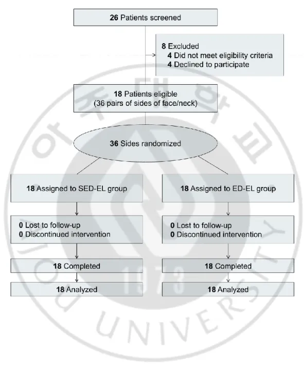
Figure 1. Flow diagram of the study. ... 7
Figure 2. Repigmentation of vitiligo after sub-erythema dose and conventional erythema dose of excimer laser treatment. ... 10
Figure 3. Rate of repigmentation in monthly taken photo... 11
Figure 4. Repigmentation rate using conventional DSLR and UV camera. ... 12
Figure 5. Less repigmentated case ... 13
vi
LIST OF TABLES
Table 1. Demographics of the enrolled patients in this study ... 8
- 1 -
INTRODUCTION
A. Vitiligo
Vitiligo is a common autoimmune disease of the skin that causes depigmentation through T-cell–mediated destruction of melanocytes. According to a recent international consensus conference, vitiligo can be classified into two major forms—namely, non-segmental vitiligo, also known as vitiligo, and non-segmental vitiligo (Ezzedine et al., 2012). Non-segmental vitiligo, the commonest form of this unpredictable disease, is characterized by symmetrical and bilateral white patches. Different clinical subtypes have been described, including generalized, acrofacial, and universalis types, all with a bilateral distribution. Segmental vitiligo is less common than non-segmental vitiligo and usually has a unilateral distribution. Overall, progressive patchy loss of pigmentation from skin, overlying hair, and sometimes mucosa remains the basis of diagnosis of vitiligo (Ezzedine et al., 2015).
Histological examination and immunohistochemical studies with a large panel of antibodies generally show an absence of melanocytes in lesional skin, although sometimes an occasional melanocyte can be seen (Le Poole et al., 1993).
Various theories have been suggested for the cause of melanocyte loss in vitiligo; some have proposed that vitiligo is a multifactorial disease, with both genetic and environmental factors implicated in its initiation. The autoimmune or autoinflammatory theory is the leading hypothesis for causation and is supported by strong evidence (Ezzedine et al., 2015).
B. Excimer laser
Excimer laser in which “excimer” is a terminological reference of “excited dimer”, composed of a noble gas and halide which repel each other, has turned into a prevalent mode of action in confronting numerous skin disorders (Spencer et al., 2004). Of these ultraviolet B rays which comprise beams of varied wavelengths, the 308nm xenon-chloride is the most practical in dermatology (Spencer et al., 2004). The advantages of
- 2 -
monochromatic excimer laser over phototherapies of other kinds have been depicted as lower UV dose exposure, shorter course of therapy and for the most part, the possibility of being directed at distinct sites of skin rather than compromising the adjacent normal skin (Grema et al., 2004; Mavilia et al., 2008; Morita et al., 2008).
Medium doses of the 308-nm excimer laser has proved to be effective in the treatment of limited vitiligo, however, the rate and speed of repigmentation is highly associated with the site and duration of disease as the face and neck (UV sensitive areas) are the highly respondent areas along with an earlier resolution of the lesions while the joints and extremities (UV resistant areas) exhibit the slightest response to therapy (Wang et al., 2009; Shen et al., 2007; Ostovari et al., 2004). Also, the shorter disease course is more promising in the treatment results (Zhang et al., 2010). Additionally, trials have demonstrated further efficacy of treatment in Fitzpatrick skin types other than I and II (Hadi et al., 2004; Al-Otaibi et al., 2009). Combination therapy of the 308-nm excimer laser and calcineurin inhibitors, topical tacrolimus in particular, is regarded to be more beneficial than excimer laser alone (Goldinger et al., 2007).
C. Background and objective
308-nm xenon chloride excimer laser (EL) has been widely used in treating localized vitiligo. The EL treatment is usually given two or three times weekly as long as ongoing repigmentation is observed.
Vitiligo is a common depigmenting skin disorder affecting 1% of the population, and profoundly impacts patients’ quality of life (Lee et al., 2015; Ezzedine et al., 2015; Bae et al., 2018). The 308-nm excimer laser (EL) has been widely used in treating localized vitiligo in a targeted way, preventing unnecessary UV irradiation of uninvolved skin (Passeron et al., 2006). The EL treatment is usually given twice or thrice weekly with the dose that make lesions pink erythema for 24 hours life (Hofer et al., 2005; Lim et al., 2007). However, there is largely lack of evidence for the optimal dosimetry of EL treatment for vitiligo yet (Sung et al., 2018).
- 3 -
and rarely unintended burn (Bae et al., 2016). Recently, Chiu et al. demonstrated the low-dose phototherapy not associated with erythema, could induce successful treatment response in patients with vitiligo in their pilot study (Chiu et al., 2018). Low-dose phototherapy would be associated with fewer unwanted adverse events. In the present study, we conducted a randomized controlled trial to compare the efficacy and safety of sub-erythema dose (SED) with the conventional erythema dose (ED) of EL treatment for stable vitiligo.
- 4 -
MATERIALS AND METHODS
A. Study design and population
A randomized controlled, split-body, non-inferiority trial was performed between April 2018 and December 2018. Adult patients aged 18 years or older with stable vitiligo that was symmetrically distributed over their face and neck were recruited from our clinics. All participants provided written informed consent. The stable vitiligo was defined as the absence of new lesions and no increase in the size of existing lesions for 6 months prior to the enrollment. The exclusion criteria included the patients taking systemic corticosteroids or immunosuppressants for last 4 weeks; having chemotherapy or immunotherapy for last 4 weeks; having phototherapy including EL for last 4 weeks. The two sides of the face or neck were randomly assigned to either SED or ED group of EL treatment. This study was approved by the institutional review board of Ajou
university hospital (AJIRB-MED-OBS-17-369) and St. Vincent’s hospital
(VC17DEDI0209).
B. Intervention
The ED was defined as the dose to produce pinkish erythema lasting less than 24 hours, and the SED as the half of the ED. The 308-nm EL treatment was conducted twice weekly on both sides for 3 months (EXL-440, Jetema, Seoul, Korea). Initial irradiation dose was 50 mJ/cm2 and 100 mJ/cm2 on the face, and 100 mJ/cm2 and 200 mJ/cm2 on the neck for SED and ED group, respectively (Lee et al., 2017). If there had not been pinkish erythema lasting less than 24 hours after treatment in the ED side, the dose was escalated by 25 and 50 mJ/cm2 in subsequent treatment session for SED and ED group, respectively. Topical tacrolimus 0.1% ointment (LEO Pharma Inc, Ballerup, Denmark) was applied twice daily in both groups for study period. Photographic documentation was conducted at baseline (M0), and after 4 (M1), 8 (M2), and 12 weeks (M3, end of study) using conventional DSLR and UV camera (Bae et al., 2017).
- 5 -
C. Treatment outcome
The primary outcome was degree of repigmentation (%) from baseline in each side, and which was assessed using Vitiligo Extent Score for a Target Area (VESTA) by blinded evaluators who did not know about the groups (Bae et al., 2019). The degree of perilesional hyperpigmentation was assessed as 4 grades by blinded evaluators: ‘no’, ‘somewhat’, ‘obvious’, and ‘pronounced.’ The preference of patient between the two treatments was asked as no preference, SED side, and ED side. For safety surveillance, any adverse event such as prolonged erythema lasting 3 days, symptomatic erythema (i.e., pain) and blistering was also monitored at every visit.
D. Statistical analysis
The mean differences and 95% confidence interval (95% CI) of the repigmentation rate between the two groups were calculated; the non-inferiority margin was set as 10%. Intention-to-treat (ITT) analyses using the last-observation-carried-forward principle were the primary outcomes. Per-protocol (PP) analyses were also planned for the supplementary. All statistical analyses were conducted using R 3.6.1 (R Foundation for Statistical Computing, Austria).
- 6 -
RESULTS
A. Study population
Total 12 patients (9 females and 3 males) were enrolled. The median age was 54.5-year-old (range 14-72) and the median disease duration was 17 months (range 1-58). The total number of paired vitiliginous lesions was 32 (16 pairs) and those were most frequently located on face (43.75%), followed by trunk (18.75%) and extremities
(37.5%). Twenty-six patients were screened for inclusion: of these, 4 did not meet
inclusion criteria and 4 declined to participate. Eighteen patients (12 females and 6 males; 15 on the face and 3 on the neck) were enrolled and a total of 36 left or right sides of them were randomly assigned to either SED-EL or ED-EL group (Figure 1). The median age was 48 year old (range 21 – 71) and the median disease duration was 1 year (range 0.6 – 17). Among them, 11 (61.1%) had Fitzpatrick skin type III, and the other 7 (38.9%) had type IV (Table 1).
- 7 -
- 8 -
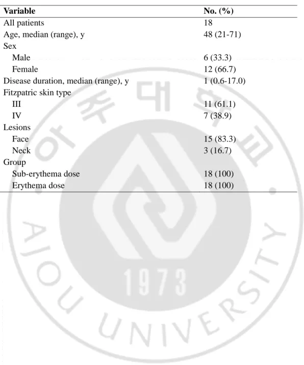
Table 1. Demographics of the enrolled patients in this study
Variable No. (%)
All patients 18
Age, median (range), y 48 (21-71)
Sex
Male 6 (33.3)
Female 12 (66.7)
Disease duration, median (range), y 1 (0.6-17.0)
Fitzpatric skin type
III 11 (61.1) IV 7 (38.9) Lesions Face 15 (83.3) Neck 3 (16.7) Group Sub-erythema dose 18 (100) Erythema dose 18 (100)
- 9 -
B. Treatment parameters
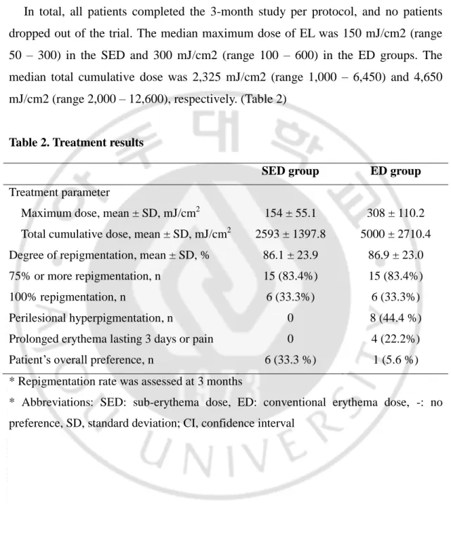
In total, all patients completed the 3-month study per protocol, and no patients dropped out of the trial. The median maximum dose of EL was 150 mJ/cm2 (range 50 – 300) in the SED and 300 mJ/cm2 (range 100 – 600) in the ED groups. The median total cumulative dose was 2,325 mJ/cm2 (range 1,000 – 6,450) and 4,650 mJ/cm2 (range 2,000 – 12,600), respectively. (Table 2)
Table 2. Treatment results
SED group ED group
Treatment parameter
Maximum dose, mean ± SD, mJ/cm2 154 ± 55.1 308 ± 110.2
Total cumulative dose, mean ± SD, mJ/cm2 2593 ± 1397.8 5000 ± 2710.4
Degree of repigmentation, mean ± SD, % 86.1 ± 23.9 86.9 ± 23.0
75% or more repigmentation, n 15 (83.4%) 15 (83.4%)
100% repigmentation, n 6 (33.3%) 6 (33.3%)
Perilesional hyperpigmentation, n 0 8 (44.4 %)
Prolonged erythema lasting 3 days or pain 0 4 (22.2%)
Patient’s overall preference, n 6 (33.3 %) 1 (5.6 %)
* Repigmentation rate was assessed at 3 months
* Abbreviations: SED: sub-erythema dose, ED: conventional erythema dose, -: no preference, SD, standard deviation; CI, confidence interval
- 10 -
C. Degree of repigmentation
The degree of repigmentation was 86.1% ± 23.9% in the SED group and 86.9% ± 23.0% in the ED group. The mean difference between the two groups was -0.8% (95% confidence interval, -1.9% to 0.2%), which was less than the noninferiority margin. In both group, 15 (83.4%) of the patients achieved 75% or more repigmentation, respectively; 6 (33.3%) showed 100% repigmentation (Figure 2,3,4 and 5).
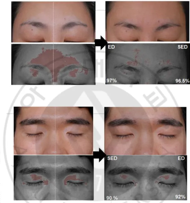
Figure 2. Repigmentation of vitiligo after sub-erythema dose and conventional erythema dose of excimer laser treatment. Patients 17 had vitiligo on the face before
treatment and showed repigmentation rate of 96.5% in SED-EL treatment and 97% in ED-EL treatment at the end of the 3-month trial (a and d). Patient 4 had vitiliginous patches on the face symmetrically and showed repigmentation rate of 98 % in SED-EL treatment and 98 % in ED-EL treatment at the end of the trial (b and e).
- 11 -
Figure 3. Rate of repigmentation in monthly taken photo. Patient 18 showed that rate
of repigmentation was not different between SED-EL treatment and- ED-EL treatment side.
- 12 -
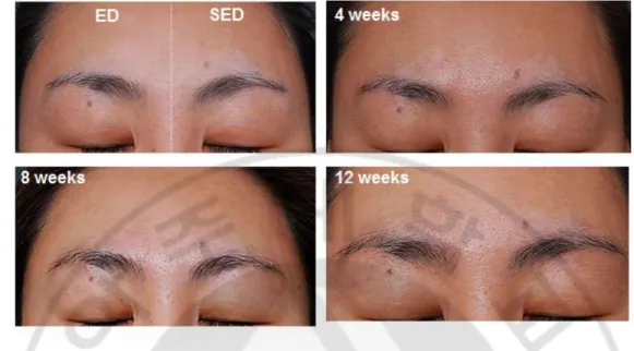
Figure 4. Repigmentation rate using conventional DSLR and UV camera.
Patients 18 had vitiligo on the forehead before treatment and showed repigmentation rate of 96.5% in SED-EL treatment and 97% in ED-EL treatment at the end of the 3-month trial.
Patient 16 had vitiliginous patches on the upper eyelid symmetrically and showed repigmentation rate of 90 % in SED-EL treatment and 92 % in ED-EL treatment at the end of the trial
- 13 -
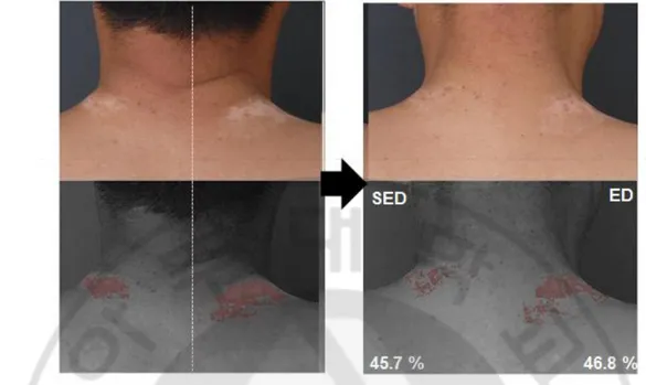
Figure 5. Less repigmentated case. Patient 13 on the nape showed repigmentation rate
of 45.7% in SED-EL treatment and 46.8% in ED-EL treatment at the end of the 3-month trial. It showed similar amount of change between the two lesions.
- 14 -
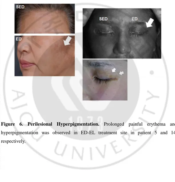
D. Perilesional Hyperpigmentation
In the ED group, 8 (44.4%) showed obvious or pronounced perilesional hyperpigmentation, whereas no one showed in the SED group (Figure 6).
Figure 6. Perilesional Hyperpigmentation. Prolonged painful erythema and
hyperpigmentation was observed in ED-EL treatment site in patient 5 and 14, respectively.
- 15 -
E. Preference of Patients
For the preference of each group, 6 (33.3%) preferred the SED side, 1 (5.6 %) ED, and remaining 11 (61.1%) no preference.
F. Adverse Events
All patients had no serious adverse event to stop the treatment in both groups. Four patients (22.2%) complained of prolonged erythema lasting 3 days or pain on the ED side at least once during the 3-month period, but not on the SED side.
- 16 -
DISCUSSION
According to a recent recommendation of narrowband UV-B (NB-UVB) phototherapy for vitiligo, the desired response to phototherapy is pink asymptomatic erythema lasting <24 hours (Mohammad et al., 2017). Because there have not been much studies for EL treatment of vitiligo, the basic principles of NB-UVB phototherapy are largely followed. However, several adverse events such as perilesional hyperpigmentation, blistering, and skin hypersensitivity are often problematic, especially on the face and neck.
In the present study, we demonstrated that SED-EL treatment is as effective as ED-EL treatment for achieving repigmentation of stable vitiligo. After the treatment was completed, 33.3% preferred SEL over EL treatment, while 5.6% preferred ED-EL treatment. The remaining 61.1% claimed no preference, probably because the treatment response did not differ from each other. Among the 18 patients, 44.4% complained of perilesional hyperpigmentation on the ED side, while none on the SED side. In addition, adverse events such as prolonged erythema lasting 3 days or pain were observed only on the ED side as well. The higher patients’ preference for SED-EL treatment may be due to better cosmetic results, i.e., less hyperpigmentation, and fewer adverse events.
Although the mechanism of action of EL treatment in vitiligo is not yet fully understood yet, a dual mechanism of action would be proposed in the treatment of vitiligo: cutaneous immunosuppression and repigmentation induction. In studies of
patients with psoriasis, supra-erythemogenic dose EL treatment showed
immunosuppressive effects through apoptosis of T-cells, which is expected to inhibit autoreactive T cells in patients with vitiligo (Levin et al., 2015; Gattu et al., 2010). Meanwhile, stimulation of melanoblast migration and melanocyte proliferation would be the major effect of UVB for repigmentation in stable vitiligo. This bio-stimulatory dosage of UVB might differ from the conventional UVB dosage that makes erythema in the skin. This suggests that the cytokines responsible for vasodilation in the skin (i.e.,
- 17 -
erythema response) would differ from those responsible for melanocyte induction, and the latter may be induced with a lower dose of EL treatment.
Our findings are in line with recent reports using Janus kinase (JAK) inhibitors in patients with vitiligo. Rothstein et al. described topical ruxolitinib 1.5% cream provided significant repigmentation only in facial vitiligo, not in truncal lesions in their open-label study (Rothstein et al., 2017). Liu et al. also reported some reversals of vitiligo only in sun-exposed areas in patients receiving both oral tofacitib (5 to 10 mg daily or twice daily) and sunlight exposure or low-dose NB-UVB phototherapy (360 – 500 mJ/cm2) (Liu et al., 2017). They suggested that natural sunlight would be sufficient to induce repigmentation in patients with vitiligo, when autoimmune response was inhibited by the JAK inhibitors. Our findings were also consistent with the pilot study by Chiu et al., which showed that stable vitiligo could be successfully treated with low-dose NB-UVB phototherapy or excimer light therapy: the medians of maximum dose were 245 mJ/cm2 (range: 210 – 420 mJ/cm2) and 325 mJ/cm2 (range: 300 – 400 mJ/cm2), respectively (Chiu et al., 2018). For stable vitiligo that do not require immunosuppression exogenously, SED-EL treatment would be sufficient to induce repigmentation in vitiligo patches.
Low-dose EL treatment may have additional benefits to avoid the chronic phototoxic changes of the skin in long-term high-dose EL treatment. Repetitive high-dose UV irradiation thickens the stratum corneum through photoadaptation, increasing the minimal erythema dose of UV, and consequently leading a higher dose of EL treatment (Rivard et al., 2007; Shin et al., 2016). Furthermore, some physicians often raise the dose when expected results are not observed, resulting in a hardened dermis due to repeated burns. However, we found that SED-EL treatment could achieved the equivalent therapeutic outcomes with half of the cumulative dose of conventional ED-EL treatment.
The present study has some limitations. First, the sample population was small. Second, the split-body study hardly excluded the paracrine effects. Third, treatment was performed only on the face and neck areas which normally shows better prognosis than
- 18 -
other areas. However, the treatment response was comparable in three months, because the face and neck areas were targeted. Further research will be needed for other body parts. Fourth, as we used tacrolimus ointment for both groups, our result may not reflect the effect of EL solely. But, the use of tacrolimus ointment better reflects real clinical practice. Lastly, we did not provide a practical protocol for determining the appropriate SED when the clinical indicator of erythema is not available. Nevertheless, we first revealed that stable vitiligo can be treated without the erythema response by a randomized controlled trial.
- 19 -
CONCLUSION
We demonstrated the SED-EL treatment would be sufficient for achieving repigmentation in stable vitiligo in the randomized clinical trial. Our findings suggested that low-dose EL treatment could induce repigmentation probably by stimulating melanocyte migration and melanoblast differentiation in patients with stable vitiligo. We do not oppose the existing ED-EL treatment protocol, but our findings would add new perspectives to clinicians in treating a diversity of vitiligo patients. More controlled trials are needed to find optimal dosimetry of EL treatment for vitiligo.
- 20 -
REFERENCES
1. Ezzedine K, Lim HW, Suzuki T, et al. Revised classifi cation/nomenclature of vitiligo and related issues: the Vitiligo Global Issues Consensus Conference. Pigment Cell Melanoma Res. 2012;25: E1–13.
2. Ezzedine K, Eleftheriadou V, Whitton M, van Geel N. Vitiligo. Lancet. 2015;386(9988):74-84.
3. Le Poole IC, Das PK, van den Wijngaard RM, Bos JD, Westerhof W. Review of the etiopathomechanism of vitiligo: a convergence theory. Exp Dermatol. 1993;2:145– 153.
4. Spencer JM, Hadi SM. The excimer lasers. J Drugs Dermatol. 2004;3:522–5
5. Grema H, Raulin C. The excimer laser in dermatology and esthetic medicine. Hautarzt. 2004;55:48–57.
6. Mavilia L, Mori M, Rossi R, Campolmi P, Puglisi Guerra A, Lotti T. 308 nm monochromatic excimer light in dermatology: personal experience and review of the literature. G Ital Dermatol Venereol. 2008;143:329–37.
7. Morita A, Weiss M, Maeda A. Recent developments in phototherapy: treatment methods and devices. Recent Pat Inflamm Allergy Drug Discov. 2008;2:105–8. 8. Wang HW, Zuo YG, Jin HZ, Liu YH, Ma DL, Jiang GT. et al. Efficacy and safety of
308 nm excimer laser for vitiligo. Zhongguo Yi Xue Ke Xue Yuan Xue Bao. 2009;31:34–6.
9. Shen Z, Gao TW, Chen L, Yang L, Wang YC, Sun LC. et al. Optimal frequency of treatment with the 308-nm excimer laser for vitiligo on the face and neck. Photomed Laser Surg. 2007;25:418–27.
- 21 -
10. Ostovari N, Passeron T, Zakaria W, Fontas E, Larouy JC, Blot JF. et al. Treatment of vitiligo by 308-nm excimer laser: an evaluation of variables affecting treatment response. Lasers Surg Med. 2004;35:152–6.
11. Zhang XY, He YL, Dong J, Xu JZ, Wang J. Clinical efficacy of a 308 nm excimer laser in the treatment of vitiligo. Photodermatol Photoimmunol Photomed. 2010;26(3):138–42.
12. Hadi SM, Spencer JM, Lebwohl M. The use of the 308- nm excimer laser for the treatment of vitiligo. Dermatol Surg. 2004;30:983–6.
13. Al-Otaibi SR, Zadeh VB, Al-Abdulrazzaq AH, Tarrab SM, Al-Owaidi HA, Mahrous R. et al. Using a 308- nm excimer laser to treat vitiligo in Asians. Acta Dermatovenerol Alp Panonica Adriat. 2009;18:13–9.
14. Goldinger SM, Dummer R, Schmid P, Burg G, Seifert B, Läuchli S. Combination of 308-nm xenon chloride excimer laser and topical calcipotriol in vitiligo. J Eur Acad Dermatol Venereol. 2007;21:504–8.
15. Lee H, Lee MH, Lee DY, et al. Prevalence of vitiligo and associated comorbidities in Korea. Yonsei Med J. 2015;56(3):719-725.
16. Ezzedine K, Sheth V, Rodrigues M, et al. Vitiligo is not a cosmetic disease. J Am Acad Dermatol. 2015;73(5):883-885.
17. Bae JM, Lee SC, Kim TH, et al. Factors affecting quality of life in patients with vitiligo: a nationwide study. Br J Dermatol. 2018;178(1):238-244.
18. Passeron T, Ortonne JP. Use of the 308-nm excimer laser for psoriasis and vitiligo. Clin Dermatol. 2006;24(1):33-42.
- 22 -
19. Hofer A, Hassan AS, Legat FJ, Kerl H, Wolf P. Optimal weekly frequency of 308-nm excimer laser treatment in vitiligo patients. Br J Dermatol. 2005;152(5):981-985. 20. Lim HW, Hexsel CL. Vitiligo: to treat or not to treat. Arch Dermatol.
2007;143(5):643-646.
21. Sung JM, Bae JM, Kang HY. Comparison of cyclic and continuous 308-nm excimer laser treatments for vitiligo: A randomized controlled noninferiority trial. J Am Acad Dermatol. 2018;78(3):605-607.e601.
22. Bae JM, Hann S-K. Laser Treatments for Vitiligo. Medical Lasers. 2016;5(2):63-70. 23. Chiu SH, Liu IL, Chen YW, Lan CE. Low-dose UVB therapy is comparable with
conventional UVB phototherapy for treatment of vitiligo: A pilot study. J Dermatol Sci. 2018;92(2):218-220.
24. Lee H, Chu H, Oh SH. Investigation of suitable starting doses of narrowband UVB in Asian vitiligo patients: a pilot study. J Eur Acad Dermatol Venereol. 2017;31(5):894-897.
25. Bae JM, Han TY. A grayscale photograph with high dynamic range taken under a Wood's lamp for better recognition of vitiligo lesions. J Am Acad Dermatol. 2017;76(3):e89-e90.
26. Bae JM, Oh SH, Kang HY, et al. Development and validation of the Vitiligo Extent Score for a Target Area (VESTA) to assess the treatment response of a target lesion. Pigment Cell Melanoma Res. 2019;32(2):315-319.
27. Mohammad TF, Al-Jamal M, Hamzavi IH, et al. The Vitiligo Working Group recommendations for narrowband ultraviolet B light phototherapy treatment of vitiligo. J Am Acad Dermatol. 2017;76(5):879-888.
- 23 -
28. Levin E, Debbaneh M, Malakouti M, et al. Supraerythemogenic excimer laser in combination with clobetasol spray and calcitriol ointment for the treatment of generalized plaque psoriasis: Interim results of an open label pilot study. J Dermatolog Treat. 2015;26(1):16-18.
29. Gattu S, Pang ML, Pugashetti R, et al. Pilot evaluation of supra-erythemogenic phototherapy with excimer laser in the treatment of patients with moderate to severe plaque psoriasis. J Dermatolog Treat. 2010;21(1):54-60.
30. Rothstein B, Joshipura D, Saraiya A, et al. Treatment of vitiligo with the topical Janus kinase inhibitor ruxolitinib. J Am Acad Dermatol. 2017;76(6):1054-1060.e1051.
31. Liu LY, Strassner JP, Refat MA, Harris JE, King BA. Repigmentation in vitiligo using the Janus kinase inhibitor tofacitinib may require concomitant light exposure. J Am Acad Dermatol. 2017;77(4):675-682 e671.
32. Rivard J, Hexsel C, Owen M, Strickland FM, Lim HW, Hamzavi I. Photoadaptation of vitiliginous skin to targeted ultraviolet B phototherapy. Photodermatol Photoimmunol Photomed. 2007;23(6):258-260.
33. Shin S, Hann SK, Oh SH. Combination treatment with excimer laser and narrowband UVB light in vitiligo patients. Photodermatol Photoimmunol Photomed. 2016;32(1):28-33.
- 24 - - 국문요약 -
백반증 환자에서 엑시머 레이저 치료의 저용량과 홍반용량의
치료 효과 비교 연구
아주대학교 대학원 의학과 권 수 현 (지도교수: 강 희 영) 연구배경: 국한된 백반증의 치료에는 엑시머 레이저를 이용한 치료가 가장 흔히 사용되고 있다. 그러나 엑시머 레이저 치료의 적절한 용량에 대한 증거는 아직 불충분하다. 연구목적: 따라서 본 연구에서는 백반증 환자의 치료에서 엑시머 레이저를 홍반 용량을 사용하는 기존의 치료 방법과, 저용량의 치료 효과를 비교해 보고자 하였 다. 연구방법: 연구는 분할 신체(Split body) 무작위 대조 시험으로, 18명의 얼굴과 목에 대칭적 분포를 보이는 백반증을 가진 환자들이 포함되었다. 연구에 포함된 병변은 총 36개(18쌍)으로 각 쌍의 병변들은 홍반용량과 저용량으로 무작위화 되어 배정되었다. 총 치료기간은 3개월로 주 2회 엑시머 레이저 치료를 시행하 며, 홍반용량은 24시간동안 분홍빛의 홍반이 유지되는 용량으로 정하였고 저용 량은 홍반용량의 반으로 정의하였다. 양측 치료군 모두에서 타크로리무스 국소 제제를 동일하게 사용하였다. 치료 효과는, 치료 시작 전 사진과 치료 완료 후 사진을 비교하여 색소 재침착률(%)로 평가하였다. 연구결과: 홍반용량의 평균 색소 재침착률은 86.9%, 저용량의 평균 색소 재침착 률은 86.1% 이었으며 두 그룹 사이의 색소 재침착률의 평균차이는 0.8%로 연 구자들이 정한 비열등성 경계(Non-inferiority margin)인 10% 보다 낮았다.- 25 -
결론: 저용량 엑시머 레이저는 백반증 치료에 있어서 충분히 효과가 있으며 안전 한 용량으로 백반증 환자들을 치료하는 새로운 치료법으로 적용될 수 있을 것이 다.