Computer-Navigated Versus Conventional
Total Knee Arthroplasty
A Prospective Randomized Trial
Young-Hoo Kim, MD, Jang-Won Park, MD, and Jun-Shik Kim, MD
Investigation performed at the Joint Replacement Center, Ewha Womans University School of Medicine, Seoul, Republic of Korea
Background: The literature lacks studies that confirm whether the improved radiographic alignment that can be achieved with computer-navigated total knee arthroplasty improves patients’ activities of daily living or the durability of total knee prostheses. The purpose of this study was to determine whether computer-navigated total knee arthroplasty improves the clinical function, alignment, and survivorship of the components.
Methods: We prospectively compared the results of 520 patients with osteoarthritis who underwent computer-navigated total knee arthroplasty for one knee and conventional total knee arthroplasty for the other. The assignment of the knee to navigation or not was done randomly. There were 452 women (904 knees) and sixty-eight men (136 knees) with a mean age of sixty-eight years (range, forty-nine to eighty-eight years) at the time of the index arthroplasty. The mean follow-up period was 10.8 years (range, ten to twelve years). The patients were assessed clinically and radiographically with the rating system of the Knee Society and with the Western Ontario and McMaster Universities Osteoarthritis Index (WOMAC) score at three months, one year, and annually thereafter.
Results: Total knee scores, knee function scores, pain scores, WOMAC scores, knee motion, and activity scores did not show statistically significant differences between the two groups preoperatively or at the time of the final follow-up. Alignment and the survivorship of the components were not significantly different between the two groups. The Kaplan-Meier survi-vorship with revision as the end point at 10.8 years was 98.8% (95% confidence interval [CI], 0.96 to 1.00) in the computer-navigated total knee arthroplasty group and 99.2% (95% CI, 0.96 to 1.00) in the conventional total knee arthroplasty group. Conclusions: Our data demonstrated no difference in clinical function or alignment and survivorship of the components between the knees that underwent computer-navigated total knee arthroplasty and those that underwent conventional total knee arthroplasty.
Level of Evidence: Therapeutic Level I. See Instructions for Authors for a complete description of levels of evidence.
I
nterest in the accurate positioning and alignment of total knee arthroplasty components has been the subject of controversy, particularly following the development of computer-navigatedsurgery1
. Computer-navigated total knee arthroplasty is reported to improve the overall accuracy of tibial and femoral component positioning2-4
. However, an acceptable target for alignment remains
Disclosure: None of the authors received payments or services, ei-ther directly or indirectly (i.e., via his or her institution), from a third party in support of any aspect of this work. None of the authors, or their institution(s), have had any financial relationship, in the thirty-six months prior to submission of this work, with any entity in the biomedical arena that could be perceived to influence or have the potential to influence what is written in this work. Also, no author has had any other relationships, or has engaged in any other activities, that could be perceived to influence or have the potential to influence what is written in this work. The complete Disclosures of Potential Conflicts of Interest submitted by authors are always provided with the online version of the article.
This article was chosen to appear electronically on October 10, 2012, in advance of publication in a regu-larly scheduled issue.
A commentary by Thomas J. Blumenfeld, MD, is linked to the online version of this article at jbjs.org.
a matter for debate. A mechanical axis within 3° of neutral has been used as the primary outcome measure in many clinical trials comparing computer-navigated and conventional total knee arthroplasty5-7
. However, the evidence supporting this arbitrary value is somewhat unreliable because previous reports have been limited by small sample size, inadequate radiographic follow-up, short follow-up, and lack of clarity when defining a margin of accuracy8-10
.
Advocates of computer-navigated total knee arthroplasty suggest that improved placement of the total knee components will lead to better midterm and long-term function and sur-vival11
, although the literature lacks studies that confirm that the improved radiographic alignment achieved with computer-navigated total knee arthroplasty improves patient’s activities of daily living or the durability of the total knee components. Available comparative studies of the two techniques had only short follow-up periods and used different assessment scales8-10
. The purpose of this study was to address the following questions: (1) Is computer-navigated total knee arthroplasty
associated with better clinical function as compared with the conventional procedure? (2) Is computer-navigated total knee arthroplasty associated with better alignment and longer sur-vivorship of the implant? (3) Does a deviation of >3° from the mechanical axis of the lower limb increase the rate of aseptic loosening and failure?
Materials and Methods
Demographics
T
he senior author (Y.-H.K.) performed bilateral simultaneous primary total knee arthroplasty in 536 consecutive patients. In each patient, the bilateral procedure was performed sequentially during the same anesthetic session. Five patients died from causes unrelated to surgery and eleven were lost to follow-up less than two years after the operation. Therefore, 520 patients (1040 knees) were included in the study (see Fig. E-1 in Appendix). The study was registered in the ClinicalTrials.gov Protocol Registration System (trial number, NCT 01422642). The study was approved by our institutional review board, and all patients signed and provided informed consent. Two hundred patients (400 knees) received a press-fit condylar posterior cruciate-retaining mobile-bearing knee prosthesis (PFC Sigma posterior CR mobile-bearing knee; DePuy, Warsaw, Indiana) with an all-polyethylene patellar component. Three-hundred twenty patients (640 knees) received aTABLE I Demographic Data of the 1040 Total Knee Arthroplasties*
Study Groups
Parameters PFC Sigma CR-Mobile-Bearing NexGen LPS-Flex Fixed-Bearing P Value
Number of patients (knees) 200 (400) 320 (640) 0.511
Number of males/females 30/170 38/282 0.468
Age* (yr) 67.4 (49-88) 68.7 (50-86) 0.897
Height* (cm) 151.3 (141-168) 151.2 (140-181) 0.498
Weight* (kg) 63.1 (41-91) 64.0 (38-108) 0.721
BMI* (kg/m2) 27.6 (21-32.3) 27.9 (19.4-32.9) 0.812
Diagnosis of osteoarthritis 400 knees 640 knees 0.125
Follow-up* (yr) 11.1 (10-12) 10.5 (10-12) 0.119
*Values are given as the mean, with the range in parentheses. BMI= body mass index.
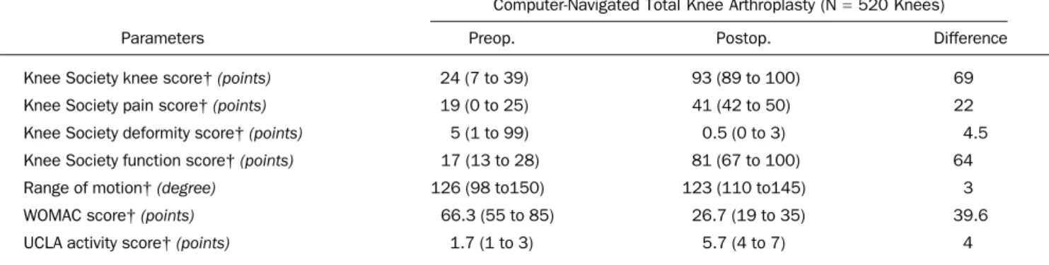
TABLE II Comparison of Knee Scores at Preoperative and Mean 10.8-Year Postoperative Evaluations* ä
Computer-Navigated Total Knee Arthroplasty (N= 520 Knees)
Parameters Preop. Postop. Difference
Knee Society knee score† (points) 24 (7 to 39) 93 (89 to 100) 69
Knee Society pain score† (points) 19 (0 to 25) 41 (42 to 50) 22
Knee Society deformity score† (points) 5 (1 to 99) 0.5 (0 to 3) 4.5
Knee Society function score† (points) 17 (13 to 28) 81 (67 to 100) 64
Range of motion† (degree) 126 (98 to150) 123 (110 to145) 3
WOMAC score† (points) 66.3 (55 to 85) 26.7 (19 to 35) 39.6
UCLA activity score† (points) 1.7 (1 to 3) 5.7 (4 to 7) 4
*Preop.= preoperative, Postop. = postoperative, CI = confidence interval, WOMAC = Western Ontario and McMaster Universities Osteoarthritis Index, UCLA= University of California, Los Angeles. †The values are given as the mean, with the range in parentheses. ‡Paired t test. §Chi-square test.
posterior-stabilized high-flexion fixed-bearing knee prosthesis (NexGen LPS-Flex; Zimmer, Warsaw, Indiana) with an all-polyethylene patellar component. We used the mobile-bearing prosthesis in some patients and the fixed-bearing prosthesis in others so that our results would be applicable to both designs. There was no significant difference, based on the numbers, between the two cohorts in terms of preoperative conditions, including the extent of the index disease, pain, functional disability, deformity, knee motion, bone loss, and prior surgical treatments.
Randomization between the use of computer-navigated and conventional total knee arthroplasty was determined from a sequential pool on the basis of a table of random numbers. Each of the 520 patients received a computer-navigated total knee arthroplasty on one side and a conventional total knee arthroplasty on the contralateral side. Of the 520 patients, 200 had the press-fit condylar posterior cruciate-retaining mobile-bearing prosthesis implanted in both knees, and 320 had the posterior-stabilized high-flexion fixed-bearing prosthesis implanted in both knees. There were 452 women and sixty-eight men with a mean age of sixty-eight years (range, forty-nine to eighty-eight years) at the time of the index arthroplasty. The mean body mass index was 27.8 kg/m2(range, 19.4 to 32.9 kg/m2). The mean duration of follow-up was 10.8 years (range, ten to twelve years) (Table I).
Surgical Technique
The procedure was carried out through a midline skin incision of 10 cm to 18 cm in length, with the knee extended and with use of a medial parapatellar ar-throtomy. In all of the conventional total knee arthroplasties, extramedullary instrumentation was used for the tibial component and intramedullary instru-mentation for the femoral side. The computer-assisted surgical navigation system (VectorVision CT-free knee; BrainLAB, Munich, Germany) had an optical tracking unit that detected reflecting marker spheres with the aid of an infrared camera. In all knees, femoral preparation was performed first. Ten millimeters of tibial bone was resected, as referenced from the least-involved tibial plateau, to achieve a surface perpendicular to the axis of the tibia in the coronal plane. A 3° to 5° posterior slope was prepared in the sagittal plane for the knees in the PFC Sigma CR-mobile-bearing group, and a 0° to 3° posterior slope was pre-pared for the knees in the NexGen LPS-Flex group. Nine millimeters of the distal part of the femur was resected. Anterior cortical reference was used for the anterior-posterior cut of the distal part of the femur. Femoral component rota-tion was determined with use of three reference axes: (1) the transepicondylar axis, (2) the midtrochlear (Whiteside) line12, and (3) 3° of external rotation relative to the posterior aspect of the condyles. Ligamentous balance was estab-lished, first with the knee in extension and then with the knee in flexion, with use of a tensor. All patellae were resurfaced with a polyethylene implant. All implants were cemented after pulsed lavage, drying, and pressurization of cement. All implants were fixed with use of CMW cement without antibiotics (DePuy).
Rehabilitation
Starting with the second postoperative day, patients used a continuous passive-motion machine for passive range-of-passive-motion exercises twice daily (thirty minutes per exercise period). On the same day, under the supervision of a physical therapist, they started active knee-motion exercise and began standing at the bedside or walking with crutches or a walker twice daily for thirty minutes per period. Patients used crutches or a walker with full weight-bearing for six weeks and a cane when needed thereafter.
Clinical Evaluation
Clinical evaluations were done at three months after the operation, at one year, and yearly thereafter. All clinical data at the time of each follow-up were re-corded and compiled by a research associate (S.-M.L.) who was not part of the operative team and was blinded to allocation. We obtained the Knee Society knee score13and the Western Ontario and McMaster Universities Osteoarthritis Index (WOMAC) score14separately for each knee.
Active knee motion, with the patient in the supine position, was de-termined with use of a standard (60 cm) clinical goniometer before surgery and at the time of the review. The patients were asked to extend the knees fully while lying in a supine position so that flexion contracture could be measured. The patients were told to bend the knees maximally while lying in a supine position so that flexion could be measured. Knee motion was measured for all patients on two occasions by two of the authors (J.-W.P. and J.-S.K.), both of whom were blinded to the type of implanted prosthesis. When the measured ranges of motion were different (i.e., a difference of >5°) between the two observers, the values were averaged and that number was reported. Interobserver agreement of the range of motion was 0.94 to 0.99. The level of activity was assessed with use of the University of California, Los Angeles (UCLA) activity score15.
Radiographic Evaluation
Anteroposterior hip-to-ankle radiographs (made with the patient standing as well as with the patient lying supine), lateral radiographs, and skyline patellar radiographs were made preoperatively and at each follow-up time and were assessed for the alignment of the limb (tibiofemoral angle), the position of the components, the posterior slope of the tibial components, the level of the joint line, and the presence and location of radiolucent lines at the bone-cement interface in accordance with the recommendations of the Knee Society13. Anteroposterior standing radiographs were used to determine the sequential change in the alignment of the limb as a result of polyethylene wear and/or loosening of the implant. Supine anteroposterior radiographs were used to determine the presence of a radiolucent line more precisely. Skyline patellar radiographs were examined to determine the presence of patellar tilt, sublux-ation, or dislocation. All radiographs were made under fluoroscopic guidance
TABLE II (continued)
Conventional Total Knee Arthroplasty (N= 520 Knees) P Value
Preop. Postop. Difference Difference of difference Preop. Postop.
26 (7 to 41) 92 (91 to 100) 66 3 (95% CI, 2.1 to 4.2) 0.719‡ 0.8531‡
22 (6 to 31) 42 (41 to 50) 20 2 (95% CI, 1.1 to 2.6) 0.912§ 0.832§
4 (6 to 15) 0.4 (0 to 4) 3.6 0.9 (95% CI, 0.5 to 1.3) 0.141‡ 0.134‡
19 (15 to 31) 83 (71 to 100) 64 0 0.831‡ 0.825‡
127 (90 to150) 125 (105 to140) 2 1 (95% CI, 1.1 to 2.6) 0.935‡ 0.918‡
65.8 (51 to 81) 28.1 (21 to 39) 37.7 1.9 (95% CI, 0.9 to 2.4) 0.912‡ 0.928‡
to control rotation of the knee. Radiographic data at the time of each follow-up were analyzed and recorded by a research associate (S.-M.L.) who was not part of the operative team. This assessment was blinded to technique allocation.
Computed Tomography Evaluation3
At the latest follow-up, all patients underwent a computed tomography (CT) scan with use of a multislice scanner (General Electric LightSpeed Plus; GE Medical Systems, Milwaukee, Wisconsin) to determine the rotational alignment of the component and osteolysis. The scan sequence was between 10 cm proximal to the superior pole of the patella and 10 cm distal to the tibial tuberosity and was made in contiguous 2.5 mm slices. Rotational alignment of the femoral component was determined by measuring the angle between the line joining the medial and lateral epicondyles of the femur and that joining the posterior margins of the femoral component. Rotational alignment of the tibial component was assessed by measuring the angle between the line connecting the tibial tuberosity anteriorly and the site of insertion of the posterior cruciate ligament posteriorly and the anteroposterior line passing through the center of the anterior and posterior margins of the tibial component. Osteolysis was defined as a nonlinear region of periprosthetic cancellous bone loss with delineable margins. A research asso-ciate (S.-M.L.) examined all CT scans.
Statistical Analysis
To minimize the chance of type-2 error and increase the power of our study, we assumed the difference in the Knee Society knee score to be 1.5 points with a power of 0.99, which revealed that a total of 468 patients would be needed in each group. We recruited approximately 10% more patients to account for possible dropouts. Intraobserver reliability was almost perfect for both the computer-navigated total knee arthroplasties and the conventional total knee arthroplasties. The value of kappa was 0.96 for the computer-navigated total knee arthroplasties and 0.94 for conventional total knee arthroplasties.
With the Bonferroni method16, the alpha level of each individual test was adjusted downward to ensure that the overall risk for a number of tests remained 0.05. For our study to reach significance, the alpha level needed to be <0.00129 after thirty-seven outcome measures.
The Kolmogorov-Smirnov test17was used to evaluate whether the axial alignment followed a normal (Gaussian) distribution, and the Levene test18was used to assess the homogeneity of variance (constant variance). The alignment of the limb and the duration of the operation were compared with use of an unpaired Student t test, with the assumption of homogeneity of variance used as appropriate. Box-and-whisker plots were used to compare the postoperative alignment of the limb with median quartile and interquartile ranges, and deviations were
compared with use of the nonparametric Mann-Whitney U test. For continuous variables and differences between two means, 95% confidence intervals (CIs) were calculated. Two-tailed values of p < 0.05 were considered to be significant. Survivorship analysis was performed to determine the cumulative rate of survival of the implant during the period of the study19. A comparative analysis was performed between an aligned group (i.e., in which the neutral mechanical axis was ± 3°) and a malaligned group (i.e., in which the neutral mechanical axis was >3°).
Source of Funding
There was no external funding source for this study. Results
Clinical Results
T
here was no difference in clinical function for patients who received computer-navigated total knee arthroplasty as com-pared with those who received conventional total knee arthro-plasty. The mean preoperative and postoperative Knee Society knee and functional scores in both groups were similar. Also, the preoperative and postoperative ranges of knee motion were similar. All clinical data showed no difference between groups at a mean of 10.8 years after the operation (Table II). With use of the Bonferroni method for multiple comparison correction, the mean operative and tourniquet times were significantly longer in the computer-navigated total knee arthroplasty group than in the conventional total knee arthroplasty group. The length of the incision, the in-traoperative blood loss, the duration and volume of drainage, and the transfusion volume were not significantly different (p > 0.05) (Table III).The Kolmogorov-Smirnov test revealed that the groups arose from the same population distributions (p = 0.05). The samples in our study had equal variances as determined with use of the Levene test.
Radiographic Results
There was no difference in the alignment of the components between the computer-navigated total knee arthroplasty group and the conventional total knee arthroplasty group. Radiographic
TABLE III Operative Data*
Parameters
Computer-Navigated Total Knee Arthroplasty (N= 520 Knees)
Conventional Total Knee
Arthroplasty (N= 520 Knees) P Value
Operative time (min) 88 (67 to 109) 76 (54 to 87) <0.001
Tourniquet time (min) 59 (45 to 82) 42 (31 to 61) <0.001
Mean length of incision (cm)
Extension 14.8 (13 to 17.8) 12.8 (10 to 14) 0.823
Flexion 16.3 (14 to 18.6) 13.9 (12 to 16.7) 0.894
Intraoperative blood loss (mL) 241 (71 to 530) 238.6 (96 to 596) 0.812
Drainage volume (mL) 761.8 (140 to 1280) 718.6 (67 to 1390) 0.519
Drainage duration (days) 3.9 (2 to 5) 3.1 (3 to 6) 0.176
Volume of transfusion (mL) 1678.4 (150 to 2670) 1785.5 (190 to 2540) 0.078
results were similar in both the computer-navigated and the conventional total knee arthroplasty groups with regard to the alignment of the knee and the position of the femoral and tibial components in the coronal and sagittal planes. If one assumes a tolerance level of 3°, the prevalence of outliers ranged from 9% to 13% for all parameters in the computer-navigated total knee arthroplasty group and from 9% to 15% in the conven-tional total knee arthroplasty group. These differences between the groups were not significant (p > 0.05) (Table IV). No knee in either group had osteolysis around the components (see Figs. E-2 and E-3 in Appendix).
Computed Tomographic Results
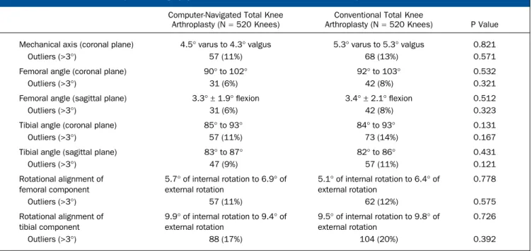
Results of three-dimensional CT evaluations were similar in both the computer-navigated total knee arthroplasty group and the conventional total knee arthroplasty group with regard to alignment of the knee and position of the femoral and tibial components in the coronal, sagittal, and axial planes (Table V). If one assumes a tolerance level of 3°, the prevalence of outliers was between 6% and 17% in the computer-navigated total knee arthroplasty group and between 8% and 20% in the conven-tional total knee arthroplasty group. These differences were not significant between the two groups (p > 0.05). Rotational
TABLE IV Radiographic Results at a Mean of 10.8 Years of Follow-up*
Computer-Navigated Total Knee Arthroplasty (N= 520)
Conventional Total Knee
Arthroplasty (N= 520) P Value Mechanical axis (coronal plane) 5.3° of varus to 4.8° of valgus alignment 5.1° of varus to 5.1° of valgus alignment 0.912
Outliers (>3°) 57 (11%) 67 (13%) 0.673
Femoral angle (coronal plane) 92°-101° 90°-103° 0.746
Outliers (>3°) 48 (9%) 53 (10%) 0.704
Femoral angle (sagittal plane) 2.1° ± 1.9° 2.8° ± 2.1° 0.132
Outliers (>3°) 31 (6%) 47 (9%) 0.231
Tibial angle (coronal plane) 86°-93° 84°-95° 0.121
Outliers (>3°) 57 (11%) 78 (15%) 0.133
Tibial angle (sagittal plane) 75°-93° 74°-91° 0.379
Outliers (>3°) 68 (13%) 78 (15%) 0.496
*Outlier values are given as the number of knees, with the percentage of the 520 total knees in parentheses.
TABLE V Three-Dimensional Computed Tomography Results at a Mean of 10.8 Years of Follow-up*
Computer-Navigated Total Knee Arthroplasty (N= 520 Knees)
Conventional Total Knee
Arthroplasty (N= 520 Knees) P Value Mechanical axis (coronal plane) 4.5° varus to 4.3° valgus 5.3° varus to 5.3° valgus 0.821
Outliers (>3°) 57 (11%) 68 (13%) 0.571
Femoral angle (coronal plane) 90° to 102° 92° to 103° 0.532
Outliers (>3°) 31 (6%) 42 (8%) 0.321
Femoral angle (sagittal plane) 3.3° ± 1.9° flexion 3.4° ± 2.1° flexion 0.512
Outliers (>3°) 31 (6%) 42 (8%) 0.323
Tibial angle (coronal plane) 85° to 93° 84° to 93° 0.131
Outliers (>3°) 57 (11%) 73 (14%) 0.167
Tibial angle (sagittal plane) 83° to 87° 82° to 86° 0.431
Outliers (>3°) 47 (9%) 57 (11%) 0.121 Rotational alignment of femoral component 5.7° of internal rotation to 6.9° of external rotation 5.1° of internal rotation to 6.4° of external rotation 0.778 Outliers (>3°) 57 (11%) 62 (12%) 0.575 Rotational alignment of tibial component 9.9° of internal rotation to 9.4° of external rotation 9.5° of internal rotation to 9.8° of external rotation 0.726 Outliers (>3°) 88 (17%) 104 (20%) 0.392
alignment of the femoral component was from 5.7° of internal rotation to 6.9° of external rotation for knees that underwent computer-navigated total knee arthroplasty and from 5.1° of internal rotation to 6.4° of external rotation for knees that had conventional total knee arthroplasty. The rotational alignment of the tibial component was from 9.9° of internal rotation to 9.4° of external rotation for the knees that underwent computer-navigated total knee arthroplasty and from 9.5° of internal ro-tation to 9.8° of external roro-tation for the knees that received conventional total knee arthroplasty. These differences were not significant between the two groups (p > 0.05). No knee in either group had osteolysis adjacent to the components. Complications
Six knees (1%) that received a NexGen LPS-Flex prosthesis (four knees with the computer-navigated technique and two knees with the conventional technique) were revised as a result of aseptic loosening of the femoral component. Four knees (1%) with a PFC Sigma CR-mobile-bearing prosthesis (two knees with the computer-navigated technique and two knees with the conventional technique) were revised for aseptic loosening of the tibial component.
Twenty-six knees (5%) had anterior femoral notching in the navigation group and six (1%) in the conventional group. Five knees (1%) in the navigation group had excessive resection of the tibia and required a tibial insert of 14 mm. The cause of anterior femoral notching and excessive tibial resection was not certain. We speculate that a registration error of computer navigation might have been the cause. Two knees (0.4%) in the navigation group had a deep wound infection; both knees were managed with open debridements followed by intrave-nous antibiotics for six weeks with no further evidence of infection.
Survival of the Implants
Survivorship of the implants at 10.8 years after the operation was not significantly different. Kaplan-Meier survival analysis, with revision used as the end point for failure, revealed a 10.8-year rate of survival of 98.8% (95% CI, 0.96 to 1.00) in the computer-navigated total knee arthroplasty group and 99.2% (95% CI, 0.96 to 1.00) in the conventional total knee arthroplasty group. Survival of the Implants in the Aligned
and Malaligned Group
A deviation of >3° from the mechanical axis of the lower limb did not increase the rate of aseptic loosening and failure. The prevalence of revision due to aseptic loosening at 10.8 years after the operation was 0.9% (one of 124 knees) in the mal-aligned group (>3° from neutral axis) and 1.0% (nine of 916 knees) in the aligned group (Fisher exact test, p = 0.38).
Discussion
P
roper alignment of the prosthesis during total knee arthro-plasty is critical in maximizing implant survival9,20-22. It has been claimed that computer-navigated total knee arthroplasty allows a higher degree of accuracy in component alignment2-4
.
Nevertheless, we are aware of no literature that documents whether or not radiographic improvement leads to an im-provement in clinical and functional scores or implant survival at midterm and long term follow-up. We found that computer-navigated total knee arthroplasty did not improve clinical func-tion or the alignment and survivorship of the components as compared with conventional total knee arthroplasty.
Several studies have shown that computer-navigated total knee arthroplasty improved the overall accuracy of positioning of the femoral and tibial components2-4
. Choong et al.23 con-cluded that the use of computer-navigated total knee arthro-plasty resulted in greater accuracy of implant alignment, better knee function, and improved quality of life than that achieved with conventional total knee arthroplasty. By contrast, studies comparing the clinical and functional results of total knee ar-throplasty performed with or without computer navigation have found no differences, even in the short term24-29
. In the current study, Knee Society scores (knee and functional scores) and WOMAC scores were not significantly different between the two groups at 10.8 years after the operation. The absence of severe malalignment (‡6°) in the conventional total knee arthroplasty group may explain these findings.
In the current study, the postoperative mechanical axis of the limb was not significantly better in the patients who had computer-navigated total knee arthroplasty than it was in those who had conventional total knee arthroplasty. This result is consistent with the results of Bauwens et al.7
, Mielke et al.30 , and Jenny and Boeri31
, who found no significant difference in the postoperative mechanical axis alignment of the limb between patients who underwent conventional or computer-navigated total knee arthroplasty. By contrast, our findings were not in agreement with the results of the many investigators who have demonstrated that, in comparison with conventional total knee arthroplasty, computer-navigated total knee arthroplasty is as-sociated with more accurate alignment on radiographs4,32-34
. However, the improvement in accuracy through computer-navigated total knee arthroplasty is a few degrees, which is within the margin of error produced by projection-related errors in standing radiographs35
.
With regard to the effect of postoperative alignment of the mechanical axis (measured with use of long-leg radiographs and three-dimensional CT scans) on the midterm risk of revision surgery, our results demonstrate only a weak relationship be-tween alignment and the need for revision surgery. Survival analysis reveals a tendency toward improved implant survival with accurate alignment, but the validity of this result is limited by the lack of significance. This may be explained by the rela-tively small number of total knee arthroplasties in the malaligned group compared with the numbers in previous reports20,36
. Our study, along with other reports, suggests that the relationship between the postoperative mechanical axis and implant survival is marginal21,22
. Although the neutral mechanical axis is a valu-able intraoperative target, its achievement does not necessarily confer satisfactory kinematics and implant survival29,37
.
Alignment in the coronal plane does not guarantee the accurate position of each component in flexion and extension,
valgus and varus, or balanced tibiofemoral rotation. The effect on implant survival of accurate positioning of each individual component in six degrees of freedom is not clearly established. There is anecdotal evidence that early mechanical failure is more likely when there is a mismatch of the femoral and tibial com-ponent in rotation38
. However, reliable evidence of the effect of rotational alignment on implant survival is limited because intraoperative and postoperative measurement techniques are often inaccurate and the optimal rotational alignment target has not been defined. However, our study and other studies2,26,27,39 do not demonstrate any difference in rotational alignment with computer-navigated or conventional total knee arthroplasties. We used the same landmarks for rotational alignment of the femoral and tibial components in both computer-navigated and conventional total knee arthroplasties. Although the trend was toward a greater outlier for each category in the conventional total knee arthroplasty group, there was no significant difference in rotational alignment between the computer-navigated or conventional total knee arthroplasty groups.
There are several strengths of this study. First, drawing a large series of patients from a single surgeon and single center allows specific coordination of surgical technique or implants used in the study. Second, the follow-up was long enough to determine functional outcome, survivorship of the implants, and prevalence of osteolysis and loosening. Third, data regarding the activity level of the patients were collected and can be analyzed as a risk factor for failure. Finally, three-dimensional CT was ob-tained to measure more accurately the mechanical axis of the limb, the rotational alignment of the component, and osteolysis. There are some limitations of this study. First, we did not perform a side-by-side comparison of each patient to deter-mine whether or not the alignment of the conventional total knee arthroplasty, if performed after the computer-navigated total knee arthroplasty, was improved by feedback gained by the surgeon during the performance of the first surgery. Sec-ond, the fact that all arthroplasties were performed by an
ex-perienced surgeon may have limited the number of outliers. Third, while our data support the contention that, at 10.8 years after the procedure, computer navigation does not offer an advantage over conventional total knee arthroplasty relative to survivorship and alignment, the data do not support an ex-trapolation past this time frame. Finally, it is frequently difficult for a patient who has undergone a bilateral total knee arthro-plasty procedure to determine which knee is functioning better than the other. Therefore, the WOMAC function scores should be interpreted with caution because it is difficult for patients to attribute functional status to a particular knee.
Our data demonstrated that there was no difference in clinical function, alignment, and survivorship of the com-ponents between the computer-navigated and conventional total knee arthroplasties. In our study, the effect of computer-navigated total knee arthroplasty compared with conventional total knee arthroplasty on long-term implant survival remains unproven.
Appendix
Figures showing the postoperative radiographic results in knees after conventional total knee arthroplasty or computer-navigated total knee arthroplasty are available with the online version of this article as a data supplement at jbjs.org.n
NOTE: The authors thank Sang-Mi Lee, BA, for her analysis of clinical, radiographic, and CT data.
Young-Hoo Kim, MD Jang-Won Park, MD Jun-Shik Kim, MD
The Joint Replacement Center at Ewha Womans University MokDong Hospital, 911-1,
MokDong, YangChun-Ku, Seoul, Republic of Korea (158-710).
E-mail address for Y.-H. Kim: [email protected]
References
1. Laskin RS, Beksacx B. Computer-assisted navigation in TKA: where we are and where we are going. Clin Orthop Relat Res. 2006 Nov;452:127-31.
2. St¨ockl B, Nogler M, Rosiek R, Fischer M, Krismer M, Kessler O. Navigation im-proves accuracy of rotational alignment in total knee arthroplasty. Clin Orthop Relat Res. 2004 Sep;(426):180-6.
3. Matziolis G, Krocker D, Weiss U, Tohtz S, Perka C. A prospective, randomized study of computer-assisted and conventional total knee arthroplasty. Three-dimensional evaluation of implant alignment and rotation. J Bone Joint Surg Am. 2007 Feb;89(2):236-43.
4. B¨athis H, Perlick L, Tingart M, L¨uring C, Zurakowski D, Grifka J. Alignment in total knee arthroplasty. A comparison of computer-assisted surgery with the conventional technique. J Bone Joint Surg Br. 2004 Jul;86(5):682-7.
5. Ensini A, Catani F, Leardini A, Romagnoli M, Giannini S. Alignments and clinical results in conventional and navigated total knee arthroplasty. Clin Orthop Relat Res. 2007 Apr;457:156-62.
6. Chauhan SK, Scott RG, Breidahl W, Beaver RJ. Computer-assisted knee arthro-plasty versus a conventional jig-based technique. A randomised, prospective trial. J Bone Joint Surg Br. 2004 Apr;86(3):372-7.
7. Bauwens K, Matthes G, Wich M, Gebhard F, Hanson B, Ekkernkamp A, Stengel D. Navigated total knee replacement. A meta-analysis. J Bone Joint Surg Am. 2007 Feb;89(2):261-9.
8. Jeffery RS, Morris RW, Denham RA. Coronal alignment after total knee replace-ment. J Bone Joint Surg Br. 1991 Sep;73(5):709-14.
9. Lotke PA, Ecker ML. Influence of positioning of prosthesis in total knee replace-ment. J Bone Joint Surg Am. 1977 Jan;59(1):77-9.
10. Hvid I, Nielsen S. Total condylar knee arthroplasty. Prosthetic component po-sitioning and radiolucent lines. Acta Orthop Scand. 1984 Apr;55(2):160-5. 11. Insall JN, Scuderi GR, Komistek RD, Math K, Dennis DA, Anderson DT. Corre-lation between condylar lift-off and femoral component alignment. Clin Orthop Relat Res. 2002 Oct;(403):143-52.
12. Whiteside LA, Arima J. The anteroposterior axis for femoral rotational align-ment in valgus total knee arthroplasty. Clin Orthop Relat Res. 1995 Dec;(321): 168-72.
13. Insall JN, Dorr LD, Scott RD, Scott WN. Rationale of the Knee Society clinical rating system. Clin Orthop Relat Res. 1989 Nov;(248):13-4.
14. Bellamy N, Buchanan WW, Goldsmith CH, Campbell J, Stitt LW. Validation study of WOMAC: a health status instrument for measuring clinically important patient relevant outcomes to antirheumatic drug therapy in patients with osteoarthritis of the hip or knee. J Rheumatol. 1988 Dec;15(12):1833-40.
15. Zahiri CA, Schmalzried TP, Szuszczewicz ES, Amstutz HC. Assessing activity in joint replacement patients. J Arthroplasty. 1998 Dec;13(8):890-5.
16. Rosner B. Fundamentals of biostatistics. 5th ed. Pacific Grove, CA: Duxbury Press; 2000. p 527-30.
17. Riffenburgh RH. Tests on the distribution shape of continuous data. In: Riffenburgh RH, ed. Statistics in medicine. Amsterdam: Elsevier; 2006. p 370-80.
18. National Institute of Standards and Technology. Statistical Engineering Divi-sion. Levene test. Dataplot. http://www.itl.nist.gov/div898/software/dataplot/ refman1/auxillar/levetest.htm. Accessed 2008 Sept 12.
19. Kaplan EL, Meier P. Nonparametric estimation from incomplete observations. J Am Statist Assoc. 1958;53:457-81.
20. Ritter MA, Faris PM, Keating EM, Meding JB. Postoperative alignment of total knee replacement. Its effect on survival. Clin Orthop Relat Res. 1994 Feb; (299):153-6.
21. Parratte S, Pagnano MW, Trousdale RT, Berry DJ. Effect of postoperative me-chanical axis alignment on the fifteen-year survival of modern, cemented total knee replacements. J Bone Joint Surg Am. 2010 Sep 15;92(12):2143-9.
22. Bonner TJ, Eardley WG, Patterson P, Gregg PJ. The effect of post-operative mechanical axis alignment on the survival of primary total knee replacements after a follow-up of 15 years. J Bone Joint Surg Br. 2011 Sep;93(9): 1217-22.
23. Choong PF, Dowsey MM, Stoney JD. Does accurate anatomical alignment result in better function and quality of life? Comparing conventional and computer-assisted total knee arthroplasty. J Arthroplasty. 2009 Jun;24(4):560-9. Epub 2008 May 19.
24. Kamat YD, Aurakzai KM, Adhikari AR, Matthews D, Kalairajah Y, Field RE. Does computer navigation in total knee arthroplasty improve patient out-come at midterm follow-up? Int Orthop. 2009 Dec;33(6):1567-70. Epub 2008 Nov 26.
25. Seon JK, Park SJ, Lee KB, Li G, Kozanek M, Song EK. Functional comparison of total knee arthroplasty performed with and without a navigation system. Int Orthop. 2009 Aug;33(4):987-90. Epub 2008 Jun 28.
26. Spencer JM, Chauhan SK, Sloan K, Taylor A, Beaver RJ. Computer navigation versus conventional total knee replacement: no difference in functional results at two years. J Bone Joint Surg Br. 2007 Apr;89(4):477-80.
27. Kim YH, Kim JS, Choi Y, Kwon OR. Computer-assisted surgical navigation does not improve the alignment and orientation of the components in total knee arthro-plasty. J Bone Joint Surg Am. 2009 Jan;91(1):14-9.
28. Kim YH, Kim JS, Yoon SH. Alignment and orientation of the components in total knee replacement with and without navigation support: a prospective, randomised study. J Bone Joint Surg Br. 2007 Apr;89(4):471-6.
29. Hern´andez-Vaquero D, Suarez-Vazquez A, Iglesias-Fernandez S. Can computer assistance improve the clinical and functional scores in total knee arthroplasty? Clin Orthop Relat Res. 2011 Dec;469(12):3436-42. Epub 2011 Aug 27. 30. Mielke RK, Clemens U, Jens JH, Kershally S. [Navigation in knee endopros-thesis implantation—preliminary experiences and prospective comparative study with conventional implantation technique]. Z Orthop Ihre Grenzgeb. 2001 Mar-Apr; 139(2):109-16. German.
31. Jenny JY, Boeri C. Computer-assisted implantation of total knee prostheses: a case-control comparative study with classical instrumentation. Comput Aided Surg. 2001;6(4):217-20.
32. Haaker RG, Stockheim M, Kamp M, Proff G, Breitenfelder J, Ottersbach A. Computer-assisted navigation increases precision of component placement in total knee arthroplasty. Clin Orthop Relat Res. 2005 Apr;(433):152-9.
33. Sparmann M, Wolke B, Czupalla H, Banzer D, Zink A. Positioning of total knee arthroplasty with and without navigation support. A prospective, randomised study. J Bone Joint Surg Br. 2003 Aug;85(6):830-5.
34. Stulberg SD, Loan P, Sarin V. Computer-assisted navigation in total knee re-placement: results of an initial experience in thirty-five patients. J Bone Joint Surg Am. 2002;84-A Suppl 2:90-8.
35. Krackow KA, Pepe CL, Galloway EJ. A mathematical analysis of the effect of flexion and rotation on apparent varus/valgus alignment at the knee. Orthopedics. 1990 Aug;13(8):861-8.
36. Vince KG, Insall JN, Kelly MA. The total condylar prosthesis. 10- to 12-year results of a cemented knee replacement. J Bone Joint Surg Br. 1989 Nov;71(5):793-7. 37. Kalairajah Y, Simpson D, Cossey AJ, Verrall GM, Spriggins AJ. Blood loss after total knee replacement: effects of computer-assisted surgery. J Bone Joint Surg Br. 2005 Nov;87(11):1480-2.
38. Sikorski JM. Alignment in total knee replacement. J Bone Joint Surg Br. 2008 Sep;90(9):1121-7.
39. Oswald MH, Jakob RP, Schneider E, Hoogewoud HM. Radiological analysis of normal axial alignment of femur and tibia in view of total knee arthroplasty. J Arthroplasty. 1993 Aug;8(4):419-26.