저작자표시-비영리-변경금지 2.0 대한민국 이용자는 아래의 조건을 따르는 경우에 한하여 자유롭게 l 이 저작물을 복제, 배포, 전송, 전시, 공연 및 방송할 수 있습니다. 다음과 같은 조건을 따라야 합니다: l 귀하는, 이 저작물의 재이용이나 배포의 경우, 이 저작물에 적용된 이용허락조건 을 명확하게 나타내어야 합니다. l 저작권자로부터 별도의 허가를 받으면 이러한 조건들은 적용되지 않습니다. 저작권법에 따른 이용자의 권리는 위의 내용에 의하여 영향을 받지 않습니다. 이것은 이용허락규약(Legal Code)을 이해하기 쉽게 요약한 것입니다. Disclaimer 저작자표시. 귀하는 원저작자를 표시하여야 합니다. 비영리. 귀하는 이 저작물을 영리 목적으로 이용할 수 없습니다. 변경금지. 귀하는 이 저작물을 개작, 변형 또는 가공할 수 없습니다.
A Thesis for the Degree of Doctor of Philosophy
Characterization and Attenuation of
Regulatory Network of Vibrio vulnificus rtxA
Encoding a MARTX Toxin
MARTX 독소를 발현하는 패혈증 비브리오균
rtxA
유전자 조절 기전의 특성 규명 및 제어 연구
August, 2020
Zee-Won Lee
Department of Agricultural Biotechnology
The Graduate School
Characterization and Attenuation of
Regulatory Network of Vibrio vulnificus rtxA
Encoding a MARTX Toxin
MARTX 독소를 발현하는 패혈증 비브리오균
rtxA
유전자 조절 기전의 특성 규명 및 제어 연구
지도교수 최 상 호
이 논문을 농학박사학위논문으로 제출함
2020 년 5 월
서울대학교 대학원
농생명공학부
이 지 원
이지원의 박사학위논문을 인준함
2020 년 7 월
위 원 장 ___________________(인)
부위원장 ___________________(인)
위 원 ___________________(인)
위 원 ___________________(인)
위 원 ___________________(인)
I
Abstract
Characterization and Attenuation of
Regulatory Network of Vibrio vulnificus rtxA
Encoding a MARTX Toxin
Zee-Won Lee
Department of Agricultural Biotechnology
The Graduate School
Seoul National University
Bacterial pathogens have evolved the ability to survive and develop diseases in several different environments within the host. The ability requires the production of various virulence factors whose expressions are coordinately controlled by regulatory networks in response to environmental changes. The opportunistic human pathogen Vibrio vulnificus can cause food-borne diseases from gastroenteritis to life-threatening septicemia. Among a wide array of virulence factors produced by V. vulnificus, a multifunctional-autoprocessing repeats-in-toxin (MARTX) toxin RtxA encoded by the rtxA gene plays an essential role in the virulence of the pathogen. It has been previously reported that the expression of rtxA is negatively and positively regulated by direct binding of H-NS and HlyU to the rtxA promoter, PrtxA,
II
respectively. In the present study, I have further examined additional regulatory proteins as well as environmental signals involved in the rtxA expression and identified a small-molecule inhibitor that attenuates the virulence of V. vulnificus. As a result, a leucine-responsive regulatory protein (Lrp) was found as a positive regulator of rtxA. Electrophoretic mobility shift and DNase I protection assays revealed that Lrp activates the rtxA expression by binding directly and specifically to PrtxA. Notably, DNase I cleavage of the PrtxA regulatory region showed phased hypersensitivity, suggesting that Lrp probably induces the DNA bending in PrtxA. Lrp activates rtxA in an independent manner with H-NS and HlyU, and leucine inhibits the binding of Lrp to PrtxA and thus decreases the Lrp-mediated activation. Moreover, a cyclic AMP receptor protein (CRP) acts as a negative regulator of the rtxA transcription, and exogenous glucose relieves the CRP-mediated repression. Interestingly, biochemical and mutational analyses demonstrated that CRP binds directly and specifically to the upstream regions of PrtxA, which presumably changes the DNA conformation in PrtxA and represses rtxA. Furthermore, CRP represses the expressions of lrp and hlyU by directly binding to their upstream regions, forming coherent feedforward loops with Lrp and HlyU. Taken together, a regulatory network comprising CRP, Lrp, H-NS, and HlyU coordinates the expression of rtxA in response to changes in host environmental signals such as leucine and glucose. This collaborative regulation will contribute to the precise expression of rtxA during the pathogenesis of V. vulnificus.
III
My next concern was about new approaches called anti-virulence strategies that target virulence of bacterial pathogens in an attempt to control the virulence of V. vulnificus. Anti-virulence strategies have the advantage of less selective pressure for inducing resistance than conventional strategies that target viability of the pathogens. Therefore, I performed a high-throughput screening of the small-molecule library containing 8,385 compounds to inhibit HlyU, a transcriptional activator essential for the expression of V. vulnificus rtxA. A small molecule [N-(4-oxo-4H-thieno[3,4-c]chromen-3-yl)-3-phenylprop-2-ynamide] was identified as an inhibitor of the HlyU activity and named CM14. CM14 reduces HlyU-dependent virulence gene expression in V. vulnificus, but does not suppress the bacterial growth or cause host cell death. Treatment of CM14 decreases hemolysis of human erythrocytes and impedes host cell rounding and lysis caused by V. vulnificus. Remarkably, co-administration of CM14 improves the survival of mice infected with V. vulnificus by alleviating hepatic and renal dysfunction and systemic inflammation. As revealed by biochemical, mass spectrometric, and mutational analyses, CM14 covalently modifies the Cys30 residue of HlyU to prevent the protein from binding to the target DNA. Based on these results, a possible molecular mechanism is proposed for the covalent modification of HlyU by CM14. Because HlyU is a conserved transcriptional activator of virulence genes in Vibrio species, CM14 is also capable of reducing the expressions of multiple virulence genes in other Vibrio species and attenuating their virulence-related phenotypes. The combined results suggest that
IV
this small-molecule could be an anti-virulence agent against HlyU-harboring pathogenic Vibrio species with a low possibility of developing resistance.
Keywords: Vibrio vulnificus, MARTX toxin, RtxA, Virulence gene
regulation, CRP, Lrp, HlyU, Small-molecule inhibitor, Anti-virulence
agent
V
Contents
Abstract ... I Contents ... V List of Figures ... VIII List of Tables ... X
Chapter I. ... 1
I-1. Vibrio vulnificus ... 2
I-1-1. Disease caused by V. vulnificus ... 3
I-1-2. Virulence factors of V. vulnificus ... 4
I-1-3. Regulation of virulence genes in V. vulnificus ... 19
I-2. Objective of this study ... 26
Chapter II... 28
II-1. Introduction ... 29
II-2. Materials and Methods ... 34
II-2-1. Strains, plasmids, and culture conditions ... 34
II-2-2. Generation and complementation of deletion mutants ... 38
II-2-3. RNA purification and transcript analysis ... 45
II-2-4. Western blot analysis... 46
II-2-5. Protein purification ... 46
II-2-6. EMSA and DNase I protection assay ... 47
II-2-7. Construction of an rtxA-lacZ transcriptional fusion reporter and β-galactosidase activity assay ... 49
VI
II-2-8. Site-directed mutagenesis of CRP-binding sequences ... 49
II-2-9. Data analyses ... 50
II-3. Results ... 51
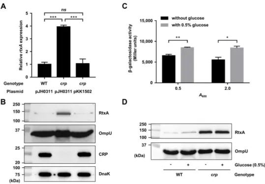
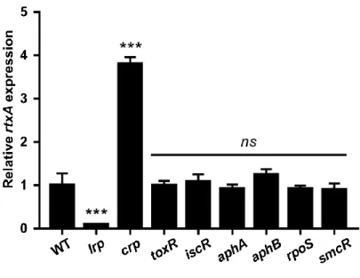
II-3-1. Lrp and CRP affect the rtxA transcription ... 51
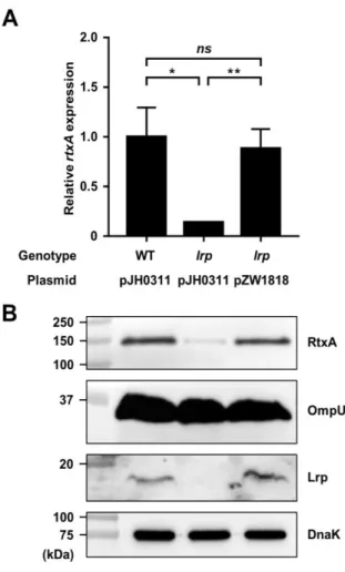
II-3-2. Lrp activates the rtxA expression by directly binding to PrtxA ... 53
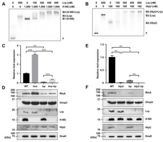
II-3-3. Lrp activates rtxA in an independent manner with H-NS and HlyU ... 59
II-3-4. Leucine inhibits Lrp binding to PrtxA and activation of rtxA ... 65
II-3-5. CRP represses but glucose derepresses the rtxA expression ... 69
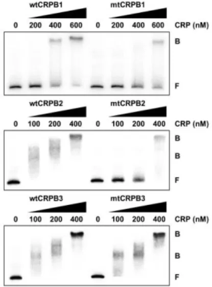
II-3-6. CRP represses rtxA by directly binding to the upstream regions of PrtxA ... 72
II-3-7. Mutational analyses of the CRP-binding sequences of PrtxA ... 76
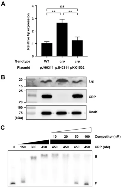
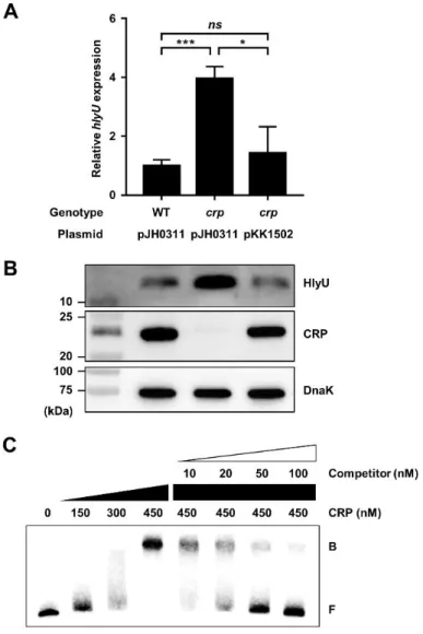
II-3-8. CRP directly represses the lrp and hlyU expression ... 80
II-4. Discussion ... 87
Chapter III. ... 94
III-1. Introduction ... 95
III-2. Materials and Methods ... 99
III-2-1. Strains, plasmids, and culture conditions ... 99
III-2-2. Transcriptome analyses ... 100
III-2-3. Construction of an E. coli reporter strain and HTS ... 102
III-2-4. Verification, structural confirmation, and determination of half maximal effective concentration (EC50) of CM14... 104
III-2-5. Western blot and transcript analyses ... 106
VII
III-2-7. Mouse infection assays ... 109
III-2-8. Protein purification, site-directed mutagenesis, and EMSA ... 111
III-2-9. Mass spectrometric analysis of the HlyU modification ... 113
III-2-10. Crystallization, structure determination, and refinement ... 115
III-2-11. Data analyses ... 117
III-2-12. Data availability ... 117
III-3. Results ... 118
III-3-1. Identification of CM14 as an inhibitor of HlyU activity ... 118
III-3-2. CM14 reduces the HlyU-dependent virulence gene expression in vitro ... 125
III-3-3. CM14 attenuates the virulence of V. vulnificus ex vivo ... 126
III-3-4. CM14 impedes the pathogenesis of V. vulnificus in mice ... 129
III-3-5. CM14 inhibits the HlyU binding to its target promoter DNA .. 134
III-3-6. CM14 leads to chemical modification of HlyU ... 136
III-3-7. CM14 exhibits anti-virulence effects against other Vibrio species ... 143
III-4. Discussion ... 149
Chapter IV. ... 159
References ... 163
VIII
List of Figures
Figure I-1. Regulation of virulence genes in V. vulnificus...25
Figure II-1. Expression of rtxA in V. vulnificus with different genetic background...52
Figure II-2. Effects of the lrp mutation on the rtxA expression...55
Figure II-3. Specific bindings of Lrp to the PrtxA regulatory region...58
Figure II-4. Sequence analysis of the PrtxA regulatory region...61
Figure II-5. Lrp activates the rtxA expression independently of H-NS and HlyU...63
Figure II-6. Effects of L-leucine on Lrp binding to and activation of PrtxA...66
Figure II-7. Effects of various amino acids on Lrp binding to PrtxA...68
Figure II-8. Effects of the crp mutation and glucose on the rtxA expression...70
Figure II-9. Specific bindings of CRP to the PrtxA regulatory region...74
Figure II-10. Mutations in the CRP-binding sequences affect the binding of CRP to and activity of PrtxA...78
Figure II-11. CRP represses the lrp expression by directly binding to the upstream region...82
Figure II-12. CRP represses the hlyU expression by directly binding to the upstream region...84
Figure II-13. Expression of crp is not affected by hns, lrp, or hlyU mutations...86 Figure II-14. A regulatory network controlling the rtxA expression and a
IX
...92
Figure III-1. Genes regulated by HlyU...120
Figure III-2. High-throughput screening for HlyU inhibitors...122
Figure III-3. CM14 inhibits the HlyU activity without affecting V. vulnificus growth...123
Figure III-4. Effects of CM14 on virulence-related phenotypes of V. vulnificus...127
Figure III-5. Effects of CM14 on the survival, pathophysiological changes, and inflammatory responsesofmice infectedwithV.vulnificus...132
Figure III-6. EMSA of HlyU and the rtxA regulatory DNA complexes...135
Figure III-7. Mass spectrum of HlyU and HlyUC30S in the presence of CM14...140
Figure III-8. Chemical modification of the Cys30 residue of HlyU by CM14...141
Figure III-9. Sequence alignment of HlyU from different Vibrio species...145
Figure III-10. CM14 is effective in attenuating virulence of other Vibrio species...146
Figure III-11. Effects of CM14 on the growth of other Vibrio species...148
Figure III-12. Proposed molecular mechanism underlying the CM14-mediated inhibition ofHlyUbindingtotargetDNA...155
Figure III-13. Effects of CM14 on HlyU regulon expression...156
Figure III-14. Effects of CM14 treatment after infection of V. vulnificus to host cells...158
X
List of Tables
Table I-1. List of V. vulnificus virulence factors...17
Table II-1. Bacterial strains and plasmids used in this study...35
Table II-2. Oligonucleotides used in this study...40
Table III-1. Small molecule screening information...103
1
Chapter I.
2
I-1. Vibrio vulnificus
Vibrio vulnificus is a gram-negative, motile, and curved-rod shaped bacterium with a single polar flagellum, which belongs to Vibrio genus in Vibrionaceae. V. vulnificus is distinguished from other members of the Vibrio genus in its ability to ferment lactose (Baumman et al., 1981; Strom and Paranjpye, 2000). The bacterium inhabits estuarine and marine environments around the world and proliferates in areas or seasons where water temperature exceeds 18°C and salinity is between 15 to 25 parts per thousand (ppt) (Horseman and Surani, 2011). However, it has been reported that salinities greater than 30 ppt negatively affect the survival of V. vulnificus regardless of water temperature (Motes et al., 1998). Moreover, at temperatures below 10°C, V. vulnificus enters a viable but non-culturable (VBNC) state, in which it becomes dormant and fails to grow even in the rich medium. The bacterium is resuscitated from the VNBC state by a gradual increase in temperature in a nutrient-free medium (Oliver, 2005a).
V. vulnificus can naturally exist in a free-living state or be associated with zooplankton and other aquatic biological flora. When V. vulnificus is taken up by filter-feeding mollusks such as oysters, clams, and mussels, the bacterium becomes concentrated in their gut and other tissues. V. vulnificus can also be found in the intestines of various fish species that consume the plankton and mollusks probably containing the bacterium. Consequently, these marine organisms such as oysters and
3
fish may serve as an environmental reservoir or source for the transmission of V. vulnificus (DePaola et al., 1994; Strom and Paranjpye, 2000).
I-1-1. Disease caused by V. vulnificus
V. vulnificus is an opportunistic human pathogen capable of causing foodborne diseases from gastroenteritis to primary septicemia (Baker-Austin and Oliver, 2018). Consumption of contaminated seafood may result in V. vulnificus infection, with the clinical symptoms including fever, chills, nausea, and abdominal pain. V. vulnificus infection is characterized by rapid onset of symptoms and can progress to fatal systemic infection within a few days, and the mortality rate has been reported to exceed 50% in primary septicemia. People at high risk of V. vulnificus infection involve males, older persons, and individuals with underlying conditions such as chronic liver diseases and compromised immune systems (Jones and Oliver, 2009; Oliver, 2015; Horseman and Surani, 2011). In addition, infection by V. vulnificus may also result from exposing open wound to water containing the pathogen during swimming, fishing, or seafood handling. These wound infections develop swelling, erythema, and cellulitis along with fever and chills, which can progress necrotizing fasciitis at the infection site. Compared to septicemia, wound infections result in a lower mortality rate of about 25%, and are restricted to the primary infection site without spreading to other areas of body (Oliver, 2005b; Horseman and Surani, 2011). Furthermore, the underlying conditions that can cause primary septicemia in the
4
ingestion cases do not appear to be a prerequisite for the wound infections (Gulig et al., 2005; Oliver, 2015).
In South Korea, V. vulnificus sepsis is classified as the Group 3 legal infectious disease, which is required to be reported within 24 hours in case of an outbreak for the monitoring. In the last 8 years (from 2011 to 2018), a total number of 418 infections by V. vulnificus has been reported, and 203 of them (49%) were dead
(Korea Centers for Disease Control and Prevention, KCDC;
http://www.cdc.go.kr/npt). In the United States, V. vulnificus accounts for over 95% of seafood-related death and has the highest mortality rate among foodborne pathogens (Baker-Austin and Oliver, 2018). Recently, the geographical distribution of V. vulnificus has expanded due to global warming and rising seawater temperature, which may increase the incidence of V. vulnificus infections (Oliver, 2015; Phillips and Satchell, 2017).
I-1-2. Virulence factors of V. vulnificus
In an effort to understand the pathogenesis of V. vulnificus infection, considerable works have been conducted to identify and characterize the virulence factors of the pathogen (Jones and Oliver, 2009). For this purpose, various approaches have been developed, including in vivo-induced antigen technology (IVIAT), in vivo expression technology (IVET), and random transposon mutagenesis (RTM). These studies successfully achieved the list of virulence genes, such as pyrH, hlyU, tolC, wbpP,
5
and rtxA, responsible for the cytotoxicity of V. vulnificus to host cells (Kim et al., 2003; Park et al., 2006b; Lee et al., 2007a; Lee et al., 2007c). In addition, proteomic analysis has led to identification and characterization of multiple genes, purH, trpD, tsaA, and groEL2, which are induced upon exposure to human epithelial INT-407 cells (Oh et al., 2008). Transcriptome analyses, such as microarray and RNA sequencing, have also identified a number of genes, including vvhA and plpA, which are differentially or preferentially expressed in in vivo-like conditions (Kim et al., 2011b; Bisharat et al., 2013; Jang et al., 2017; Choi et al., 2020). More recently, signature-tagged mutagenesis (STM) has been applied to search the neuC, ask, flgK, vuuA, and mukB genes that are active in vivo or are required for the wound infection (Yamamoto et al., 2015; Yamazaki et al., 2019). Among these, the virulence factors that have been well characterized in the pathogenesis of V. vulnificus will be further described in the following sections, as classified by capsular polysaccharide, adhesion factors, iron uptake systems, and exotoxins.
Capsular polysaccharide (CPS)
V. vulnificus produces a firmly linked form of extracellular polysaccharide capsule on its cell surface, which is called capsular polysaccharide (CPS) (Strom and Paranjpye, 2000; Chatzidaki-Livanis et al., 2006). The presence of CPS is correlated with colony morphology of V. vulnificus; encapsulated strains form opaque colonies, while colonies that undergo phase variation form translucent colonies with reduced
6
amount of CPS (Wright et al., 1999). Encapsulation by CPS protects V. vulnificus by conferring resistance to bactericidal effects of serum and phagocytosis by macrophages (Strom and Paranjpye, 2000; Williams et al., 2014). Therefore, encapsulated V. vulnificus cells are more slowly cleared out from the bloodstream and more invasive to subcutaneous tissue than unencapsulated cells in the host (Yoshida et al., 1985). Indeed, a 50% lethal dose (LD50) of the encapsulated cells is
lower than that of the unencapsulated cells in a mouse model (Simpson et al., 1987). Inactivation of the wbpP and wza genes related to the CPS biosynthesis and transport, respectively, reduces CPS expression and attenuates virulence of V. vulnificus (Park et al., 2006b; Wright et al., 2001).
Adhesion factors
Adhesion to host cell surfaces is an important step in the early stage of bacterial infection. In addition to CPS, surface structures, such as pili and flagella, and outer membrane proteins assist in the adhesion of V. vulnificus to host cells (Strom and Paranjpye, 2000; Srivastava et al., 2009).
Pili. Pili are adhesive hair-like appendages composed of a scaffold-like rod and an
adhesin attached upon the scaffold, which protrude from the surface of bacteria (Pizarro-Cerda and Cossart, 2006). In V. vulnificus, piliation is correlated with adhesion to human epithelial cells (Gander and LaRocco, 1989), and mutations in
7
genes encoding pili components decrease attachment to the epithelial cells and virulence in mice (Paranjpye et al., 1998; Paranjpye and Strom, 2005). Recently, thin fimbrial projections from the surface of V. vulnificus, called Flp pili encoded by the tad operons, are identified as another adhesion factors responsible for invasion of host tissue, survival in the blood, and resistance to complement system (Duong-Nu et al., 2019).
GbpA. Mucins are highly glycosylated large glycoproteins which constitute the
mucosal surface of the intestine (McGuckin et al., 2011). V. vulnificus secretes a mucin-binding protein GbpA, an adhesin required for adhering to host cells. Accordingly, the gbpA mutant of V. vulnificus shows impaired binding to mucin and mucin-secreting human colonic cells and attenuated virulence in mice. The amino acid sequence of V. vulnificus GbpA (VvGbpA) is 80% identical to that of V. cholerae GbpA (VcGbpA). VvGbpA also exhibits a four domain modular structure consisting of two chitin-binding domains and two bacterial surface binding domains as observed in VcGbpA (Jang et al., 2016). These suggest that VvGbpA may bind to N-acetyl-D-glucosamine residues of mucin, as VcGbpA does (Kirn et al., 2005; Wong et al., 2012).
Flagellum. A flagellum is a locomotive organelle composed of the basal body, hook,
8
V. vulnificus possesses a single polar flagellum that plays an important role in the pathogenesis. Loss of flgC and flgE, encoding the flagellar basal body and hook protein, respectively, results in a significant decrease in bacterial motility, adhesion and cytotoxicity to human epithelial cells, and lethality to mice (Kim and Rhee, 2003; Lee et al., 2004). Disruption of the genes encoding the flagellar filament components, flagellins, also reduces motility of V. vulnificus as well as its ability to adhere to human epithelial cells and invade the bloodstream of mice (Kim et al., 2014b). Intriguingly, flagellin-homologous proteins FlaE and FlaF are not involved in filament formation and motility, but are related to biofilm formation by directly interacting with exopolysaccharides, the essential constituents for biofilm maturation (Jung et al., 2019).
Outer membrane proteins. V. vulnificus produces a membrane-bound lipoprotein
IlpA and an outer membrane protein OmpU (Goo et al., 2007; Goo et al., 2006). IlpA directly binds to human epithelial cells and also stimulates the production of pro-inflammatory cytokines in human immune cells (Goo et al., 2007; Lee et al., 2011). OmpU binds to fibronectin, one of the main components comprising extracellular matrix of mammalian cells, thereby helping V. vulnificus adhere to host cells (Goo et al., 2006). Both ilpA and ompU mutants of V. vulnificus exhibit decreased adhesion and cytotoxicity to human epithelial cells and reduced lethality to mice (Goo et al., 2007; Lee et al., 2010; Goo et al., 2006).
9
Iron uptake systems
Iron is an essential nutrient for almost all organisms, including humans and bacterial pathogens. In healthy individuals, most iron in serum is bound to the iron-binding glycoproteins transferrin and lactoferrin, leading to iron-limited conditions to invading bacteria (Weinberg, 1978; Cassat and Skaar, 2013). For acquisition of iron, V. vulnificus utilizes multiple iron uptake systems such as siderophores and a heme receptor protein (Simpson and Oliver, 1987; Oh et al., 2009). Siderophores are small secreted iron-chelating compounds that bind to iron with high affinity and reenter the bacterial cell through their specific transporters (Cassat and Skaar, 2013). V. vulnificus produces two types of siderophores that use distinct ligands, catechol and hydroxamate, to chelate the iron, respectively (Simpson and Oliver, 1983). It has been reported that the catechol siderophore, called vulnibactin, captures iron from transferrin (Kim et al., 2006), while little is known about the iron uptake mechanism of the hydroxamate siderophore (Alice et al., 2008). Mutations in the venB, vvsA, vvsB, and vuuA genes involved in the vulnibactin synthesis and transport result in decreased virulence in mice (Litwin et al., 1996; Kim et al., 2008a; Webster and Litwin, 2000). V. vulnificus also produces a heme receptor protein HupA to use heme as an iron source. The hupA mutant shows impaired growth during infection of human epithelial cells and reduced virulence in mice, suggesting that HupA is required for survival and multiplication of V. vulnificus in the host (Oh et al., 2009).
10
Interestingly, V. vulnificus can obtain iron not only from its own siderophores but also from exogenous siderophores of other bacteria. This ability is attributed to the outer membrane receptor proteins DesA and IutA that bind to a deferoxamine produced by Streptomyces pilosus and an aerobactin produced by Escherichia coli, respectively (Kim et al., 2007b; Tanabe et al., 2005).
Exotoxins
V. vulnificus secretes a variety of toxins which contribute to invasiveness and tissue damaging ability of the pathogen toward host cells. These exotoxins include cytotoxins such as hemolysin VvhA and a multifunctional-autoprocessing repeats-in-toxin (MARTX) toxin RtxA, and enzymes such as an elastolytic protease VvpE and a phospholipase PlpA (Wright and Morris, 1991; Lee et al., 2007a; Kothary and Kreger, 1987; Jang et al., 2017).
Cytolysin/hemolysin VvhA. VvhA is an extracellular cytolytic hemolysin encoded
by vvhA and confers powerful hemolytic and cytolytic activities to V. vulnificus (Yamamoto et al., 1990; Wright and Morris, 1991). Secreted VvhA monomers are delivered by outer membrane vesicles (OMVs) to target cells (Kim et al., 2010b). The delivered VvhA monomers bind to the target cell membrane, oligomerize into tetramers in a cholesterol-dependent manner, and form small transmembrane pores (Kim et al., 1993; Kim and Kim, 2002a; Park et al., 2005; Yu et al., 2007). A
C-11
terminal β-trefoil lectin domain of VvhA can facilitate VvhA binding to galactosyl-terminating groups, a common structural motif found on cell-surface glycans, with a wide range of affinity (Kaus et al., 2014).
Purified VvhA lyses erythrocytes of various animal species and causes a variety of pathological manifestations in mice, including extensive extracellular edema, severe tissue damage, and inflammation (Gray and Kreger, 1985; 1987; Qin et al., 2019). VvhA also induces apoptosis by generation of superoxide and elevation of cytosolic Ca2+ level (Kwon et al., 2001; Rho et al., 2002; Park et al., 2009; Zhao et al., 2009),
and increases vascular permeability and neutrophil sequestration (Park et al., 1996 ; Kim et al., 1998; Kim and Kim, 2002b). In addition, VvhA produces nitric oxide (NO) by increasing inducible NO synthase (iNOS) expression through an interferon-γ signaling pathway (Kang et al., 2002). VvhA triggers NF-κB-dependent mitochondrial and autophagy-related cell death (Lee et al., 2015b; Song et al., 2016). Despite these various effects of the purified VvhA in the pathogenicity of V. vulnificus, the vvhA mutant shows no significant changes in tissue damage and mortality in mice compared with wild type (Wright and Morris, 1991; Fan et al., 2001). Therefore, this observation suggested that VvhA would exert its effect in combination with other virulence factors, rather than acting alone in the pathogenesis of V. vulnificus.
12
diverse proteolytic activities (Kothary and Kreger, 1987; Jeong et al., 2000; Chang et al., 2005; Miyoshi, 2006). VvpE contains two functional domains, an N-terminal domain mediating the proteolytic action and a C-terminal domain mediating attachment to the substrate (Miyoshi et al., 1997). Injection of purified VvpE into mice results in many pathological aspects shown in the V. vulnificus infection, including hemorrhagic and edematous tissue damage (Miyoshi et al., 1998; Jeong et al., 2000). VvpE enhances vascular permeability in mammalian dorsal skin and forms cutaneous lesions through generation of inflammatory mediators such as bradykinin and histamine (Miyoshi and Shinoda, 1988; 1992; 1997). The production of bradykinin can facilitate the intravascular dissemination of V. vulnificus from the peritoneal cavity to the bloodstream in mice (Maruo et al., 1998). Moreover, VvpE degrades fibrin and activates prothrombin and procaspase-3, leading to blood coagulation and apoptosis (Kim et al., 2007c; Kwon et al., 2007; Park et al., 2014). VvpE can also modulate intestinal barrier function by inhibiting mucin 2 expression, stimulating interleukin (IL)-1β production, and disrupting tight junctions (Lee et al., 2015a; Lee et al., 2015c; Lee et al., 2016b). The mutation in vvpE impairs swarming ability of V. vulnificus on semisolid agars and attenuates the virulence of the pathogen in invertebrates (Kim et al., 2007a; Ha et al., 2014). However, the vvpE mutant shows no difference from the wild type with regard to cytotoxicity to human epithelial cells and lethality to mice (Jeong et al., 2000; Shao and Hor, 2000). Furthermore, even the vvhA vvpE double mutant remains highly virulent to human epithelial cells and mice
13
(Fan et al., 2001; Kim et al., 2008b). These results strongly suggested the presence of another toxin(s) that contribute to the virulence of V. vulnificus.
MARTX toxin RtxA. The rtx gene cluster, encoding RtxA and its associated
secretion system, has been identified as a potent virulence factor of V. vulnificus from screening of a random transposon mutant library (Lee et al., 2007a; Kim et al., 2008b). RtxA belongs to a multifunctional-autoprocessing repeats-in-toxin (MARTX) toxin produced by various bacterial genera including Aeromonas, Xenorhabdus, Photorhabdus, and Vibrio (Satchell, 2007). In V. vulnificus, the rtx gene cluster consists of rtxHCA and rtxBDE operons that are divergently transcribed (Park et al., 2012; Lee et al., 2008b). The rtxHCA operon encodes the MARTX toxin RtxA, a putative acyl-transferase RtxC, and a conserved hypothetical protein RtxH. The rtxBDE operon encodes a trans-membrane linker RtxD and ATPases RtxB and RtxE, which constitute the type I secretion system (T1SS) along with RtxC, for the secretion of RtxA (Boardman and Satchell, 2004; Lee et al., 2008b). Indeed, the mutation in rtxE inhibits the secretion of RtxA and reduces cytotoxicity to human epithelial cells and lethality to mice (Lee et al., 2008b).
In general, MARTX toxins are composed of central effector domains and conserved repeat-containing regions at the N- and C-terminus (Satchell, 2011; 2015). Once secreted, the repeat regions are proposed to bind to host cell membrane and form a pore-like structure for translocation of the central effector domains into the host cell
14
cytoplasm (Satchell, 2007; Kim et al., 2015). Effector domains inside the host cell are first processed by an internal cysteine protease domain (CPD) which is activated upon binding of inositol hexakisphosphate (InsP6), a signal molecule found in
eukaryotic cells (Prochazkova and Satchell, 2008; Egerer and Satchell, 2010). Then, the effector domains could be further processed and fully activated by a makes caterpillars floppy-like effector domain (MCF) which functions by interacting with ADP-ribosylation factor (ARF) proteins of the host cells (Lee et al., 2019a; Herrera et al., 2020). Consequently, each of the released effector domains exhibits diverse cytopathic and/or cytotoxic activities to host cells (Gavin and Satchell, 2015; Kim, 2018).
The most remarkable characteristic of RtxA is that this toxin can cause cytolysis of a wide range of eukaryotic cell types, including erythrocytes, epithelial cells, and macrophages (Kim et al., 2008b; Lee et al., 2007a; Lo et al., 2011). In addition to pore formation in host cell membrane, RtxA triggers apoptotic and necrotic cell death by dysregulating host cell functions (Lee et al., 2008a; Kim et al., 2008b; Jeong and Satchell, 2012). These include cytoskeletal rearrangement, bleb formation, generation of excess reactive oxygen species (ROS), and mitochondrial dysfunction of the host cells (Kim et al., 2008b; Chung et al., 2010; Kim et al., 2013b).
RtxA induces intestinal barrier disruption and increases paracellular permeability, promoting rapid growth and dissemination of V. vulnificus from the intestine to the other organs in mice (Jeong and Satchell, 2012; Gavin et al., 2017). RtxA also
15
contributes to the survival of the pathogen by antagonizing and inhibiting the phagocytic activity of host immune cells (Lo et al., 2011; Chen et al., 2017; Gavin and Satchell, 2019). Consistent with this, the rtxA mutant exhibits impaired colonization at the infection site, limited systemic spread, and significantly reduced lethality to mice (Lee et al., 2007a; Kim et al., 2008b; Lo et al., 2011; Jeong and Satchell, 2012). Besides, RtxA and VvhA play an additive role in the pathogenesis of V. vulnificus, causing intestinal tissue damage and inducing inflammation such as caspase-1 activation, IL-1β production, and Th17 cell responses (Toma et al., 2010; Jeong and Satchell, 2012; Lee et al., 2018).
Phospholipase A2 PlpA. Phospholipases cleave phospholipids in the host cell
membrane and cause the membrane destruction and cell lysis (Schmiel and Miller, 1999; Ghannoum, 2000). Although V. vulnificus has been reported to produce extracellular phospholipase(s) that is important for the virulence of the pathogen (Testa et al., 1984; Koo et al., 2007), the corresponding gene had not been yet addressed. Recently, a transcriptome analysis identified the plpA gene which is preferentially expressed in V. vulnificus exposed to human intestinal cells. A phospholipase PlpA encoded by plpA exhibits a phospholipase A2 activity that
hydrolyzes phospholipids at the sn-2 position to produce fatty acid and glycerol moiety (Jang et al., 2017). Structural analysis revealed that PlpA consists of an N-terminal domain of unknown function and a C-N-terminal phospholipase domain,
16
together with a hydrophobic substrate-binding pocket (Wan et al., 2019). Inactivation of plpA results in lower cytotoxicity toward the human epithelial cells than wild type, indicating that PlpA is essential for the lysis and necrotic death of host cells. Consistent with this, the plpA mutant shows reduced inflammation, systemic infection, and mortality in mice (Jang et al., 2017).
The virulence factors of V. vulnificus discussed above are summarized in Table I-1 with descriptions and references.
17
Table I-1. List of V. vulnificus virulence factors
Virulence factor Description Reference
CPS Extracellular polysaccharide capsule on bacterial cell surface
Confers resistance to bactericidal effects of serum and phagocytosis by macrophages
(Strom and Paranjpye, 2000; Chatzidaki-Livanis et al., 2006; Williams et al., 2014)
Adhesion factors
Pili Adhesive hair-like appendages protruding from the surface of bacteria
Contributes to attachment and adhesion to host cells
(Gander and LaRocco, 1989; Paranjpye and Strom, 2005)
GbpA Mucin-binding protein required for adhesion to host cells (Jang et al., 2016)
Flagellum Locomotive organelle conferring motility to bacteria (Kim and Rhee, 2003; Lee et
al., 2004; Kim et al., 2014b)
Outer membrane proteins Help adhesion of bacteria to host cells (Goo et al., 2006; Goo et al.,
2007; Lee et al., 2011)
Iron uptake systems
Siderophores Small chelating compounds for acquisition of iron from host
iron-binding proteins
(Simpson and Oliver, 1983; Kim et al., 2006)
18
Exotoxins
VvhA Extracellular cytolytic hemolysin
Lyses erythrocytes of various animal species
Causes edema, tissue damage, inflammation, and cell death by increasing vascular permeability and neutrophil sequestration
(Gray and Kreger, 1985; Wright and Morris, 1991; Park et al., 1996; Lee et al., 2015b; Song et al., 2016)
VvpE Extracellular zinc metalloprotease with diverse proteolytic activities
Triggers enhanced vascular permeability, hemorrhage, and edematous tissue damage
Modulates intestinal barrier functions for bacterial invasion and colonization
(Kothary and Kreger, 1987; Jeong et al., 2000; Miyoshi, 2006; Lee et al., 2015a; Lee et al., 2016b)
RtxA Multifunctional-autoprocessing repeat-in-toxin (MARTX) toxin
Causes cytolysis of various eukaryotic cells by pore formation
Dysregulates host cell functions such as cytoskeletal rearrangement, bleb formation, generation of excess reactive oxygen species (ROS), and mitochondrial dysfunction
Induces intestinal barrier disruption and increases paracellular permeability Contributes to bacterial survival, colonization, and systemic spread
(Lee et al., 2007a; Kim et al., 2008b; Lee et al., 2008a; Lo et al., 2011; Chung et al., 2010; Jeong and Satchell, 2012; Gavin et al., 2017; Chen et al., 2017)
PlpA Cleaves phospholipids in host cell membrane and causes the membrane
destruction and host cell lysis
(Testa et al., 1984; Koo et al., 2007; Jang et al., 2017)
19
I-1-3. Regulation of virulence genes in V. vulnificus
To obtain maximum efficiency during infection, the expressions of many virulence factors are coordinately regulated by common regulatory proteins in pathogens under changing environmental conditions within the host. This coordinate regulation facilitates the cooperation of virulence factors for the successful pathogenesis of invading bacteria, including V. vulnificus (Miller et al., 1989; Cotter and DiRita, 2000). Extensive studies have revealed the roles of a lot of regulatory proteins involved in the virulence gene expression in V. vulnificus. These include transcriptional regulators such as a cAMP receptor protein (CRP), a leucine-responsive regulatory protein (Lrp), a transcriptional regulator HlyU, an iron-sulfur cluster regulator (IscR), and a quorum-sensing master regulator SmcR (Choi et al., 2002; Ho et al., 2017; Liu et al., 2007; Lim and Choi, 2014; Kim et al., 2013a).
Cyclic AMP receptor protein (CRP). A cyclic AMP (cAMP) receptor protein (CRP)
is a global regulator widely found in bacteria. CRP modulates expression of genes associated with carbon and energy metabolism, along with its effector cAMP. CRP consists of an N-terminal region comprising the cAMP-binding domain and a C-terminal region containing a helix-turn-helix DNA-binding domain. Upon binding of cAMP, CRP undergoes a conformational change that allows its binding to DNA and regulation of target genes (Green et al., 2014). In V. vulnificus, CRP plays an important role in both virulence and metabolism, leading to the production of
20
virulence factors such as the hemolysin VvhA, elastolytic protease VvpE, iron-binding protein HupA, mucin-iron-binding protein GbpA, and phospholipase PlpA (Choi et al., 2002; Jeong et al., 2003a; Oh et al., 2009; Jang et al., 2016; Jang et al., 2017). Interestingly, virulence factors whose expressions are negatively regulated by CRP have been rarely reported in V. vulnificus, although the expressions of virulence factors such as cholera toxin (CT) and toxin co-regulated pilus (TCP) of V. cholerae and type 3 fimbriae of Klebsiella pneumoniae are regulated by CRP (Skorupski and Taylor, 1997b; Lin et al., 2016).
Leucine-responsive regulatory protein (Lrp). In addition to CRP, a
leucine-responsive regulatory protein (Lrp) is another global regulator that monitors the nutritional state of bacteria and adjusts their metabolism to changing nutritional conditions (Cho et al., 2008). Lrp contains a conserved N-terminal DNA-binding domain and a C-terminal amino acid effector-binding domain (Brinkman et al., 2003). It has been reported that Lrp contributes to the survival of V. vulnificus under various stresses such as acidic pH, low temperature, and hyper-osmolarity (Jeong et al., 2003b; Rhee et al., 2008b). Lrp also regulates the expression of the TonB3 transport system that is involved in the invasion of V. vulnificus to organs in iron-overloaded mice (Alice and Crosa, 2012). Recent transcriptome study showed that Lrp is required for expression of genes related to chemotaxis, iron-acquisition, and virulence of V. vulnificus (Ho et al., 2017).
21
HlyU. HlyU is a conserved transcriptional regulator that activates various virulence
genes in Vibrio species, including V. vulnificus, V. cholerae, V. anguillarum, and V. parahaemolyticus (Williams et al., 1993; Liu et al., 2007; Li et al., 2011; Getz and Thomas, 2018). HlyU belongs to metal-responding SmtB/ArsR family and forms a dimeric structure similar with that of the proteins in the same family. However, HlyU does not act as a metal-binding protein and lacks the metal binding site(s) (Nishi et al., 2010). In V. vulnificus, HlyU induces the expressions of rtxA, vvhA, and plpA,
encoding the MARTX toxin RtxA, hemolysin VvhA, and phospholipase A2 PlpA,
respectively, by directly binding to their promoter regions (Liu et al., 2009; Choi et al., 2020; Jang et al., 2017). In the case of rtxA and vvhA, the binding of HlyU to each promoter region relieves the repression by H-NS, a histone-like nucleoid-structuring protein that silences the expression of a variety of genes in gram-negative bacteria (Liu et al., 2009; Choi et al., 2020). Accordingly, the mutation in hlyU significantly attenuates the virulence of V. vulnificus against human epithelial cells and mice (Kim et al., 2003; Liu et al., 2007).
Iron-sulfur cluster regulator (IscR). An iron-sulfur (Fe-S) cluster-containing
transcription factor IscR senses the cellular Fe-S cluster status and adjusts the Fe-S cluster biogenesis (Schwartz et al., 2001; Giel et al., 2013). Under conditions such as anaerobic growth, the amount of the Fe-S cluster is sufficient to occupy IscR,
22
which results in the [2Fe-2S]-IscR (holo-IscR). This holo-form of IscR represses the isc operon encoding IscR along with the proteins required for Fe-S cluster biogenesis. In contrast, under conditions such as oxidative stress or iron starvation, the [2Fe-2S] cluster in IscR is disrupted. As a result, the clusterless IscR (apo-IscR) relieves the repression of the isc operon and increases the cellular level of IscR, which most likely exists in its apo-form. Consequently, the increased level of IscR promotes Fe-S cluster biogenesis (Giel et al., 2013; Imlay, 2006; Outten et al., 2004; Fe-Schwartz et al., 2001; Zheng et al., 2001). In V. vulnificus, IscR activates the expression of gbpA and vvhA, encoding the mucin-binding protein GbpA and hemolysin VvhA, in response to oxidative or nitrosative stress, respectively (Jang et al., 2016; Choi et al., 2020). IscR also regulates various genes whose products are involved in motility, methyl-accepting chemotaxis, and survival under oxidative stress (Lim and Choi, 2014; Lim et al., 2014a). The iscR mutant exhibits reduced cytotoxicity to human epithelial cells and mortality in mice, indicating that IscR is essential for the virulence of V. vulnificus (Lim and Choi, 2014).
SmcR. Quorum sensing (QS) is a bacterial cell-to-cell communication process in
which bacteria secretes and detects diffusible signaling molecules called autoinducers (Ng and Bassler, 2009). In addition to monitoring the cell population density, this communication process also acts as a global regulatory system which controls the expression of numerous virulence factors in pathogens, including V.
23
vulnificus (Antunes et al., 2010; Rutherford and Bassler, 2012). V. vulnificus possesses a QS master regulator SmcR, a homologue of V. harveyi LuxR that regulates diverse bacterial phenotypes (Rutherford and Bassler, 2012). SmcR directly controls the expressions of vvpE and flhF encoding the elastolytic protease VvpE and flagellar regulator FlhF (Jeong et al., 2003a; Kim et al., 2012). Furthermore, SmcR also appears to regulate a lot of genes related to the virulence, biofilm development, and survival of the pathogen under acidic pH and hyper-osmolarity conditions (Lee et al., 2007b; Lee et al., 2008c; Kim et al., 2013a; Jang et al., 2016).
OxyR1 and OxyR2. OxyR, a member of LysR-type transcriptional regulator family,
recognizes hydrogen peroxide (H2O2) above a certain threshold concentration and
regulates the expression of antioxidant genes, including catalase-peroxidase katG and peroxiredoxin (prx) genes (Aslund et al., 1999; Jo et al., 2017). V. vulnificus has two OxyR proteins, OxyR1 and OxyR2, which induce the expressions of prx1 and prx2, respectively, in response to oxidative stress (Bang et al., 2012; Kim et al., 2014a). Prx1 decomposes large amounts of H2O2 rapidly with high turnover rate
(Baek et al., 2009b). In contrast, Prx2 scavenges small amounts of H2O2 effectively
with high affinity (Bang et al., 2012). Consistent with this, OxyR1 activates prx1 upon exposure to exogenous H2O2, while OxyR2 activates prx2 during aerobic
24
to OxyR1 (Bang et al., 2016). Employing two OxyRs and two Prxs may provide V. vulnificus with the ability to cope with various ranges of oxidative stress that the pathogen might encounter during infection (Kim et al., 2014a).
In summary, Figure I-1 shows the regulation of virulence genes in V. vulnificus discussed above.
25
Figure I-1. Regulation of virulence genes in V. vulnificus. Details of the regulatory
proteins are described in the following references: OxyR1 and OxyR2 (Kim et al., 2014a), IscR (Lim and Choi, 2014; Jang et al., 2016; Choi et al., 2020), CRP (Choi et al., 2002; Jeong et al., 2003a; Jang et al., 2016; Jang et al., 2017), HlyU (Liu et al., 2007; Jang et al., 2017 ; Choi et al., 2020), and SmcR (Jeong et al., 2003a; Lee et al., 2008c; Kim et al., 2013a; Jang et al., 2016). T1SS, type 1 secretion system; T2SS, type 2 secretion system.
26
I-2. Objective of this study
The pathogenicity of V. vulnificus comes from the production of numerous virulence factors during host infection. Many of these virulence factors are coordinately expressed by a common global regulatory network in response to various environmental signals. This coordinate regulation facilitates cooperation of the virulence factors for the overall success of V. vulnificus within the host. Therefore, it is necessary to elucidate the regulatory mechanisms of virulence gene expression for understanding the molecular pathogenesis of V. vulnificus. This will contribute to the development of advanced treatment and prevention of V. vulnificus, as well as the discovery of novel approaches for the control of the pathogen. Considerable works have been conducted to investigate the virulence gene regulation in V. vulnificus, including the rtxA gene encoding an essential virulence factor MARTX toxin RtxA. However, studies focusing on regulatory proteins and environmental signals involved in the expression of rtxA are still limited. In the present study, I identified a regulatory network coordinating the rtxA expression in response to environmental signals that V. vulnificus may encounter within the host during infection. These findings led to the development of the anti-virulence strategy that controls the virulence of V. vulnificus by targeting HlyU, a transcriptional activator of rtxA. Compared to strategies that impede viability, this anti-virulence strategy may exert less selective pressure for the emergence of resistant strains. Accordingly, a
small-27
molecule inhibitor of HlyU was found from a high-throughput screening of 8,385 compounds using the Escherichia coli reporter strain. Effects of the HlyU inhibitor on the virulence of V. vulnificus were assessed and evaluated in vitro, ex vivo, and in vivo. The possible molecular mechanism of the HlyU inhibitor was proposed by biochemical, mass spectrometric, and crystallographic analyses.
28
Chapter II.
Regulatory Network of Vibrio vulnificus rtxA
Encoding a MARTX Toxin
Part of this work in Chapter II was published in mBio in 2020, as an article entitled “A MARTX toxin rtxA gene is controlled by host environmental signals through a CRP-coordinated regulatory network in Vibrio vulnificus”.
29
II-1. Introduction
For establishing a successful infection, bacterial pathogens recognize environmental changes and produce virulence factors appropriately to survive and multiply within the host (Cotter and DiRita, 2000). These environmental signals that the pathogens may encounter in the host include pH, temperature, osmolarity, iron levels, types and levels of nutrients, and concentrations of various ions (Mekalanos, 1992). Accordingly, the pathogens have evolved mechanisms to regulate the expression of virulence genes in response to the various environmental signals (Miller et al., 1989; Fang et al., 2016). Numerous transcriptional regulatory proteins have been identified and characterized in an effort to understand the virulence gene regulation (Cotter and DiRita, 2000; Fang et al., 2016). Integration of the signals by the multiple transcriptional regulators may allow the pathogens to fine-tune the expression of virulence factors during all stages of infection (Miller et al., 1989; Skorupski and Taylor, 1997a). The different types of global regulatory proteins cooperate to regulate the expression of many virulence genes, resulting in the coordinated production of virulence factors for the overall success of the pathogens during host infection (Cotter and DiRita, 2000; Miller et al., 1989).
A leucine-responsive regulatory protein (Lrp) is a global regulator which participates in the regulation of a large number of genes, including the genes involved in amino acid biosynthesis and degradation, small molecule transport, pili synthesis, and stress
30
tolerance (Cho et al., 2008; Ho et al., 2017). Lrp also regulates the expression of virulence factors in pathogens such as Salmonella enterica Serovar Typhimurium, X. nematophila, and V. cholerae (Baek et al., 2009a; Cowles et al., 2007; Lin et al., 2007). As a small nucleoid-structuring protein, Lrp binds DNA and induces the bending or wrapping of DNA (Rhee et al., 2005; Pul et al., 2007). The transcriptional regulatory action of Lrp on target genes may be modulated by binding of leucine, a small effector molecule known to affect the multimeric state of the protein (Cho et al., 2008; Deng et al., 2011). Upon addition of leucine, the regulatory activity of Lrp can be enhanced, reversed, or unaffected, as represented in different regulatory modes of Lrp for individual genes (Cho et al., 2008). As a result, Lrp regulates a variety of genes in response to changing conditions such as nutritional state of the bacteria and host environments, and coordinates the gene expression in cooperation with other regulatory proteins (Baek et al., 2009a; Lin et al., 2007; Cho et al., 2008). A cyclic AMP receptor protein (CRP) is a central regulator of carbon and energy metabolism which makes the expression of virulence factors metabolically coordinated (Skorupski and Taylor, 1997a; Jeong et al., 2003a). The availability of carbon and energy sources in the environment is sensed by the carbohydrate phosphotransferase system (PTS). In the absence of glucose, the enzyme IIAglu of
PTS remains phosphorylated and activates the adenylate cyclase which synthesizes cAMP, resulting in the increase of intracellular cAMP levels (Skorupski and Taylor, 1997a). CRP forms a complex with cAMP and then binds DNA to control the gene
31
expression. Therefore, the genes regulated by cAMP-CRP signaling pathway are expressed in response to nutrient availability (Manneh-Roussel et al., 2018). In this way, CRP coordinates the expression of genes involved in metabolism and virulence, and thus assures optimal growth and virulence factor production in bacteria under changing environmental conditions. Accordingly, the expressions of virulence factors, such as cholera toxin (CT) and toxin co-regulated pilus (TCP) of V. cholerae, type 3 fimbriae of Klebsiella pneumoniae, and plasminogen activator protease of Yersinia pestis, are regulated by CRP and affected by exogenous glucose (Kühn et al., 2014; Lin et al., 2016; Kim et al., 2007d).
The opportunistic human pathogen, Vibrio vulnificus, is a causative agent of foodborne diseases from mild gastroenteritis to primary septicemia (Baker-Austin and Oliver, 2018; Jones and Oliver, 2009). Infection by V. vulnificus is characterized by rapid dissemination and severe tissue destruction, leading to high mortality rates. The pathogenicity of V. vulnificus results from numerous virulence factors produced by the bacteria. The virulence factors include a carbohydrate capsule, a lipopolysaccharide, a metalloprotease, a cytolysin/hemolysin, and a large pore-forming toxin RtxA which is also referred to as a multifunctional-autoprocessing repeats-in-toxin (MARTX) toxin (Baker-Austin and Oliver, 2018; Jang et al., 2017). Notably, the ability of V. vulnificus to cause diseases is strongly linked to the production of RtxA, encoded by the rtxA gene in the rtxHCA operon (Park et al., 2012). RtxA is composed of the effector domains that exhibit cytopathic effects to
32
host cells and the N-terminal and C-terminal repeat-containing regions that form a pore in the host cell membrane for the translocation of effector domains (Kim et al., 2015; Satchell, 2015). Consequently, RtxA triggers cytoskeletal rearrangement, bleb formation, and actin aggregation of host cells (Kim et al., 2008b). Such changes result in apoptotic and necrotic cell death and enable V. vulnificus to invade the host bloodstream (Kim et al., 2008b; Lee et al., 2008a; Jeong and Satchell, 2012). Furthermore, RtxA contributes to the survival of the pathogen during infection by antagonizing phagocytic activity of host immune cells (Lo et al., 2011; Gavin and Satchell, 2019).
A histone-like nucleoid-structuring protein (H-NS) represses the expression of rtxA by directly binding to multiple AT-rich regions in the rtxA promoter, PrtxA. HlyU directly binds to PrtxA and induces the rtxA expression by relieving the binding of H-NS (Liu et al., 2009). Although those two regulatory proteins have been reported to control the rtxA transcription, very little is known about the environmental signals involved in the rtxA expression. In the present study, Lrp was identified as a positive regulator of the rtxA transcription that directly binds to the specific sequences in the PrtxA regulatory region. Molecular genetic analyses revealed that Lrp activates rtxA in an independent manner with H-NS and HlyU. The effect of leucine on the regulatory mode of Lrp was investigated, and leucine acts as an antagonist of the PrtxA activation by Lrp. Furthermore, CRP represses the rtxA expression, and glucose alleviates the repression of rtxA caused by CRP. Biochemical and mutational
33
analyses demonstrated that CRP directly and specifically binds to upstream regions in the PrtxA regulatory region, which results in the repression of rtxA. Interestingly, CRP also represses both lrp and hlyU by directly binding to their upstream regions, forming coherent feedforward loops with Lrp and HlyU to regulate rtxA. Taken together, this study suggested that CRP coordinately regulates the expression of rtxA in an elaborate regulatory network comprising Lrp and HlyU for the overall success of V. vulnificus during infection.
34
II-2. Materials and Methods
II-2-1. Strains, plasmids, and culture conditions
The strains and plasmids used in this study are listed in Table II-1. Unless stated otherwise, the V. vulnificus and Escherichia coli strains were grown at 30°C in Heart Infusion (HI) medium supplemented with 2.5% (w/v) NaCl and at 37°C in Luria-Bertani (LB) medium, respectively. Growth of the V. vulnificus strains was monitored spectrophotometrically at 600 nm (A600). When necessary, antibiotics were added to
the medium at the following concentrations: 3 μg/mL chloramphenicol and 2 μg/mL tetracycline for V. vulnificus, and 20 μg/mL chloramphenicol and 10 μg/mL tetracycline for E. coli.
35
Table II-1. Bacterial strains and plasmids used in this study
Strain or plasmid Relevant characteristicsa Reference or source
Bacterial strains V. vulnificus
MO6-24/O Wild type; clinical isolate; virulent Laboratory collection
ZW181 MO6-24/O with Δlrp This study
EJ151 MO6-24/O with Δhns (Choi et al., 2020)
ZW141 MO6-24/O with ΔhlyU (Jang et al., 2017)
MO6ΔlacZ MO6-24/O with ΔlacZ (Baek and Kim,
2003)
DI0201 MO6-24/O with Δcrp (Choi et al., 2002)
ZW191 MO6-24/O with Δhns Δlrp This study
ZW192 MO6-24/O with ΔhlyU Δlrp This study
ZW193 MO6-24/O with Δlrp ΔlacZ This study
ZW194 MO6-24/O with Δcrp ΔlacZ This study
GR192 MO6-24/O with ΔtoxR This study
JK093 MO6-24/O with ΔiscR (Lim and Choi,
2014)
JK131 MO6-24/O with ΔaphA (Lim et al., 2014b)
JR312 MO6-24/O with ΔaphB (Jeong and Choi,
2008)
MO6_rpoS MO6-24/O with ΔrpoS (Kim et al., 2018b)
HS03 MO6-24/O with ΔsmcR::nptI; Kmr (Kim et al., 2018a)
V. parahaemolyticus
FORC_008 Wild type; clinical isolate; virulent (Kim et al., 2016)
V. alginolyticus
ATCC17749 Wild type; virulent Korean Collection
for Type Cultures V. cholerae
El Tor N16961 Wild type; clinical isolate; virulent (Fullner and
36 E. coli
DH5α
supE44 ΔlacU169 (Φ80 lacZ ΔM15) hsdR17 recA1 endA1 gyrA96 thi-1 relAI
Laboratory collection
BL21 (DE3) F
-, ompT, hsdS (r
B-, mB-), gal dcm
(DE3) Laboratory collection
C43 (DE3) F -, ompT, hsdS (r B-, mB-), gal dcm (DE3) (Dumon-Seignovert et al., 2004) S17-1 λ pir Tc::Mu-Km::Tn7;Tp r Smr; host for
π-requiring plasmids (Simon et al., 1983)
Plasmids
pDM4 Suicide vector; R6K γ ori sacB;
oriT of RP4; Cmr (Milton et al., 1996)
pBS0907 pDM4 with Δcrp; Cmr (Kim et al., 2011a)
pZW1817 pDM4 with Δlrp; Cmr This study
pGR1907 pDM4 with ΔtoxR; Cmr This study
pJH0311 0.3-kb MCS of pUC19 cloned into
pCOS5; Apr, Cmr (Goo et al., 2006)
pKK1502 pJH0311 with crp; Apr, Cmr (Jang et al., 2017)
pZW1818 pJH0311 with lrp; Apr, Cmr This study
pHK0201 pRSET A with crp; Apr (Choi et al., 2002)
pET-28a(+) His6-tag fusion protein expression
vector; Kmr Novagen
pKK1636 pET-28a(+) with hns; Kmr (Choi et al., 2020)
pZW1903 pET-28a(+) with lrp; Kmr This study
pRKΩlacZ pRK415 derivative containing
promoterless lacZ; Tcr (Park et al., 2006a)
pZW1517 pRKΩlacZ with PrtxA; Tcr This study
pZW1930
pRKΩlacZ with PrtxA carrying mutated CRP-binding sequence 1; Tcr
This study
pZW1931
pRKΩlacZ with PrtxA carrying mutated CRP-binding sequence 2; Tcr
This study
37
mutated CRP-binding sequence 3; Tcr
pBBR_lux Broad host range vector with
promoterless luxCDABE; Cmr (Lenz et al., 2004)
pZW1608 pBBR_lux with PVVMO6_00539; Cmr This study
pZW1609 pBBR_lux with PrtxA; Cmr This study
pBAD24 Expression vector with the PBAD
promoter; Apr
(Guzman et al., 1995)
pKK1306 pBAD24 with hlyU; Apr This study
pProEX-HTa His6-tag fusion protein expression
vector; Apr Invitrogen
pZW1610 pProEX-HTa with hlyU; Apr This study
pZW1611 pProEX-HTa with mutant hlyU
encoding HlyU-C30S; Apr This study
pZW1612 pProEX-HTa with mutant hlyU
encoding HlyU-C96S; Apr This study
pZW1510 pJH0311 with hlyU; Apr, Cmr (Jang et al., 2017)
pZW1511 pJH0311 with mutant hlyU
encoding HlyU-C30S; Apr, Cmr This study
pZW1512 pJH0311 with mutant hlyU
encoding HlyU-C96S; Apr, Cmr This study
a Kmr, kanamycin-resistant; Tpr, trimethoprim resistant; Smr, streptomycin resistant;
38
II-2-2. Generation and complementation of deletion mutants
The isogenic hns mutant EJ151, hlyU mutant ZW141, lacZ mutant MO6ΔlacZ, and crp mutant DI0201 were constructed previously (Choi et al., 2020; Jang et al., 2017; Baek and Kim, 2003; Choi et al., 2002) and used in this study (Table II-1). For construction of an isogenic lrp mutant, the lrp gene was inactivated in vitro by deletion of the ORF of lrp (324 of 495 bp) using the PCR-mediated linker-scanning method as described previously (Jang et al., 2016). Briefly, pairs of primers, LRPD-F1 and -R1 or LRPD-F2 and -R2, were used for amplification of the 5’ amplicon and 3’ amplicon, respectively (Table II-2). The 324-bp deleted lrp was amplified by PCR using the mixture of both amplicons as the templates and LRPD-F1 and -R2 as primers. The resulting Δlrp was ligated into SpeI-SphI-digested pDM4 (Milton et al., 1996) to create pZW1817 (Table II-1). Similarly, pGR1907 carrying the 501-bp deleted toxR on pDM4 was constructed using the primers TOXRD-F1 and -R1 or TOXRD-F2 and -R2 (Table II-2). E. coli S17-1 λpir strain (Simon et al., 1983) containing pZW1817 was used as a conjugal donor to the V. vulnificus MO6-24/O wild type and to the hns, hlyU, or lacZ mutant to generate the lrp mutant ZW181, hns lrp double mutant ZW191, hlyU lrp double mutant ZW192, or lrp lacZ double mutant ZW193, respectively (Table II-1). Similarly, E. coli S17-1 λpir strain containing pGR1907 or pBS0907, which was constructed previously to carry a mutant allele of V. vulnificus crp on pDM4 (Kim et al., 2011a), was used as a conjugal donor to the wild type or the lacZ mutant to generate the toxR mutant GR192, or crp
39
lacZ double mutant ZW194 (Table II-1). The conjugation and isolation of the transconjugants were conducted using the method as described previously (Bang et al., 2016).
To complement the mutations, pKK1502 carrying the crp gene on the broad-host-range vector pJH0311 (Goo et al., 2006) was used in this study (Table II-1) (Jang et al., 2017). Similarly, the lrp gene was amplified by PCR using a pair of specific primers LRPC-F and -R listed in Table II-2 and cloned into pJH0311 to create pZW1818 (Table II-1). The plasmids were transferred into the appropriate mutants by conjugation as described above.
40
Table II-2. Oligonucleotides used in this study
Name Oligonucleotide sequence, 5’ →3’a, b Use
For mutant construction
LRPD-F1 AGCTCAGGTTACCCGCATGCCAGCTTGAGGTTCTTTTACC Deletion of lrp ORF LRPD-R1 CAACGTAGGATCCCACGCGCTTAGAGAGTTC LRPD-F2 GCGCGTGGGATCCTACGTTGTAATGGAAGAAG LRPD-R2 CTCGAGTACGCGTCACTAGTATATCTCCACCCCATGAGG TOXRD-F1 GAGCTCAGGTTACCCGCATGGAGATGTTGGTCTAAGCG
Deletion of toxR ORF
TOXRD-R1 ATCCGGATCCCGTTACGAGTTAACACCTC
TOXRD-F2 ACTCGTAACGGGATCCGGATGCCTTCTATTAGGC
TOXRD-R2 CGACCCTCGAGTACGCGTCAGTGATGACTGTCACCATATAG
For mutant complementation
LRPC-F GAGGATCCCCGGGTACCTTGGTGACCATGTGAGATA
Amplification of lrp ORF
LRPC-R CATGATTACGAATTCGAGCTCAGTAACTGAAACATTCCGAG
For protein overexpression
LRPP-F GTTTAACTTTAAGAAGGAGATATACCATGGTAGATAACTACAAAAAG
CC Amplification of lrp ORF
LRPP-R CAGTGGTGGTGGTGGTGGTGACGTGTTTTAATCACAAGTTG
HLYUP-F GGTGGATCCAATGAACTTAAAAGATATGG
Amplification of hlyU ORF
41
For EMSA and DNase I protection assay
PrtxA_P1-F GTTAAGTTCGTGATAAGAGACCAC Amplification of PrtxA regulatory
region, Probe 1
PrtxA_P1-R CACACAATGAAGACCAATAAACG
PrtxA_P2-F CGTTTATTGGTCTTCATTGTGTG Amplification of PrtxA regulatory
region, Probe 2
PrtxA_P2-R TTTCAGCCATTACGCCATT
Plrp-F AATGAGCTCTGAAAAACCGATGCCT Amplification of Plrp regulatory
region
Plrp-R TTTACTAGTTGGAGAAAGCCCCACG
PhlyU-F CAAGAGCTCGACTCGACACAAAGT Amplification of PhlyU regulatory
region
PhlyU-R AGACTAGTTCATGTGTTGGTCCTCTAG
PrtxA-EMSA-F TCAAATAAAATGGCGGGTGT Amplification of 264-bp PrtxA
regulatory region
PrtxA-EMSA-R CCTTCAAAAACGCTGCAAT
For rtxA-lacZ reporter construction
PrtxAZ-F CTGCAGGAATCAAATAAAATGGCGG Amplification of PrtxA regulatory
region
PrtxAZ-R GGATCCATTTTTTTGATCCTGGCCTAC
For site-directed mutagenesis
CRPB1_mt-F AATACAAAACCGCGTCAAGCGTTCATTGCCGTCCATAATGAAATTA Site-directed mutagenesis of
CRP-binding sequence 1
CRPB1_mt-R TAATTTCATTATGGACGGCAATGAACGCTTGACGCGGTTTTGTATT
CRPB2_mt-F CAAATGAATGATGCAGCATTCGTTAAGATGTAATCAAGGT Site-directed mutagenesis of
CRP-binding sequence 2
CRPB2_mt-R ACCTTGATTACATCTTAACGAATGCTGCATCATTCATTTG
CRPB3_mt-F TCAAGGGCCTACGTCCATGAAGATGGAATTGAG Site-directed mutagenesis of
CRP-binding sequence 3
42
HLYUC30S-F GACGCCTGCAAATCTTATCCATGCTACACAATCAAGAG Construction of HlyU-C30S
mutant
HLYUC30S-R CTCTTGATTGTGTAGCATGGATAAGATTTGCAGGCGTC
HLYUC96S-F GCACAGTCTTTATTCCGAAGAATAATGCTTTTGCGTGCC Construction of HlyU-C96S
mutant
HLYUC96S-R GGCACGCAAAAGCATTATTCTTCGGAATAAAGACTGTGC
For E. coli reporter strain construction
HLYUS-F GCTAGCTAGAGGACCAACACATG Amplification of hlyU ORF for
reporter strain construction
HLYUS-R GGTACCTTATTCTTCGCAATAAAG
00539S-F GAGCTCATTTGTTTAAGCGTGTAAAGC Amplification of VVMO6_00539
upstream region
00539S-R ACTAGTTTTTCGTAGCTGCTCAATTTGTAAT
03281S-F GGCCAAGTAATTTTATCGTTTTCATGATAC Amplification of VVMO6_03281
upstream region 03281S-R ACTAGTAAAGGGGTTGTGAGTCGATAATCA PrtxA-F GAGCTCGAATCAAATAAAATGGC Amplification of PrtxA PrtxA-R ACTAGTTATTTTTTTGATCCTGGCCTAC For qRT-PCR analysis RTXA_qRT-F TAGCGGCGACAATGAAACCT RTXA_qRT-R CCCATCACCGCAAGGGTATT LRP_qRT-F GGGGCTTTCTCCAACTCCAT LRP_qRT-R GCAACGAGGCGTCTAGGTAT HLYU_qRT-F CATGGCCAATGAAAGACGCC HLYU_qRT-R ACCATGCCAGATGCTGAGAC VVHA-qRT-F ACAGCTGGTTCCAGAGTTGG

![Figure II-6. Effects of L-leucine on Lrp binding to and activation of P rtxA . (A and B) Each 452-bp DNA probe of the PrtxA regulatory region [Probe 1 for (A) and Probe 2 for (B); 5 nM] was radioactively labeled and then incubated with increa](https://thumb-ap.123doks.com/thumbv2/123dokinfo/4718777.8883/79.808.199.614.95.685/figure-effects-leucine-activation-regulatory-radioactively-labeled-incubated.webp)
![Figure II-7. Effects of various amino acids on Lrp binding to P rtxA . (A and B) Each 452-bp DNA probe of the PrtxA regulatory region [Probe 1 for (A) and Probe 2 for (B); 5 nM] was radioactively labeled and then incubated with 400 nM (A) or 100 nM (B)](https://thumb-ap.123doks.com/thumbv2/123dokinfo/4718777.8883/81.808.135.673.118.247/figure-effects-various-binding-regulatory-radioactively-labeled-incubated.webp)