Usefulness of multidetector computed
tomography for the evaluation of
coronary vascular lesions assessed
by intravascular ultrasound
Jong Kwan Park
Department of Medicine
Usefulness of multidetector computed
tomography for the evaluation of
coronary vascular lesions assessed
by intravascular ultrasound
Jong Kwan Park
Department of Medicine
Usefulness of multidetector computed
tomography for the evaluation of
coronary vascular lesions assessed
by intravascular ultrasound
Directed by Professor Hyuck Moon Kwon
The Master’s Thesis submitted to the Department
of Medicine, the Graduate School of Yonsei
University in partial fulfillment of the requirements
for the degree of Master of Medical Science
Jong Kwan Park
This certifies that the Master’s Thesis
of Jong Kwan Park is approved.
---Thesis Supervisor : Hyuck Moon Kwon
---Thesis Committee Member : Byoung Kwon Lee
---Thesis Committee Member : Tae Hoon Kim
The Graduate School
Yonsei University
ACKNOWLEDGEMENTS
I am grateful to Professor Hyuck Moon Kwon for
meticulous guidance and critical comments during the
fulfillment of this dissertation. He was the supervisor of
my life, job, and the doctoral course, granted me the title
of master’s degree, and has led me in my right position
until now.
Special thanks to Dr. Byoung Kwon Lee and Tae
Hoon Kim for generous advice and helpful reading for
manuscript. I sincerely appreciate Dr. Byoung Kwon Lee’s
kindness; He conducted my study with good grace.
I would like to thank the technicians of cathroom
at Gangnam Severance Hospital for their help in the data
gathering of present study.
I want to express my appreciation to my parents,
parents in law, Cecilia Hyun, my eternal loving partner,
and my daughters; Jenny Park and Sunny Park for their
love and warm encouragement.
TABLE OF CONTENTS
ABSTRACT………1
I. INTRODUCTION ………2
II. MATERIALS AND METHODS………4
1. Patients ………4
2. 64 channel-MDCT technique ………5
3. IVUS technique………8
4. Comparison of IVUS versus MDCT···………10
5. Statistical analysis···………12
III. RESULTS ………13
1. Patient characteristics………13
2. Comparison for each lesion between IVUS and MDCT··15
3. Plaque remodeling index in worst stenosis site…………19
4. Comparison of plaque characterization………20
IV. DISCUSSION………21
V. CONCLUSION………25
REFERENCES ………26
LIST OF FIGURES
Figure 1.
Coronary plaques in the mid-left anterior
descending artery of a patient presenting
with stable angina
………6
Figure 2.
Intravascular ultrasound longitudinal image
…9
Figure 3.
Linear correlations between MDCT and IVUS
measurements
………17
Figure 4.
Box-and-whisker diagram of CT density
values for the various plaque types determined
LIST OF TABLES
Table 1.
Patient characteristics of study population
…14
Table 2.
Comparison
between
IVUS
and
MDCT
measurements of the lesion
………16
Table 3.
Correlations of plaque area between IVUS
and MDCT at 162 sites for each coronary
calcium score
………18
Table 4.
Comparison
of
plaque
remodeling
index
between IVUS and MDCT at 54 worst
stenosis sites for each coronary calcium
ABSTRACT
Usefulness of multidetector computed tomography for the evaluation of coronary vascular lesions assessed
by intravascular ultrasound
Jong Kwan Park
Department of Medicine
The Graduate School, Yonsei University
(Directed by Professor Hyuck Moon Kwon)
Contrast-enhanced multidetector computed tomography (MDCT) has been used as an alternative to coronary angiography for the assessment of coronary artery disease in the intermediate risk group. Because coronary calcium is a known limiting factor for MDCT evaluation, we evaluated the accuracy of 64-channel MDCT accuracy for each coronary calcium score by comparing it with that of intravascular ultrasound (IVUS). 54 intermediate-risk subjects (Framingham point scores, 9~20) with 162 sites (worst stenosis, proximal and distal 10mm from worst stenosis) who had a suspected lesion on 64-channel MDCT before performing coronary angiography with IVUS were enrolled. External elastic membrane (EEM) cross sectional area (CSA), minimal luminal area (MLA), and stenosis area were compared between IVUS and MDCT measurements, and the population was divided into 4 groups based on coronary calcium score: group 1 (coronary calcium score, 0), group 2 (1~99), group 3 (100~399), and group 4 (>400). Within each of the 4 groups, correlations for plaque area were close (r=0.671, r=0.623, r=0.562, r=0.571, respectively). Plaque character, categorized into pathologic intimal thickening, fibroatheroma, and fibrocalcific plaque, was compared to MDCT density. there was significant overlap of MDCT density values among plaque groups. These results show that the intra/extra luminal CSA of MDCT are well correlated with those of IVUS despite increasing of coronary calcium score among groups.
Therefore, MDCT is reliable enough for evaluation of coronary stenosis severity in the intermediate-risk group with coronary calcium. However, 64-channel MDCT currently has a limited role currently for plaque characterization and further study is needed.
---Key words : coronary artery disease, coronary calcium score, computed tomography, plaque, intravascular ultrasound
Usefulness of multidetector computed tomography for the evaluation of coronary vascular lesions assessed
by intravascular ultrasound
Jong Kwan Park
Department of Medicine
The Graduate School, Yonsei University
(Directed by Professor Hyuck Moon Kwon)
I. INTRODUCTION
In the majority of asymptomatic individuals, acute coronary syndrome (ACS) is the first clinical event of coronary atherosclerosis.1 Although detection of subclinical coronary atherosclerosis is the main concern,2 in clinical practice, it is not easy to predict of clinical events in asymptomatic individuals. The current guidelines in primary prevention recommend initial assessment and risk stratification based on traditional risk factors (e.g., hypertension, hypercholesterolemia, cigarette smoking, diabetes mellitus and Framingham risk scores3,4), followed by goal-directed therapy when necessary. In the high-risk patient, conventional coronary angiography was a gold standard for confirming the disease severity; it plays however a limited role as a screening test due to its invasiveness, and it detects only intravascular luminal narrowing. In fact, although the demonstration of luminal obstruction or consequent reduction in blood flow may underlie the symptomatic
status of coronary disease, it does not accurately determine the likelihood of acute coronary syndrome.5,6,7
Conventional coronary angiography provides excellent data on luminal size; however, it is limited to the detection of plaques in remodeling vessels and also it is not useful for the evaluation of the extra luminal features of lesions, such as wall thickness or plaque composition. Evaluation of the latter features has required the additional use of intravascular ultrasound (IVUS) has been needed. In particular, virtual histology (VH) of IVUS provides a good information on tissue characterization,8 and currently it is used as reference standard for plaque characterization.
Currently, the increased accuracy of multidetector computed tomography (MDCT) permits detection of coronary calcification and imaging of coronary artery stenosis after only one peripheral injection of a contrast agent.9 In contrast to conventional coronary angiography, MDCT can evaluate not only intraluminal narrowing but also extraluminal plaque and surrounding structures. Many studies have reported that non-invasive cardiac MDCT has appropriate for sensitivity and negative predictive value compared with conventional percutaneous coronary angiography.10,11,12,13
Although advances in CT imaging technology have made imaging of the heart and the coronary arteries feasible, calcified vessels continue to represent a strong limitation for most MDCT.14
This study was aimed at determining the accuracy of MDCT for the assessment of intra/extra luminal coronary lesions compared to IVUS-VH in patients with intermediate coronary artery disease risk and classified by coronary calcium score (CCs).
II. MATERIALS AND METHODS
1. Patients
Through review of medical records between October 2007 and February 2010, we found 69 patients with suspected coronary atherosclerotic disease who underwent MDCT before conventional coronary angiography with IVUS-VH at Gangnam Severance Hospital, Yonsei University. Of these 69 patients, we excluded 15 patients because they had medical history of previous coronary artery bypass surgery, stent insertion, or a period longer than 180 days from MDCT to IVUS. The patients included 43 men and 11 women, aged from 46 to 75 years (mean age, 60 years). The mean time interval between MDCT and IVUS was 36±29 days. The 10-year risk of major coronary events of all subjects was calculated for all subjects using the Framingham scoring system based on the Framingham Heart Study,3,4 and the intermediate-risk group was defined as that with a 10-year risk of major coronary events of <30%. Patients with ACS were excluded.
2. 64-channel MDCT technique
Cardiac MDCT was performed using a 64-channel MDCT scanner (Philips Brilliance 64, Philips Medical System, Best, The Netherlands). The scanning parameters were as follows: step-and-shoot axial scanning direction, 420msec gantry rotation time, 120kV, 210mAs, 64 x 0.625mm slice collimation, 4cm table feed per rotation, and the center of the imaging window was set at 75% of the R-R interval.
A 70ml of iodinated contrast medium (Ioversol, Optiray 350, Tyco Health / Mallinckrodt) was administered intravenously at a rate of 5mL/sec followed by 50mL of normal saline at a rate of 5mL/sec using a power injector (Nemoto; Nemoto Kyorindo, Tokyo, Japan). Imaging was performed by using a real-time bolus tracking technique. The scans were started 7 seconds after a trigger threshold of 110 Hounsfield Unit (HU) was reached.
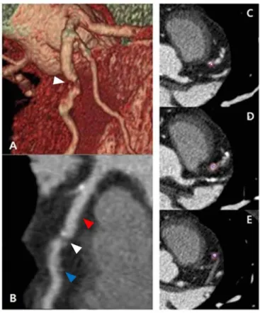
Image reconstruction was performed on the scanner’s workstation using commercially available software (Extended Brilliance Workstation, Philips Medical Systems, Best, The Netherlands). MDCT data were transferred for post processing to on an Aquarius Workstation V3.6 (TeraRecon, San Mateo, CA). Multi-planar reformatted (MPR), curved-planar reformatted (CPR), and volume rendering (VR) images with orthogonal and perpendicular projections to the vessel courses of each coronary segment were generated for the evaluation of coronary artery disease (Figure 1).
Figure 1. Coronary plaques in the mid-left anterior descending artery
(m-LAD) of a patient presenting with stable angina: (A) Volume rendering (VR) image. (B) Curved multi-planar reformatted (CPR) image. White, red, and blue arrowheads show worst stenosis, and 10 mm proximal and distal to worst stenosis, respectively. Cross sectional images at 10 mm proximal to worst stenosis (C), worst stenosis (D), and 10 mm distal to worst stenosis (E).
MDCT scan images were analyzed by two radiologists who were blinded to patients’ identity, clinical history and results of coronary angiography. Stenotic area parameters (lumen cross-sectional area (CSA), external elastic membrane (EEM) CSA, among others) were measured on a magnification view of the selected MPR and CPR images including through plane projection with an electronic ruler on an Aquarius Workstation V3.6 (TeraRecon, San Mateo, CA).
3. IVUS technique
After coronary angiography, 0.2mg of nitroglycerin was administered to all patients before introduction of the IVUS catheter. IVUS was performed using a 20-MHz, 2.9F phase-array IVUS catheter (Eagle Eye, Volcano Corporation, Rancho Cordova, CA, USA) under fluoroscopic guidance, and motorized pullback at 0.5mm/sec. All IVUS exams were digitalized for off-line analysis. Image analysis was performed with the use of dedicated software (pcVH2.1, Volcano Corporation, Rancho Cordova, CA, USA). For each region of interest, relative compositional quantitative plaque parameters were obtained. The lumen and the media-adventitia interface were defined by automatic contour detection and manual editing was performed on all individual frames. For each frame, EEM CSA and lumen CSA were calculated. All four plaque components were differentiated into different color codes (fibrotic tissue being labeled in dark green, fibro-fatty in light green, necrotic core in red and dense calcium in white), as validated previously.15
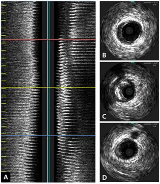
Measurements were made for the entire lesion, which was defined as the segment between the distal and proximal references.16 Plaque volume was calculated using Simson’s rule and averaged over the length of the lesion to also generate a mean plaque area. The length of the lesion was measured in the longitudinal image (Figure 2).
Figure 2. Intravascular ultrasound longitudinal image (A). Yellow
line, red line, and blue line show worst stenosis, and 10 mm proximal and distal to worst stenosis, respectively. Cross-sectional image at 10 mm proximal from worst stenosis (B), worst stenosis (C), and 10 mm distal to worst stenosis (D).
4. Comparison of IVUS versus MDCT
The location of the target vessel and worst stenosis (the stenosis with the smallest lumen size) was selected on the basis of conventional coronary angiography and IVUS. For MDCT, minimal lumen CSA site was detected on the decided target vessel by coronary angiography. The lesion length was measured from normally looking proximal to normally looking distal portion in longitudinal view of IVUS and MPR or CPR view of MDCT. Proximal reference was defined as the site with the largest lumen proximal to a stenosis but within the same segment (usually within 10 mm of the stenosis with no major intervening branches).16 Proximal reference EEM CSA, lumen CSA, lesion length, and plaque volume were measured by IVUS and MDCT for the entire lesion, and EEM CSA, lumen CSA, and plaque components were measured at 3 points (a worst stenosis and 10 mm proximal and distal to a worst stenosis as shown Figures 1 and 2) in each lesion. To evaluate vascular remodeling of the lesion, the remodeling index was calculated at the point of worst stenosis. Remodeling index was defined as EEM CSA at worst stenosis divided by reference EEM CSA, and the remodeling index was reported as positive remodeling when the CSA at the worst stenosis was larger than 1.1, and as negative remodeling when the CSA at the worst stenosis was smaller than 0.9.
In plaque evaluation of MDCT, regions of interest (ROIs) of 1.5 mm2 in area were determined inside the plaque, and tissue contrast was measured at 3 randomly selected points in HU. Based on definitions used in a previous study,17 plaque type by IVUS-VH was categorized as pathological intimal thickening (PIT; defined as a
mixture of fibrous and fibro-fatty tissues, a plaque burden ≥40% and <10% necrotic core and dense calcium), fibroatheroma (FA; defined as having a plaque burden ≥40% and a confluent necrotic core occupying 10% of the plaque area or greater in three successive frames with evidence of an overlying fibrous cap), and fibrocalcific plaque (FC; defined as a lesion with a plaque burden ≥40%, being mainly composed of fibrotic tissue, having dense calcium >10% and a confluent necrotic core of <10%. Higher amount accepted if necrotic core was located exclusively behind the accumulation of calcium).
5. Statistical analysis
Continuous values with normal distribution were expressed as means (with standard deviation) and compared with the two-tailed t test for independent samples. MDCT densities of plaques according to different plaque characteristics determined by IVUS-VH were expressed as medians (with interquartile range) and compared with the two-tailed Mann-Whitney test. Correlation of IVUS measurements with MDCT was performed using Pearson’s correlation coefficient. If the comparative parameter was too small to use the Pearson’s correlation coefficient, the Spearman’s correlation coefficient was used. Because of the small sample sizes, the Wilcoxon signed rank test was used for comparison of plaque remodeling between MDCT and IVUS. All analyses were two-tailed and p values < 0.05 were considered as statistically significant. Statistical analysis was performed using SPSS (version 15.0, Statistical Package for the Social Sciences Inc., Chicago, IL, USA) software.
III. RESULTS
1. Patient characteristics
Sixty-nine patients who underwent MDCT before IVUS-VH were initially enrolled. Among them, 15 patients were excluded due to previous stent implantation, coronary artery bypass graft, chronic total occlusion, or a period of over 180 days between MDCT and IVUS. Therefore, 54 patients (44 male, 10 female; mean age, 60±7
years), were enrolled. The location of the lesions in the 54 patients
was the left anterior descending artery (LAD) in 38 (70.3%), the left circumflex artery in 3 (5.6%), and the right coronary artery in 13 (24.1%). Framingham point scores of the 54 subjects ranged from 9 to 20, corresponding to a 10-year risk of major coronary events of 5~30%.4 Characteristics of the 54 patients enrolled are summarized in Table 1.
Characteristics Total (n=54) Male gender (%)
Age (years)
Body Mass Index (kg/m2)
Risk factors Hypertension (%) Diabetes (%) Current smoker (%) Hyperlipidemia (%) History of Angina (%) Framingham point scores
Interval from MDCT to IVUS (days) Agatston score on MDCT
Lesion vessel (LAD/LCx/RCA) (%)
44 (81.4%) 60±7 (46-75) 25.3±3.0 31 (57.4%) 26 (48.1%) 24 (44.4%) 8 (14.8%) 47 (87.0%) 13.7±2.1 (9-20) 36±29 (7-125) 224.64±286.97 38/3/13 (70.3/5.6/24.1) Table 1. Patient characteristics of study population
MDCT: Multidetector computed tomography IVUS: Intravascular ultrasound
LAD: Left anterior descending artery LCx: Left circumflex artery
2. Comparison for each lesion between IVUS and MDCT
Average lesion length (20.0±8.6 mm on IVUS, 15.3±9.6 mm on MDCT, p=0.060) and plaque volume (160.8±84.7 mm3 on IVUS, 124.4±101.7 mm3 on MDCT, p=0.132) were not significantly different as measured by IVUS and MDCT, but there were a trend toward underestimation in the MDCT measurement (Table 2).
Each lesion was measured at 3 sites: 10 mm proximal to worst stenosis, worst stenosis, and 10 mm distal to worst stenosis as shown as Figures 1 and 2. In total, 162 sites identified on MDCT and IVUS were evaluated.
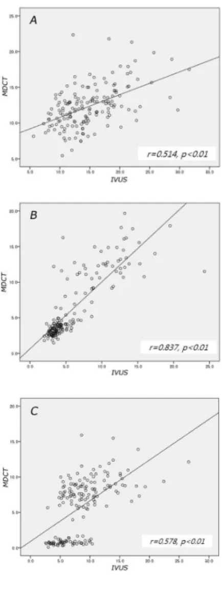
IVUS and MDCT measuredments were 14.84±4.73 mm2 versus 12.33±2.94 mm2 (p<0.01) for mean EEM CSA, 6.19±4.03 mm2 versus 6.45±4.53mm2 (p=0.188) for mean lumen CSA. Although MDCT did not yield a significantly different lumen CSA, it significantly underestimated EEM CSA and plaque area (Table 2). Correlation for those measurements were close (r=0.514, 0.837, 0.578, p<0.01) (Figure 3).
IVUS MDCT IVUS vs. MDCT Total length (mm) 20.0±8.6 15.3±9.6 p*=0.06 Plaque vol. (mm3) 160.8±84.7 124.4±101.7 p*=0.13 Agatston score (lesion vessel) 100.0±127.0 EEM CSA (mm2) 14.8±4.7 12.3±2.9 p*<0.01 Lumen CSA (mm2) 6.5±4.1 5.9±3.1 p*=0.19 Plaque area (mm2) 8.7±4.5 1.6±3.1 p*<0.01
* Two-tailed t test for independent samples
Table 2. Comparison between IVUS and MDCT measurements of the lesion
MDCT: Multidetector computed tomography, IVUS: Intravascular ultrasound
EEM: External elastic membrane CSA: Cross-sectional area
Figure 3. Linear correlations between IVUS and MDCT measurements. There are significant correlations for EEM CSA (A), lumen CSA (B), and plaque area (C) between them.
MDCT: Multidetector computed tomography IVUS: Intravascular ultrasound
EEM: External elastic membrane CSA: Cross-sectional area
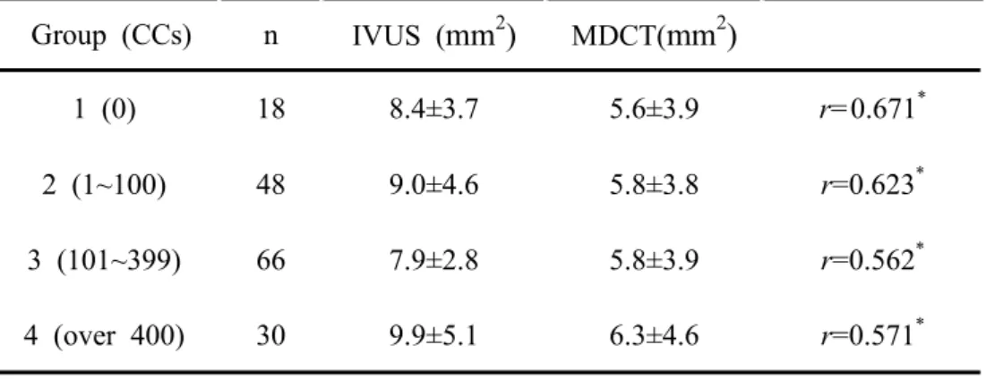
For evaluation of MDCT accuracy by coronary calcium content, 162 lesions were divided into 4 groups by CCs (Group 1, CCs 0; Group 2, CCs 1~99; Group 3, CCs 100~399; and Group 4, CCs over 400). There was also good correlation for plaque area between MDCT and IVUS, although CCs was higher (Table 3).
Table 3. Correlations of plaque area between IVUS and MDCT at
162 sites for each coronary calcium score
Group (CCs) n IVUS (mm2) MDCT(mm2) 1 (0) 18 8.4±3.7 5.6±3.9 r=0.671* 2 (1~100) 48 9.0±4.6 5.8±3.8 r=0.623* 3 (101~399) 66 7.9±2.8 5.8±3.9 r=0.562* 4 (over 400) 30 9.9±5.1 6.3±4.6 r=0.571* * p<0.05
IVUS: Intravascular ultrasound
MDCT: Multidetector computed tomography CCs: Coronary calcium score on MDCT
3. Plaque remodeling index in worst stenosis site
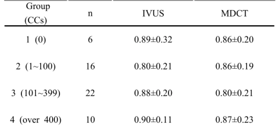
In 54 worst stenosis sites, MDCT and IVUS measurements showed negative remodeling (remodeling index of IVUS and MDCT was 0.86 and 0.84, p=0.441). There were similar results for each CCs group (Table 4).
Table 4. Plaque remodeling index comparison between IVUS and
MDCT at 54 worst stenosis sites by coronary calcium score
Group (CCs) n IVUS MDCT 1 (0) 6 0.89±0.32 0.86±0.20 2 (1~100) 16 0.80±0.21 0.86±0.19 3 (101~399) 22 0.88±0.20 0.80±0.21 4 (over 400) 10 0.90±0.11 0.87±0.23
Two-tailed t test, p>0.05 for all categories
IVUS: Intravascular ultrasound
MDCT: Multidetector computed tomography CCs: Coronary calcium score on MDCT
4. Comparison of plaque characterization
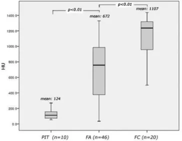
The mean CT density value for PIT, FA, and FC was 124 ± 65 HU (range, 52~270 HU), 672 ± 378 HU (range, 35 ~ 1329 HU), and 1107 ± 284 HU (range, 500 ~ 1437 HU), respectively. Although there were significant differences in HU among plaque characterization groups, there was a significant overlap of MDCT density values among plaque groups (Figure 4).
Figure 4. Box-and-whisker diagram of CT density values for the
various plaques types (pathological intimal thickening: PIT, fibroatheroma; FA, and fibrocalcific plaque; FC) determined by IVUS-VH. Each box describes the distribution of density values within 1 SD. The bold line in the box depicts the median.
IV. DISCUSSION
Coronary computed tomographic angiography has emerged as a promising noninvasive method for the detection and exclusion of obstructive coronary artery disease. For 64-channel MDCT, Leber et al. reported a sensitivity of 84% and a specificity of 91% in the detection of proximal coronary plaques.10 In the present study, 64-channel MDCT allows accurate noninvasive determination of intra/extra luminal coronary lesion characteristics in intermediate-risk individuals, as previously reported.11,12,13 There were correlations for plaque volume, EEM CSA, and lumen CSA measurements between MDCT and IVUS-VH.
On the other hand, the present study revealed that MDCT relative to IVUS particularly underestimated lesion length, EEM CSA and plaque area for several reasons. In this study, IVUS examination was performed after intracoronary nitroglycerine administration, while MDCT was not. Also, after the guidewire passed through the lesion, the IVUS catheter was inserted for measurements which straightened the vessel maybe influencing measurement of the lesion length and plaque area. The fact that the LCx and RCA are more tortuous than the LAD could explain why measurements in LAD lesions were better correlated than in other vessels.18 Lumen CSA was relatively not affected probably because of its smaller magnitude and of contrast dye actually passing through the lesion.
Coronary calcium, which provides prognostic information over traditional risk factors, is known as a limiting factor of evaluation in MDCT evaluation.14 A previous SHAPE task force report19 recommends CCs as a screening method for atherosclerosis,
and its categorization as 0, 1~99, 100~399, and over 400. The higher the CCs, the more atherosclerosis suspected. In this study, we divided all 162 lesions into 4 groups by coronary calcium score in MDCT. Even in the higher CCs categories, EEM CSA, lumen CSA, and plaque area showed good correlations between MDCT and IVUS.
The atherosclerotic plaques that are causally related to acute coronary syndromes reveal a variable extent of luminal narrowing but are almost always associated with expansive or positive vessel remodeling.20 MDCT can evaluate not only lumen stenosis, but also EEM CSA, and vessel remodeling was also evaluated in this study. However, in this study the population was composed of asymptomatic patients or patients with stable angina, not ACS, and vessel remodeling at worst stenotic site showed negative remodeling. Moreover, the location of the lesion was mainly LAD(70.3%), which might influence the measurement of vessel remodeling because the LAD and the LCx have severe vessel tapering and the location of lesions is near to major side branches.21 Also it is known that atherosclerotic plaque composition is an important predictor of plaque stability.22 Comparison of MDCT and histology in post-mortem coronary artery specimens has shown that the atherosclerotic plaques themselves undergo variable enhancement after contrast injection.23 While calcified and non-calcified plaques can be differentiated by MDCT, reliable subclassification of non-calcified plaques is not currently available.10 In this study, plaque type by IVUS-VH was categorized into PIT, FA, and FC, and MDCT attenuation was measured by HU. Although MDCT density measurements were significantly different among groups, there was significant overlap of MDCT density values between PIT
and FA, and between FA and FC. This result indicates that reliable classification of plaques based on MDCT measurement is currently limited when using 64-channel MDCT.
Detection of intraluminal stenosis and atherosclerotic plaque by MDCT would be important determinants of treatment plan in intermediate-risk patients. Several studies have reported the diagnostic accuracy of MDCT in the quantification of coronary artery plaques,8,24,25 but it is only a structural and no a fuctional evaluation. The atherosclerotic plaques that are causally related to ACS reveal a variable extent of luminal narrowing but are almost always associated with expansive or positive vessel remodeling, and soft plaque composition.26 Computed tomography coronary angiography has the potential to enable detection of not only significant luminal stenosis but also vessel remodeling or plaque composition. However, this study has shown that plaque remodeling and plaque composition characterization on 64-channel MDCT is limited. Some institutions are now also using 128-channel MDCT, and both 256- and 320-channel MDCT have been introduced.27 The latter configurations offer the potential to image the entire heart in more detailed.
This study had several limitations. This was a retrospective, single-center, and observational study. In this study, the use of IVUS-VH during conventional coronary angiography was at operator discretion, thereby possibly introducing operator bias. Also, the study population was too small to generalize this result.
Despite limitations, MDCT is superior to conventional coronary angiography because it is non-invasive; tomographic imaging allows multiplanar reconstructions of the tri-dimensional structure of the coronary arteries; and it provides a more accurate
description of eccentric stenosis and extra luminal structures such as plaque.
V. CONCLUSION
The intra luminal area and EEM CSA of MDCT are well correlated with those of IVUS even in the context of increased coronary calcium; however, MDCT currently has a limited role in plaque characterization. The information content of MDCT is broader than that of conventional coronary angiography, and the results of this study support that MDCT as being reliable enough for the evaluation of coronary stenosis severity in the intermediate-risk group independent of coronary calcium. Therefore, MDCT might be used as an alternative screening method for the intermediate CAD-risk group and for plaque burden monitoring of known coronary atherosclerosis. However, 64-channel MDCT currently has a limited role in plaque characterization and further study is needed.
REFERENCES
1. Zipes DP, Wellnes HJ. Sudden cardiac death. Circulation 1998; 98:2334-51.
2. Kunimasa T, Sato Y, Sugi K, Moroi M. Evaluation by multislice computed tomography of atherosclerotic coronary artery plaques in non-culprit, remote coronary arteries of patients with acute coronary syndrome. Circ J 2005; 69: 1346-51.
3. Anderson KM, Odell PM, Wilson PW, Kannel WB.
Cardiovascular disease risk profiles. Am Heart J 1991;121: 293-8.
4. Wilson PW, Castelli WP, Kannel WB. Coronary risk prediction in adults (The Framingham Study). Am J Cardiol 1987;59:91G-94G.
5. Falk E, Shah PK, Fuster V. Coronary plaque disruption. Circulation 1995;92:657-71.
6. Fayad ZA, Fuster V. Clinical imaging of the high-risk or vulnerable atherosclerotic plaque. Circ Res 2001;89:305-16.
7. Muller JE, Abela GS, Nesto RW, Tofler GH. Triggers, acute risk factors and vulnerable plaques: the lexicon of a new frontier. J Am Coll Cardiol 1994;23:809-13.
8. Nair A, Kuban BD, Tuzcu EM, Schoenhagen P, Nissen SE, Vince DG. Coronary plaque classification with intravascular ultrasound radiofrequency data analysis. Circulation 2002;106: 2200-6.
9. Inoue F, Sato Y, Matsumoto N, Tani S, Uchiyama T. Evaluation of plaque texture by means of multislice
computed tomography in patients with acute coronary syndrome and stable angina. Circ J 2004;68:840-4.
10. Achenbach S, Moselewski F, Ropers D, Ferencik M, Hoffmann U, MacNeill B, et al. Detection of calcified and noncalcified coronary atherosclerotic coronary plaques with multislice computed tomography. J Am Coll Cardiol 2001;37: 1430-5.
11. Leber AW, Knez A, von Ziegler F, Becker A, Nikolaou K, Paul S, et al. Quantification of obstructive and non-
obstructive coronary lesions by 64-slice computed
tomography: a comparative study with quantitative coronary angiography and intravascular ultrasound. J Am Coll Cardiol 2005;46:147-54.
12. Vanhoenacker PK, Heijenbrok-Kal MH, Van Heste R, Decramer I, Van Hoe LR, Wijns W, et al. Diagnostic performance of multidetector CT angiography for assessment of coronary artery disease: meta analysis. Radiology 2007; 244:419-28.
13. Budoff MJ, Dowe D, Jollis JG, Gitter M, Sutherland J, Halamert E, et al. Diagnostic performance of 64-multidetector row coronary computed tomographic angiography for
evaluation of coronary artery stenosis in individuals without known coronary artery disease. J Am Coll Cardiol 2008;52: 1724-32.
14. Hoffmann U, Moselewski F, Cury RC, Ferencik M, Jang IK, Diaz LJ, et al. Predictive value of 16-slice multidetector spiral computed tomography to detect significant obstructive coronary artery disease in patients at high risk for coronary artery disease: patient- versus segment-based analysis. Circulation 2004;110:2638-43.
15. Nasu K, Tsuchikane E, Katoh O, Vince DG, Virmani R, Surmely JF, et al. Accuracy of in vivo coronary plaque morphology assessment: a validation study of in vivo virtual histology compared with in vitro hitopatholgy. J Am Coll Cardiol 2006;47:2405-12.
16. Minz GS, Nissen SE, Anderson WD, Bailey SR, Erbel R, Fitzgerald PJ, et al. American College of Cardiology Clinical Expert Consensus Document on Standards for Acquisition, Measurement and Reporting of Intravascular Ultrasound Studies (IVUS). A report of the American College of Cardiology Task Force on Clinical Expert Consensus
Documents. J Am Coll Cardiol. 2001;37:1478-92.
plaque using radiofrequency ultrasound signal processing. J Nucl Cardiol 2006;21:831-40.
18. Jakob M, Spasojevic D, Krogmann ON, Wiher H, Hug R, Hess OM. Tortuosity of coronary arteries in chronic pressure and volume overload. Cathet Cardiovasc Diagn. 1996;38: 25-31.
19. Naghavi M, Falk E, Hecht HS, Jamieson MJ, Kaul S, Berman D, et al. From vulnerable plaque to vulnerable patient-Part III: Executive summary of the screening for heart attack prevention and education (SHAPE) task force report. Am J Cardiol 2006;98(suppl):2H-15H.
20. Motoyama S, Kondo T, Sarai M, et al. Multislice computed tomographic characteristics of coronary lesions in acute coronary syndromes. J Am Coll Cardiol 2007;50:319-26.
21. Javier SP, Mintz GS, Popma JJ, Pichard AD, Kent KM, Satler LF, et al. Intravascular ultrasound assessment of the magnitude and mechanism of coronary artery and lumen tapering. AM J Cardiol 1995;75:177-80.
22. Newby AC, Libby P, van der Wal AC. Plaque instability-the real challenge for atherosclerosis research in the next decade? Cardiovasc Res. 1999;41:321-2.
23. Halliburton SS, Schoenhagen P, Nair A, Stillman A, Lieber M, Murat Tuzcu E, et al. Contrast enhancement of coronary
atherosclerotic plaque: a high-resolution, multidetector row computed tomography study of pressure-perfused, human exvivo coronary arteries. Coron Artery Dis. 2006;17:553-60.
24. Moselewski F, Ropers D, Pohle K, Hoffmann U, Ferencik M, Chan RC, et al. Comparison of measurement of
cross-sectional coronary atherosclerotic plaque by contrast- enhanced, submillimeter multidetector spiral computed tomography versus intravascular ultrasound. Am J Cardiol. 2004;94:1294-7.
25. Leber AW, Becker A, Knez A, von Ziegler F, Sirol M, Nikolaou K, et al. Accuracy of 64-sclice computed
tomography to classify and quantify plaque volumes in the proximal coronary system: a comparative study using intravascular ultrasound. J Am Coll Cardiol. 2006;47:672-7.
26. Motoyama S, Sarai M, Harigaya H, Anno H, Inoue K, Hara T, et al. Computed tomographic angiography characteristics of atherosclerotic plaques subsequently resulting in acute coronary syndrome. J Am Coll Cardiol 2009;54:49-57.
27. Hein PA, Romano VC, Lembcke A, May J, Rogalla P. Initial experience with a chest pain protocol using 320-slice volume MDCT. Eur Radiol. 2009;19(5):1148-55.
ABSTRACT(IN KOREAN) 혈관내 초음파도로 평가한 관동맥 병변의 평가에 있어서의 다중검출 전산화 단층촬영의 유용성 <지도교수 권 혁 문> 연세대학교 대학원 의학과 박 종 관 최근 심장 전산화 단층촬영 (MDCT) 은 관동맥 질환의 혈관 병변의 평가에 있어서 심혈관조영술을 대치하는 검사로 사용되고 있다. 하지 만, 혈관 석회화 정도는 MDCT 평가에 제한점으로 알려져 있다. 이에 본 연구는 중등도의 위험성을 가진 관동맥질환자의 평가에 있어서의 MDCT의 유용성을 관동맥 석회화 정도에 따라 나누어 혈관내 초음파 도 (IVUS-VH) 와 비교하여 평가하고자 하였다. 관상동맥질환이 의심되 어 MDCT를 시행하고 혈관내 초음파를 시행한 54명을 대상으로 162 부분의 병변의 EEM 및 혈관내 단면적 MDCT상의 석회화 수치에 따라 4군 (0, 1~99, 100~399, 400이상)으로 나누어 비교하였으며, 병변의 죽 상경화반 성상을 IVUS-VH로 측정하여 pathologic intimal thickening, fibroatheroma, fibrocalcific plaque로 나누고 MDCT와 비교하였다. MDCT 로 측정한 죽상경화반 단면적은 IVUS로 측정한 값과 관상동맥 석회화 수치에 상관없이 양의 상관관계를 나타내었다 (r=0.671, r=0.623, r=0.562, r=0.571). 한편 죽상경화반의 조직학적 특성은 MDCT로 구분은 가능하였으나, 석회화 수치에 따라 각 군간 겹치는 부분이 많아 임상 적 유용성은 떨어졌다. 본 연구는 중등도의 위험도를 가진 관상동맥질 환자에서 MDCT가 죽상경화반의 평가에는 적절하지 못하였으나, 심혈 관조영술에 비해 비침습적으로 혈관 내/외의 병변을 평가하여 침습적 관상동맥 조영술 대신에 선별적 검사로 사용 될 수 있음을 시사한다. ---핵심되는 말 : 관상동맥질환, 관상동맥 석회화 수치, 전산화 단층촬영,