Regulation of inflammatory cell signaling
by heat shock protein 70 in cerebral
ischemia
Jong Youl Kim
Department of Medical Science
Regulation of inflammatory cell signaling
by heat shock protein 70 in cerebral
ischemia
Directed by Professor Jong Eun Lee
The Doctoral Dissertation submitted to the Department of
Medical Science, the Graduate School of Yonsei
University in partial fulfillment of the requirements for the
degree of Doctor of Philosophy of Medical Science
Jong Youl Kim
This certifies that the
Doctoral Dissertation of Jong Youl Kim
is approved.
Thesis Supervisor :
Thesis Committee Member #1 :
Thesis Committee Member #2 :
Thesis Committee Member #3 :
Thesis Committee Member #4 :
The Graduate School
Yonsei University
ACKNOWLEDGEMENT
지난 8년 동안 지내온 해부학교실을 떠날 생각을 하니 제가
이곳에서 받은 것이 너무나 많다는 생각이 듭니다. 처음 학위를
시작하면서 새로운 기대감과 막연한 두려움으로 학문의 길로 들어선
시작이 어느덧 한 편의 논문과 함께 학위를 마치게 되었습니다.
지난 날들이 기억되면서 많은 도움과 격려를 주신 모든 분께 이
자리를 빌어 감사의 마음을 전하고 싶습니다.
우선 제 인생에 새로운 전환점을 만들어 주시고 학문을 하는
사람은 어떤 마음가짐으로 살아야 하는지 가르쳐 주신 이종은
교수님께 감사 드립니다. 항상 좋은 가르침을 주시고 최종 심사까지
애써주신 박경아 교수님과 이원택 교수님께 감사 드립니다. 또한
바쁘신 중에도 저의 논문심사를 맡아주신 허만욱 교수님과 박수철
교수님께 감사 드립니다.
때론 힘들고 고된 생활이었지만 함께 실험실 생활을 하며
위로와 힘이 되어주신 실험실 선생님들 (신영호, 김재환, 최윤정,
이연옥, 손미란, 박유미, 김재영, 노윤미)께 감사 드립니다.
어느 때나 한결같이 저를 믿어 주시고 큰 힘이 되어 주신
부모님께 깊은 감사를 드리며, 묵묵히 저를 지원해준 누나 와
매형께 고마움을 전합니다. 이 분들 모두 저와 함께 해 주셨기에 이
자리가 있을 수 있음을 기억하며 감사를 드립니다.
TABLE OF CONTENTS
ABSTRACT --- 1
I.
INTRODUCTION --- 3
II.
MATERIALS AND METHODS --- 8
1.
Animal ---
--8
2.
Transient Middle Cerebral Artery Occlusion Model ---
--8
3.
Primary astrocyte culture --- 8
4.
Heat Pretreatment ---
-9
5.
Oxygen and Glucose Deprivation ---
-9
6.
Cresyl violet stain ---
-10
7.
Hoeschst-PI nuclear staining ---
-10
8.
Two-dimensional electrophoresis ---10
9.
MALDI-TOF analysis --- 12
10.
Western blot analysis --- 13
11.
Co-immunoprecipitation ---
-
14
12.
Phosphorylation ELISA assay ---
-15
13.
Electrophoretic-Mobility Shift Assay ---
-15
14.
RT-PCR Reaction ---
-16
III.
RESULTS
1.
Overexpression of Hsp70 in Transgenic Mice --- 17
2.
Overexpression of Hsp70 in Hsp70 Tg mice reduces infarct size
of ischemic brain ---
-18
3.
Different of protein expression in Wild type and Hsp70 Tg mice
---
-19
4.
Proteomic identification of differentially expressed proteins in
Hsp70 Tg mice --
---
--- 20
5.
Overexpression of Hsp70 reduces the expression and nuclear
translocation of NF-
κB ---
- 216.
Overexpression of Hsp70 inhibits I
κB phosphorylation --- 22
7.
Hsp70 interacts with NF-
κB and IκB --- 24
8.
Overexpression of Hsp70 decreases DNA binding capacity of
NF-
κB --- 24
9.
Overexpression of Hsp70 inhibits the expression of several
NF-κB-regulated genes --- 26
10.
Overexpression of Hsp70 in primary cultured astrocytes by heat
pretreatment ---
-27
11.
Protective effect of Hsp70 overexpression on OGD injury in
primary cultured astrocyte --- 28
12.
Overexpression of Hsp70 by heat pretreatment reduces
expression and activation of NF-
κB and IκB --- 29
13.
Overexpression of Hsp70 by heat pretreatment reduces
expression and activation of JNK and c-Jun ---
-30
14.
Overexpression of Hsp70 by heat pretreatment reduces
expression and activation of p38, c-Fos and STAT-1 --- 31
15.
Hsp70 overexpression inhibits phosphorylation of JNK, p38,
16.
Hsp70 overexpression by heat treatment interacts with
transcritpiton factors (NF-
κB, IκB, JNK, AP-1, p38 and STAT-1)
--- 34, 35, 36
17.
Overexpression of Hsp70 decreases DNA binding capacity of
NF-
κB, STAT-1 and AP-1 --- 38
18.
Overexpression of Hsp70 inhibits the expression of several
transcription factor-regulated genes --- 39
IV.
DISCUSSION --- 41
V.
CONCLUSION --- 44
REFERENCES ---
-46
LIST OF FIGURES
Figure 1. Schematic pathway of pro-inflammatory genes on ischemic injury ---
--- 6
Figure 2. Genotyping and overexpression of heat shock protein 70 (Hsp70)
transgenic mice--- 17
Figure 3. Brain infarct volume on cerebral ischemia --- 18
Figure 4. Two-dimensional gel image of Wt sham, Wt MCAO and Hsp70 Tg
MCAO Mice ---
-
19
Figure 5. Overexpression of Hsp70 inhibits nuclear translocation of NF-
κB ---
---
-
22
Figure 6. Less I
κB phosphorylation observed in Hsp70 transgenic mice ---
--- 23
Figure 7. Hsp70 co-immunoprecipitates with NF-
κB and IκB --- 24
Figure 8. Decreased DNA binding of NF-
κB in Hsp70 Tg mice ---
25Figure 9. Down-regulation of expression of several representative NF-
κB
dependent pro-inflammatory genes in Hsp70 Tg mice --- 26
Figure 10. Western blot analysis of Hsp70 overexpression --- 27
Figure 11. Protective effect of heat pretreatment on the primary cultured
astrocytes in OGD injury--- 28
Figure 12. Western blot of NF-
κB and pi-IκB on Hsp70 overexpression ---
---
--
30
Figure 13. Western blot of pi-JNK, and AP-1on Hsp70 overexpression
---
--
31
Figure 14. Western blot of pi-p38 and Pi-STAT-1 on Hsp70 overexpression
---
---32
Figure 15. Detection of phosphorylated JNK, p38, STAT-1 and c-Jun by
phosphor ELSA assay ---
---
33
Figure 16. Hsp70 co-immunoprecipitates with NF-
κB and IκB ---
--35
Figure 17. Hsp70 co-immunoprecipitates with JNK and AP-1---
--36
Figure 18. Hsp70 co-immunoprecipitates with p38 and STAT-1
-
--- 37
Figure 19. Decreased DNA binding of transcription factors in Hsp70 Tg mice
---
38
Figure 20. Overexpression of Hsp70 inhibits expression of several representativ
etranscription factors-dependent pro-inflammatory genes in primary cultured
astrocyte--- 39
LIST OF TABLES
Table 1. Hsp70 Tg mice brain proteins with altered levels after tMCAO injury,
identified by mass spectrometry --- 21
Abstract
Regulation of inflammatory cell signaling by heat shock
protein 70 in cerebral ischemia
Jong Youl Kim
Departmetn of Medical Science
The Graduate School, Yonsei University
(Directed by Professor Jong Eun Lee)
Cerebral ischemia triggers a complex series of biochemical and molecular mechanisms that impairs the neurologic functions through of cellular integrity mediated by oxidative stress, stress signaling, neurovascular pathophysiology and inflammation, cell death and gene expression.
Inflammation is important among cerebral ischemia events. Its reactions initiated at the neurovascular interface and alterations in the dynamic communication between the endothelial cells, astrocytes and neurons are thought to substantially contribute to the pathogenesis of the disease. This inflammation is caused by a transcription of
cytokine gene modulating nuclear factor-κB (NF-κB), activator protein-1 (AP-1), signal transducers and activator of transcription-1 (STAT-1).
The 70-kDa heat shock protein (Hsp70) is involved in protecting the brain from a variety of insults including stroke. Although the mechanism has been largely considered to be because of its chaperone functions, recent work indicates that Hsp70 also modulates inflammatory responses.
In this study, we investigated that Hsp70 overexpression regulates the transcription of NF-κB, AP-1 and STAT-1 after ischemic injury in in vivo and in vitro models using transgenic mice and primary cultured astrocyte constitutively expressed Hsp70. Hsp70 overexpression decreases the expression and phosphorylation of
NF-κB, AP-1 and STAT-1 in Hsp70 Tg mice and heat pretreated astrocyte under
ischemic conditions. Also, Hsp70 overexpression interacts with NF-κB, AP-1 and STAT-1 and interrupts DNA binding of NF-κB, AP-1 and STAT-1 in Hsp70 Tg mice and heat pretreated astrocyte. These findings produce that downregulates the expression of pro-inflammatory genes in Hsp70 Tg mice and heat pretreated astrocyte.
Take together, these results suggest that overexpression of Hsp70 protects against brain ischemia via an ant-inflammatory mechanism by transcription factor.
Key words: Cerebral ischemia, Inflammation, Heat shock protein 70, Nuclear
Regulation of inflammatory cell signaling by heat shock
protein 70 in cerebral ischemia
Jong Youl Kim
Departmetn of Medical Science
The Graduate School, Yonsei University
(Directed by Professor Jong Eun Lee)
I. INTRODUCTION
Cerebral ischemia results in a number of hemodynamic biochemical, and neurophysiologic alterations that can be linked clinically to behavioral and pathologic disturbances. With declining blood flow, the functional neuronal activity is affected first, and as the ischemia progresses, the metabolic activity gets suppressed which is required to maintain the structural integrity of the brain cells1. The event leads to glutamate mediated excitotoxicity and calcium overload, oxidative stress, stress
signaling, neurovascular pathophysiology and inflammation, cell death mode and gene expression2.
Moreover, inflammatory events, which are initiated at the blood microvessel interface few hours after the onset of ischemia, underlie the transition from ischemic to inflammatory injury. Major players in the inflammatory injury are cytokines which are produced transcription factors. NF-κB, AP-1 (i.e. c-Fos and c-Jun) and STAT-1 pathway play essential roles in transcription. The activation of NF-κB, AP-1 and STAT-1 is mediated through phosphorylation of their regulatory proteins and activation of other kinases. Transcription factors regulate the transcription of many genes in involved in immunity, inflammation, and protection from programmed cell death3.
The typical transcription factor NF-κB plays an important physiological and pathological role in a variety of tissue and cell, including brain cells4. In astrocytes, NF-κB activity is required for the inducible expression of various genes involved in the inflammatory of cerebral ischemia. NF-κB complexes are mainly composed of p65 and p50 subunits in tissue and cell5,6, and remain sequestered in the cytoplasm of resting cells by association with a family of inhibitory IκB proteins. Following the appropriate stimuli, the IκB proteins are rapidly phoshorylated by the IκB kinase complex (IKK), ubiquitinaed, and deraded by the 26 S proteasome7,8. As result,
NF-κB translocates to the nucleus to bind specific NF-NF-κB DNA motifs and promote
expression of target genes8.
expression in brain, including cytoskeletal proteins and growth factors that support regeneration and repair the destroyed brain tissues9,10. Protein members of the Fos and Jun families can form a stable c-Fos/c-Jun dimeric complex (namely AP-1). Upon binding to specific AP-1 site in the promoter region of target genes, this associated complex enhances the gene transcription including expression of diverse inflammatory proteins11. Activator protein-1 is important among cerebral ischemia events, which mediated through activation of mtiogen-activated protein kinases (MAPKs) signaling pathways leading to immediate early gene AP-1 activation (i.e. c-Fos expression and c-Jun phosphorylation) in RBA-1 cells12.
STAT proteins are latent cytoplasmic transcription factors that become activated by tyrosine phosphorylation. Subsequently, phosphorylated proteins can dimerize and translocate to the nucleus were they interact with DNA binding elements and induce transcription13,14,15 . Furthermore, recent data suggest that STAT-1 also regulates Th1 polarization in the early phase of T-cell differentiation 16,17. Stat-1 is activated downstream of p38 MAP kinase under hypoxic conditions 18.
During ischemia, the 70 kDa inducible heat shock protein (Hsp70) is thought to enhance cell survival by preventing protein aggregation or facilitating refolding of partially denatured proteins19,20. Work by our group and others showed that overexpressing Hsp70 is protective against focal and global cerebral ischemia and neurotoxicity21,22. The mechanism of protection after cerebral ischemic events is not well known, but has largely been attributed to its chaperone functions whereby Hsp70 improves cell survival by preventing protein aggregation. Recent paper has show that
Hsps are capable of modulating immune responses either by potentiating or inhibiting them in brain ischemia or injury23,24. To understand the mechanisms more completely that interact with transcription factor and Hsp70 in ischemic injury, we investigated whether and how Hsp70 modulates transcription factor in an in vivo and in vitro model of ischemic-like injury.
In this study, we hypothesized that overexpression of Hsp70 not only protects against brain ischemia but also appears to regulate transcription factor.
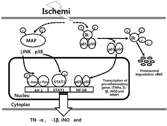
Figure 1. Schematic pathway of pro-inflammatory genes on ischemic injury. Each arrow represents a step in an activation pathway. Ischemic injury activates IkB and
(JNK p38 Proteasomal degradation of IkB Transcription of pro - inflammatory genes (TNF - α , IL - 1 β , iNOS and MMP) MAP P P P P P P P Ik Ik U U p65 p50 p65 p50 p65 p50 c-Jun STAT-1 AP-1 Cytoplas Nucleu TN -α , -1β, iNO and
Ischemi
STAT-1 NF-kB c-fos PMAPK cascade to stimulate IkB, JNK and p38 phosphorylation, leading to NF-kB (subunit p50 and p65) and AP-1 (phosphorylation of c-Jun and upregulation of c-Fos) activation and then enhances pro-inflammatory genes expression.
II. MATERIALS AND METHODS
1. Animals
Experiments were performed according to a protocol approved by the Yonsei University Animal care and Use committee in accordance with NIH guidelines. Male Hsp70 transgenic mice (Hsp70 Tg) and wild-type (Wt) littermates originally created by Dillmann et al (University of California, San Diego; Maber et al, 1995) were used.
2. Transient Middle Cerebral Artery Occlusion Model (tMCAO)
Ischemia was induced using the tMCAO model. Mice were anesthetized by face mask using 3% to 4% isofluorane followed by 1.5% to 2% for maintenance. Physiological parameters including body temperature, blood pressure, and arterial blood gases were monitored and maintained in the normal range. A 6-0 nylon monofilament suture was inserted into the common carotid artery to occlude the ostium of middle cerebral artery. After 2h, the suture was inserted of middle cerebral artery. After 2h, the suture was retracted to allow reperfusion. Twenty-four hours later, the animals were euthanized in a CO2 chamber and then perfused
transcardially with heparinized saline. Brains were harvested for different assays.
3. Primary astrocyte culture
mice and maintainedin Minimum essential medium (MEM, Gibco, USA) containing 10% fetal bovine serum and 10% equine serum (Hyclone, USA). Briefly, hemispheres of new bone ICR mice were removed aseptically from the skulls, freed of the meanings.
4. Heat Pretreatment
Astrocyte cultures were washed three times with balanced salt solution (BSS5.5)
containing 5.5mM glucose, 116mM NaCl, 1.8mM CaCl2, 0.8mM MgSO4, 5.4mM
KCl, 1mM NaH2PO4, 14.7mM NaHCO3, and HEPSE at pH7.4. The culture
medium was then exchange with BSS5.5 and incubation continued at 43℃ for 30
min.
5. Oxygen and Glucose Deprivation (OGD)
Cultures of either astrocyte were deprived of glucose and oxygen by changing the culture medium to balanced salt solution containing no glucose (BSS0.0) and
kept in an oxygen-free chamber at 37℃ for 6h. Cultures were then transferred to a 37℃ incubator with 5% CO2 and reperfused with glucose at a concentration of
5.5mmol/L (BSS5.5) at normoxia for 24h. All experiments were performed in
6. Cresyl violet stain
After animals were perfused, brains were sunk in 20% sucrose, frozen, and cut into 20㎛ sections on a cryostat. Brain sections from four consecutive coronal levels of brain were stained with cresyl violet.
7. Hoeschst-PI nuclear staining
The cell death was evaluated by staining of non-viable cell with propidium iodide (Sigma, USA), and live cell with Hoechst 33258 dye (Sigma, USA). Staining with the fluorescent dyes propidium iodide and Hoechst 33258 allows discrimination of apoptotic cells on the basis of nuclear morphology and evaluation of membrane integrity. Hoeschst dye was added to the culture medium to a final concentration of 2-5㎍/㎖ and the cultured cells were kept at 37℃ for 30min. Propidium iodide solution was the added (final concentration 2-5㎍/㎖) just before observation in a Olympus microscope equipped for epifluorescence with UV filter block.
8. Two-dimensional electrophoresis
Protein sample preparation : Cultured cell pellets were washed twice in ice-cold PBS (GIBCO, USA) and lysed in sample buffer composed with 7M urea, 2M Thiourea containing 4%(w/v) 3-[(3-cholamidopropy)dimethyammonio]-1-propanesulfonate (CHAPS,) 1%(w/v) dithiothreitol (DTT) and 2%(w/v)
pharmalyte and 1mM benzamidine. Proteins were extracted for one hour at room temperature with vortexing. After centrifugation at 15,000rpm for one hour at 15℃, insoluble material was discarded and soluble fraction was used for two-dimensional gel electrophoresis. Protein loading was normalized by Bradford assay.
A. 2D PAGE
IPG dry strips were equilibrated for 12-16h with 7M urea, thiourea containing 2% 3-[(3-cholam-idopropy) dimethyammonio]-1-propanesulfonate (CHAPS,), 1% dithiothreitol (DTT), 1% pharmalyte and respectively loaded with 200㎍ of sample. Isoelectric focusing (IEF) was performed at 20℃ using a Multiphor II electrophoresis unit and EPS 3500 XL power supply (Amersham Biosciences, Sweden) following manufacturer’s instruction. For IEF, the voltage was linearly increased from 150 to 3,500V during 3h for sample entry followed by constant 3,500V, with focusing complete after 96kVh. Prior to the second dimension, strips were incubated for 10 minutes in equilibration buffer (50mM Tris-Cl, pH6.8 containing 6M urea, 2% SDS and 30% glycerol), first with 1% DTT and second with 2.5% iodoacetamide. Equilibrated strips were inserted onto SDS-PAGE gels (20-24cm, 10-16%). SDS-PAGE was performed using Hoefer DALT 2D system (Amersham Biosciences, Sweden) following manufacture’s instruction. 2D gels were run at 20℃ for 1.7kVh. And then 2D
gels were silver stained as described by Oakley et al. but fixing and sensitization step with glutaraldehyde was omitted.
B. Image analysis
Quantitative analysis of digitized images was carried out using the PDQuest software (version 7.0, BioRad) according to the protocols provided by the manufacturer. Quantity of each spot was normalized by total valid spot intensity. Protein spot were selected for the significant expression variation deviated over two fold in its expression level compared with control or normal sample.
9. MALDI-TOF analysis
Enzymatic digestion of protein in-gel spot were enzymatically digested in-gel in a manner similar to that previously described by Shevchenko et al. and using modified porcine typsin. Gel pieces were washed with 50% acetonitrile to remove SDS, salt and stain, dried to remove solvent and then rehydrated with trypsin(8-10㎍/㎕) and incubated 8-10h at 37℃. The proteolytic reaction was terminated by addition of 5㎕ 0.5% trifluoroacetic acid. Typtic peptides were recovered by combining the aqueous phase from several extractions of gel pieces with 50% aqueous acetonitrile. After concentration the peptide mixture was desalted using C18ZipTips(Millipore, USA), and peptides eluted in 1-5㎕ of acetonitrile. An aliquot of this solution was mixed with an equal volume of a saturated solution of
cyano-4-hydroxycinnamic acid in 50% aqueous acetonitirle, and l㎕ of mixture spotted onto a target plate, and then protein analysis were performed using a Ettan MALDI-TOF (Amersham Biosciences, Sweeden).
10. Western blot analysis
Proteins were isolated from mouse brain and astrocyte and lysed in solubilizing buffer (1x PBS, 1% nonidet P-40. 0.5% sodium deoxycholate, 0.1% SDS, protease inhibitors-PMSF, aprotinin and sodium orthovandate). Equal amounts of protein extracts were separated by 10% SDS-PAGE and transferred to PVDR membrane (Millipore, USA). The membrane was blocked with 5% nonfat milk in TBS containing 0.05% and reacted with antibody mouse anti-Hsp70 (1:1000, Stressgen, USA), rabbit anti-NF-κB (1:1000, Calbiochem, Germany), mouse anti-phospho-IκB (1:1000, Santa Cruz, USA), rabbit anti-Phospho-p38 MAPK (1:1000, Cell Signaling, USA), rabbit anti-Phospho-STAT-1 (1:1000, Cell Signaling, USA), rabbit anti-Phospho-SAPK/JNK (1:1000, Cell Signaling, USA), rabbit anti-c-Fos (1:1000, Calbiochem, UK) and rabbit anti-Phospho-c-Jun (1:1000, Cell Signaling, USA). The membrane was then incubated with the secondary antibody and thoroughly washed. Immunoreactive bands were visualized with a SuperSignal (Thermo, USA).
11. Co-immunoprecipitation
This was performed by following a protocol from Stressgen Biotechnologies (Catalog and Technical Reference Guide) with minor modifications. Whole brain tissue and astrocyte cell lysates were pre-cleared by adding 50㎖ Protein A/G PLUS-Agarose (Santa Cruz, USA), 2㎎ of tissue lysate in 1mL of complete RIPA buffer. Precleared lysates (200㎖) were then incubated with 2.5㎎ of mouse monoclonal Anti-Hsp70 antibody (Stressgen, USA) or an IgG isotype control (2.5㎎ normal mouse IgG, sc-2025; Santa Cruz, USA) at 4℃ overnight. The Protein A/G PLUS-Agarose was then collected and the supernatant was aspirated off by microcentrifuging the mixture for 2min at 4,300g. After washing all reactions five times, samples were boiled for 5min and then microcentrifuged briefly to pellet Protein A/G PLUS-Agarose. Twenty microliters of the supernatants were used for Western blot analysis. It reacted with antibody mouse anti-Hsp70 (1:1000, Stressgen, USA), rabbit anti-NF-κB (1:1000, Calbiochem, Germany), mouse anti-IκB (1:1000, Santa Cruz, USA), rabbit anti-p38 MAPK (1:1000, Cell Signaling, USA), rabbit anti-Phospho-STAT-1 (1:1000, Cell Signaling, USA), rabbit anti-SAPK/JNK (1:1000, Cell Signaling, USA), rabbit anti-c-Fos (1:1000, Calbiochem, UK) and rabbit anti-c-Jun (1:1000, Cell Signaling, USA).
12. Phosphorylation ELISA assay
Cell extracts were subjected to the Immunoassay Kit (Biosource, USA) according to the manufacturer’s specifications. Add 100㎕ of the standards and samples to the appropriate microtiter wells. Tap gently on side of plate to thoroughly mix and incubated for 2h at room temperature. Pipette 100㎕ of detection antibody solution into each well except the chromogen blanks and incubate for 1h at room temperature. Add 100㎕ anti-rabbit IgG-HRP working solution to each wells and incubate for 30min at room temperature. Add 100㎕ of stabilized chromogen to each well and incubate for 30min at room temperature and in the dark. The O.D. values at 450nm can only be read after the stop solution has been added to each well.
13. Electrophoretic-Mobility Shift Assay (EMSA)
Nuclear extracts were subjected to the EMSA “Gel Shift” Kit (Panomics, CA) according to the manufacturer’s specifications. This assay enables the simultaneous detection and semiquantitative comparison of the DNA-binding activity of NF-κB AGTTGAGGGGACTTTCCCAGGC-3’), AP-1 (5’-GCCTTGATGACTCAGCCGGAA) and STAT-1 (5’-CATGTTATGCATATTC CTGTA AGTG-3’) from nuclear extracts in the mouse brain and primary cultured astroctye. Briefly, biotin-labeled DNA-binding oligonucleotides were incubated with 10㎎ of nuclear extract at 15℃ for 30min to allow the formation of
NF-κB/DNA, AP-1/DNA and STAT-1/DNA complexes. This complexes were
separated from the free probes by 6% non-denaturing gel electrophoresis in 0.5x TBE at 120V for 15min. The probes in the complexes were then extracted, ethanol precipitated and hybridized to the EMSA “Gel Shift” Kit array. Detection of signals was obtained using the SuperSignal (Thermo, USA).
14. RT-PCR Reaction
Total RNA was isolated and purified with Trizol Reagent (Invitrogen, USA) according to the protocol recommended by the manufacturer. RNA was quantified by measuring the absorbance at 260nm, and the ratio was 1.8 or higher. cDNA synthesis of mRNA was carried out by reverse transcription (RT). Normalization of the samples was accomplished using the reverse transcription-polymerase chain reaction (RT-PCR). PCR amplification for MMP-9, IL-1β, TNF- α and GAPDH were performed at 94℃ for 30 sec, at 53℃ for 30sec and at 72℃ for 30sce for 35 cycles. The sequences of the specific primers were as follows: sense, 5'-AAATGTGGGTGTACACAGGC-3' and antisense 5’ TTCACCTCATTTT GGAAACT-3' for MMP-9; sense, 5’-CTCCATTGAGCTTTGTACAAGC-3’ and antisense, 5’-GGGGTTGACCATGTAGTCGT-3’ for IL-1β; sense, 5’-TCAGCCTCTTCTCATTCCTGC-3’ and antisense, 5’-TTGGTGGTTTGCTACG ACGTG-3’ for TNF-α; sense, 5’-ACCACAGTCCATGCCATCAC-3’ and antisense, 5’-TCCACCACCC TGTTGCTGTA-3’ for GAPDH. The PCR products were separated by electrophoresis in 1.0% agarose gels with ethidium bromide.
III. RESULT
Overexpression of Hsp70 in Transgenic Mice.
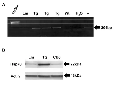
Genotyping of Hsp70 transgenic mice performed by PCR using specific primers for Hsp70 revealed a 304bp fragment in Hsp70 Tg mice (Figure 2A). To confirm the overexpression of Hsp70 protein in Hsp70 Tg mice, Western blot were performed which show more than 10-fold higher expression of Hsp70 in the brain (Figure 2B).
A B 304bp 72kDa 43kDa Hsp70 Actin Lm Tg CB6 Lm Tg Tg Tg Wt H2O +
Figure 2. Genotyping and overexpression of heat shock protein 70 (Hsp70) transgenic mice. Genotyping was performed by PCR (Figure 1A). Western blot analysis showed ~ 10 fold higher expression of Hsp70 protein in the brain of either a wild-type
littermate (LM) or background strain (CB6) (Figure 1B).
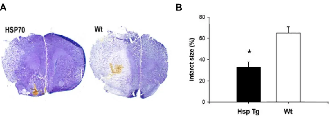
Overexpression of Hsp70 in Hsp70 Tg mice reduces infarct size of ischemic brain.
As shown in Figure 3A, a representative brain section from an Hsp70 Tg mouse had smaller infarct size than form a Wt mouse. Total infarct size among Hsp70 Tg mice was significantly reduced by about 50% compared with Wt mice (Figure 3B).
Figure 3. Brain infarct volume on crerbral ischemia. Representative cresyl violet-stained brain sections showed a smaller infarct in Hsp70 Tg mice compared with Wt mice (Figure 2A). Infarct size among Hsp70 Tg mice was reduced by ~50% compared with wt (n=6/group; *P < 0.05).
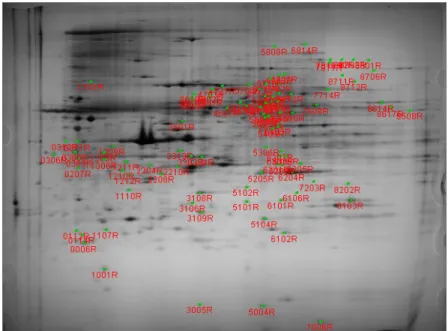
Differential protein expression in Wild type and Hsp70 Tg mice.
Proteomic studies to data, other than in biomedicine, have focused mainly on changes in genome expression that are triggered by environmental factors. As shown in 2-D gel, we could clearly separate the up-regulated and down-regulated proteins, ranging from low to high molecular weights. Most proteins were separated in the isoelectric point range of 4-7. Proteins extracted from untreated control were used as the control for comparative analysis. About 900 protein spots were detected in Hsp70 Tg and WT mice with computer-aided image analysis of 2-D gel. 114 protein spots were differently expressed when stained with silver (Figure 4).
Mice. 2D gels were generated, stained, and analyzed as described in text. Differently expressed proteins are depicted by numbers.
Proteomic identification of differentially expressed proteins in Hsp70 Tg mice.
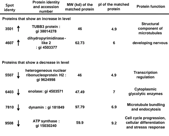
MALDI-TOF MS protein identification generates an overview of the processes occurring up to this point of cell survival and death and provides a fresh global perspective of alterations in a diverse range of proteins. Following tMCAO treatment, 6 proteins were found to be significantly different in Hsp70 Tg mice: four proteins showed a decrease in expression and two proteins showed an increase in expression. Specially, heterogenous nuclear ribonucleoprotein H2 and dynamin influenced activation of transcription factors (NF-κB and AP-1). These proteins decreased in Hsp70 Tg mice. (Table 1)
Table 1. Hsp70 Tg mice brain proteins with altered levels after tMCAO injury, identified by mass spectrometry.
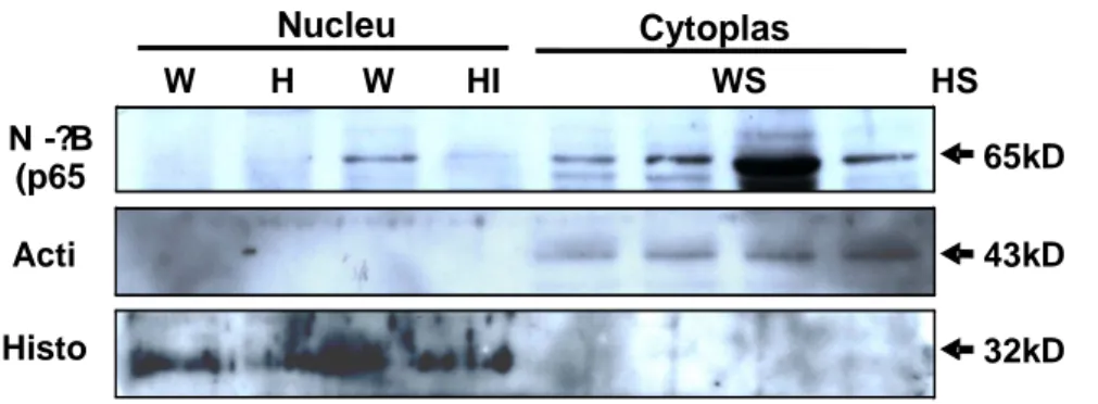
Overexpression of Hsp70 reduces the expression and nuclear translocation of NF-κB.
Expression of NF-κB proteins was confirmed by performing Western blots of cytosolic and nuclear subfractions in Hsp70 Tg and WT mice brains after tMCAO. NF-κB was found in the nuclear subfractions of ischemic Wt mice brains. In contrast,
Protein identity and accession number TUBB3 protein : gi38014278 MW (kd) of the matched protein Spot identy 3501 pIof the matched
protein Protein function
4.9 46 dihydropyrimidinase -like 2 : gi4503377 4607 62.73 6 Structural component of microtubules developing nervous 5507 46 4.9 enolase: gi4503571 6403 47.49 7 dynamin: gi181849 7810 97.79 6.9 9508 heterogeneous nuclear ribonucleoproteinH2 : gi9624998 ATP synthase: gi15030240 59.9 9.2 Microtubule bundling and endocytosis Transcription regulation Cytoplasmic glycolyticenzymes
Cell cycle progression, cellular differentiation and stresssresponse Proteins that show an increase in level
NF-κB was unchanged in ischemic Hsp70 Tg mice brains with little to no nuclear expression (Figure 5).
Figure 5. Overexpression of Hsp70 inhibits nuclear translocation of NF-κB. Western blots of cytosolic and nuclear subcellular fractions showed that NF-κB is increased in the ischemic hemispheres of Wt mice (WI) compared with the contralateral hemisphaere (WC) and both ischemic (HI) and contralateral hemisphaere (HC) of Hsp70 Tg mice. Ischemia also resulted in translocation of NF-κB to the nucleus in Wt mice, but not Hsp70 Tg mice.
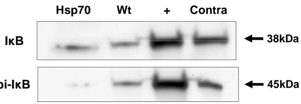
Overexpression of Hsp70 inhibits IκB phosphorylation.
Phosphorylation of IκB leads to subsequent IκB degradation, thus lierating NF-N -?B (p65 Nucleu Cytoplas Acti Histo 65kD W H W HI WS HS WI 43kD 32kD
κB to enter the nucleus. Western blot of IκB and phosphorylated IκB after tMCAO
revealed no significant difference in the amount of overall IκB protein between Hsp70 Tg and Wt mice (Figure 6). However, phosphorylated IκB levels were significantly decreased among Hsp70 Tg mice. This result suggested that overexpression of Hsp70 was somehow capable of preventing IκB phosphorylation.
Hsp70 Wt + Contra
pi-I
κ
B
I
κ
B
45kDa
38kDa
Figure 6. Less IκB phosphorylation observed in Hsp70 transgenic mice. Western blot of IκB show no difference in the amount of total IκB protein in the brain of Hsp70 Tg and Wt mice exposed to tMCAO. In contrast, phosphorylated IκB was markedly reduced in the brains of Hsp70 Tg mice after tMCAO.
Hsp70 interacts with NF-κB and IκB.
To explore further interactions between Hsp70 and NF-κB signaling in in vivo, we performed co-immunoprecipitation experiments with NF-κB and IκB. Hsp70 appeared to co-immunoprecipitate with NF-κB p65 subunit and IκB in Hsp70 Tg mouse (Figure 7).
Figure 7. Hsp70 co-immunoprecipitates with NF-κB and IκB. Mice brain tissue lysates were co-immunoprecipitated with Hsp70 antibody, followed by antibodies against NF-κB and IκB. Hsp70 was found to associate with NF-κB and IκB following tMCAO.
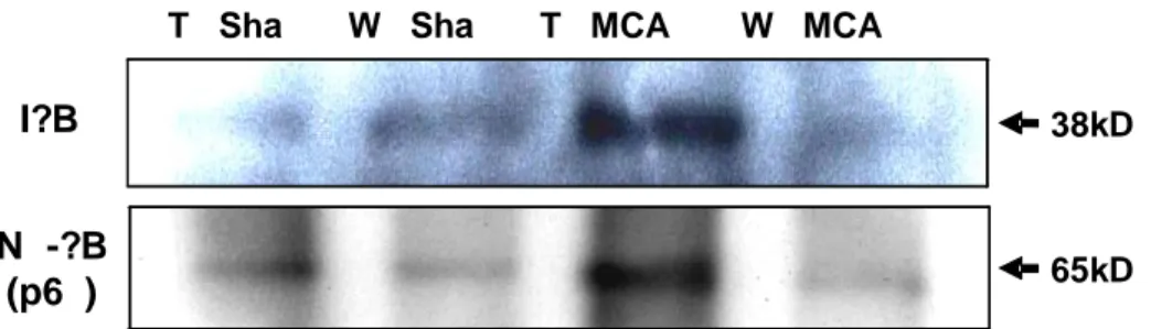
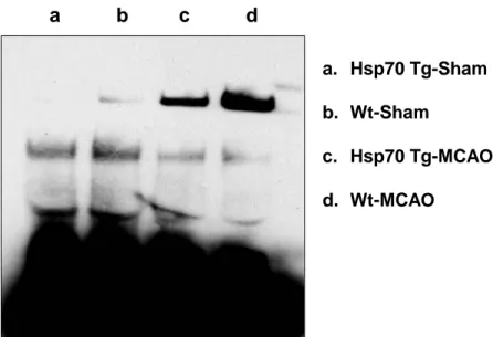
Overexpression of Hsp70 decreases DNA binding capacity of NF-κB.
To corroborate the inhibitory effects of Hsp70 on NF-κB activation, we further examined DNA binding capacity of NF-κB using EMSA assay. We show that DNA
N -?B (p6 )
I?B
T Sha W Sha T MCA W MCA
38kD a 65kD
binding of NF-κB is significantly decreased in Hsp70 Tg mouse ischemic brain (Figure 8). a. Hsp70 Tg-Sham b. Wt-Sham c. Hsp70 Tg-MCAO d. Wt-MCAO
a b c d
Figure 8. Decreased DNA binding of NF-κB in Hsp70 Tg mice. Using an EMSA assay that estimates DNA binding capacity of NF-kB activity was observed in Hsp70 Tg and Wt mice brains. Decreased NF-κB activity was observed in injured Hsp70 Transgenic mice brain compared with uninjured Wild type mice brain.
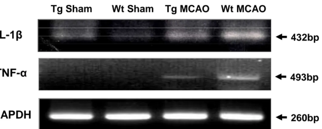
Overexpression of Hsp70 inhibits the expression of several NF-κB-regulated genes.
RT-PCR of pro-inflammatory NF-κB-regulated genes, TNF-α and IL-1β were performed from brains exposed to tMCAO in Wt and Hsp70 Tg mouse. Our results show that while ischemic hemispheres from Wt mouse have several-fold greater expression of TNF-α and IL-1β after tMCAO compared with the corresponding contralateral hemispheres, the increased expression of these genes was significantly less in the Hsp70 Tg mouse (Figure 9). Ischemic
hemispheres from the Hsp70 Tg
mouse had lower expression of all genes compared with Wt mouse.
Tg Sham Wt Sham Tg MCAO Wt MCAO
TNF-
α
GAPDH
IL-1
β
432bp493bp
260bp
Figure 9. Down-regulation of expression of several representative NF-κB-dependent pro-inflammatory genes in Hsp70 Tg mice. RT-PCR was used to estimate the
expression of NF-κB-dependent pro-inflammatory genes. Compared with Wt, expression of TNF-α and IL-1β was significantly inhibited at the mRNA level in Hsp70 Tg mice following both brain ischemia.
Overexpression of Hsp70 in primary cultured astrocytes by heat pretreatment.
Hsp70 overexpression was induced by heat pretreatment, which induced 2~3 fold higher expression in the protein level of Hsp70 compared to OGD injury (Figure 11). Hsp70 Heat OGD – + + – – – + + Actin 72kDa 43kDa
Figure 10. Western blot analysis of Hsp70 overexpression. Western blot analysis shows 2~3-fold higher expression of Hsp70 protein in the heated OGD injury compared to OGD injury.
Protective effect of Hsp70 overexpression on OGD injury in primary cultured astrocyte.
To investigate the protective effect of Hsp70 overexpression on OGD injury, primary cultured astrocytes were subjected to OGD for 4h and restoration up to 20h in the heat pretreatment. Effect of Hsp70 overexpression was analyzed by Hoechst-PI nuclear staining (Figure 10). Hoechst-stained nuclei (blue) of astrocytes after OGD injury were gradually decreased after restoration time. In contrast, PI-stained nuclei (red) increased by OGD injury. However, heat pretreated astrocytes had more Hoechst-stained nuclei in comparison with cells receiving no receiving no treatment.
NC Heat
Heat-OGD
OGD
Figure 11. Protective effect of heat pretreatment on the primary cultured astrocytes in OGD injury. Microphotographs of primary cultured astrocytes stained with Hoechst
(Blue) – Propidium iodide (Red) after heat pretreatment or OGD (Figure 9). Heat pretreatment reduced PI-positive cells induced OGD.
Overexpression of Hsp70 by heat pretreatment reduces expression and
activation of NF-
κB and IκB.
Overexpression of Hsp70 reduced protein level of NF-
κB and p-IκB and
inhibited nuclear translocation of NF–
κB and phosphorylation of IκB. There
showed by performing Western blots of cytosolic and nuclear subfractions.
Nuclear factor-
κB was found in the nuclear subfractions of OGD injury. In
contrast, NF-
κB was unchanged in the primary cultured astrocyte exposed
OGD injury after heat pretreatment with little nuclear expression. Western
blots of phosphorylated I
κB showed the amount of p-IκB protein was
increased in astrocyte exposed OGD injury. However, phosphorylated I
κB
levels were significantly decreased in astorcyte exposed OGD injury after heat
pretreatment. This result suggested that overexpression of Hsp70 inhibits
expression and activation of NF-
κB and p-IκB (Figure 12).
Figure 12. Western blot of NF-κB and pi-IκB on Hsp70 overexpression. Overexpression of Hsp70 reduces expression of NF-κB and pi-IκB and inhibits nuclear translocation of NF-κB and phosphorylation of IκB in the primary cultured astrocyte exposed OGD injury after heat pretreatment.
Overexpression of Hsp70 by heat pretreatment reduces expression and activation of JNK and c-Jun.
Overexpression of Hsp70 reduced protein level of Pi-JNK, c-Fos and Pi-c-Jun and inhibited nuclear translocation of c-Fos and Pi-c-Jun and phosphorylation of JNK and c-Jun. There showed by performing Western blots of cytosolic and nuclear subfractions. The c-Fos and p-c-Jun were found in the nuclear subfractions in the primary cultured astrocyte exposed OGD injury. In contrast, c-Fos and Pi-c-Jun were unchanged in astrocyte exposed OGD injury after heat pretreatment with little nuclear
p - IkB NF - kB (p65) Heat OGD Nucleu Cytoplas ? + + ? ? + + ? ? ? + + ? ? + + pi-I?B NF-?B (p65) Heat OGD Nucleu Cytoplas – + + – – + + – – – + + – – + + 4 kD 6 kD
expression. Phosphorylation of JNK leads activation of c-Fos and Pi-c-Jun to enter the nucleus. Western blots of phosphorylated JNK showed the amount of pi-JNK protein was increased in astrocyte exposed OGD injury. However, phosphorylated JNK levels were significantly decreased in astorcyte exposed OGD injury after heat pretreatment (Figure 13).
Figure 13. Western blot of pi-JNK, and pi-c-Jun on Hsp70 overexpression. Overexpression of Hsp70 reduces expression of pi-JNK and pi-c-Jun and inhibits nuclear translocation of pi-c-Jun and phosphorylation of JNK and c-Jun in the primary cultured astrocyte exposed OGD injury after heat pretreatment.
Overexpression of Hsp70 by heat pretreatment reduces expression and activation of p38, c-Fos and STAT-1.
Overexpression of Hsp70 reduced protein level of pi-p38, c-Fos and pi-STAT-1
pi-JNK Heat OGD Nucleu Cytoplas – + + – – + + – – – + + – – + + pi-c-Jun 5 kD 4 kD
and inhibited nuclear translocation of c-Fos and pi-STAT-1 and phosphorylation of p38 and STAT-1. There showed by performing Western blots of cytosolic and nuclear subfractions. The pi-STAT-1 was found in the nuclear subfractions in the primary cultured astrocyte exposed OGD injury. In contrast, pi-STAT-1 was unchanged in astrocyte exposed OGD injury after heat pretreatment with little nuclear expression. Phosphorylation of p38 leads activation of STAT-1 to enter the nucleus. Western blots of phosphorylated p38 showed the amount of pi-p38 protein was increased in astrocyte exposed OGD injury. However, phosphorylated p38 levels were significantly decreased in astorcyte exposed OGD injury after heat pretreatment (Figure 14).
Figure 14. Western blot of pi-p38, c-Fos and pi-STAT-1 on Hsp70 overexpression. Overexpression of Hsp70 reduces expression of pi-p38, c-Fos and pi-STAT-1 and
Heat OGD Nucleu Cytoplas pi-STAT-1 pi-p38 – + + – – + + – – – + + – – + + c-Fos 4 kD 6 kD 8 kD
inhibits nuclear translocation of pi-STAT-1 and phosphorylation of p38 in the primary cultured astrocyte exposed OGD injury after heat pretreatment.
Hsp70 overexpression inhibits phosphorylation of JNK, p38, STAT-1 and c-Jun in OGD injury after heat pretreatment.
To confirm the phosphorylation results of Western blot, we further measured the amount of phosphorylation using the phosphor ELISA assay. Phospho ELISA assay of JNK, p38, STAT-1 and c-Jun showed increase in the amount of phosphorylation in the primary cultured astrocyte exposed to OGD injury. In contrast, phosphorylated JNK, p38, STAT-1 and c-Jun were markedly reduced in the primary cultured astrocyte exposed to OGD injury after heat pretreatment (Figure 15).
Figure 15. Detection of phosphorylated JNK, p38, STAT-1 and c-Jun by phospho ELSA assay. Phosphorylation of p38 reduced in the amount of overall phosphorylation after heat pretreatment. However, this was significantly increased in OGD injury. Similar patterns were observed in phosphorylation of JNK, c-Jun and STAT-1.
Hsp70 overexpression by heat pretreatment interacts with NF-κB and IκB.
To explore further interactions between Hsp70 and NF-κB in in vitro, we -0.1 0 0.1 0.2 0.3 0.4 0.5
Con Heat Heat-OGD OGD
pi-STAT-1 n g /m 0 2 4 6 8
Con Heat Heat-OGD OGD
pi-JNK1/2 ng /m 0 0.4 0.8 1.2
Con Heat Heat-OGD OGD
pi-p38 pg/ 1 2 4 8
Con Heat Heat-OGD OGD
pi - c - Jun
performed co-immunoprecipitation experiments with NF-κB and IkB. Hsp70 appeared to co-immunoprecipitate with NF-κB p65 subunit and IκB in primary cultured astrocyte after heat pretreatment (Figure 16).
I
κ
B
NF-
κ
B
(p65)
Heat OGD–
+ + –
–
–
+ +
38kDa
65kDa
Figure 16. Hsp70 co-immunoprecipitates with NF-κB and IκB. Astrocyte lysates were co-immunoprecipitated with Hsp70 antibody, followed by antibodies against NF-κB and IκB. Hsp70 was found to associate with NF-κB and IκB in the primary cultured astrocyte after heat pretreatment.
Hsp70 overexpression by heat pretreatment interacts with JNK and c-Jun.
To explore further interactions between Hsp70 and AP-1 signaling in in vivo, we performed co-immunoprecipitation experiments with JNK and c-Jun. Hsp70 appeared to co-immunoprecipitate with p38 and c-Jun in primary cultured astrocyte after heat
pretreatment (Figure 17).
Figure 17. Hsp70 co-immunoprecipitates with JNK and c-Jun. Astrocyte lysates were co-immunoprecipitated with Hsp70 antibody, followed by antibodies against JNK and c-Jun. Hsp70 was found to associate with JNK and c-Jun in the primary cultured astrocyte after heat pretreatment.
Hsp70 overexpression by heat pretreatment interacts with p38, c-Fos and
STAT-1.
To explore further interactions between Hsp70 and STAT-1 signaling in
in vitro, we performed co-immunoprecipitation experiments with p38, c-Fos
JNK c-Jun Hea OG – + + – – – + + 54 kDa 62kDa
and STAT-1. Hsp70 appeared to co-immunoprecipitate with p38, c-Fos and
STAT-1 in primary cultured astrocyte after heat pretreatment (Figure 18).
Figure 18. Hsp70 co-immunoprecipitates with p38, c-Fos and STAT-1. Astrocyte lysates were co-immunoprecipitated with Hsp70 antibody, followed by antibodies against p38, c-Fos and STAT-1. Hsp70 was found to associate with p38, c-Fos and STAT-1 in the primary cultured astrocyte after heat pretreatment.
p3
c
-
Fo
Heat OG D–
+
+
–
–
–
+
STA -1
4 kD
4 kD
8 kD
Overexpression of Hsp70 decreases DNA binding capacity of NF-κB, STAT-1 and AP-1.
To corroborate the inhibitory effects of Hsp70 on transcription factors activation, we further examined DNA binding capacity of NF-κB, AP-1 and STAT-1 using EMSA assay. We show that DNA binding of NF-κB, AP-1 and STAT-1 is significantly decreased in the primary cultured astrocyte exposed OGD after heat treatment (Figure 19).
Figure 19. Decreased DNA binding of transcription factos in Hsp70 Tg mice. Using an EMSA assay that estimates DNA binding capacity of transcription factors activity
Heat
OGD
NF
-
kB
–
+ +
–
–
–
+ +
AP
-
1
–
+ +
–
–
–
+ +
STAT
-
1
– + +
–
– –
+ +
was observed in the primary cultured astrocytes. Decreased NF-κB, STAT-1, AP-1 activity was observed in astrocyte exposed OGD injury after heat pretreatment compared with astrocyte exposed OGD injury.
Overexpression of Hsp70 inhibits the expression of several transcription factor-regulated genes.
RT-PCR of pro-inflammatory transcription factor-regulated genes, TNF-α, IL-1β and MMP-9 were performed from primary cultured astrocyte exposed to heat pretreatment and OGD injury. RT-PCR analysis showed several-fold higher expression of TNF-α, IL-1β and MMP-9 mRNA in astrocyte exposed OGD injury compared with astrocyte exposed OGD injury after heat pretreatment (Figure 20).
Heat OGD
–
+ + –
–
–
+ +
MMP-9
IL-1
β
TNF-
α
GAPDH
300bp 432bp 493bp 260bpFigure 20. Overexpression of Hsp70 inhibits expression of several representative transcription factors-dependent pro-inflammatory genes in primary cultured astrocyte. RT-PCR was used to estimate the expression of transcription factor-dependent pro-inflammatory genes. Compared with OGD injury, expression of TNF-α, IL-1β and MMP-9 was significantly inhibited at the mRNA level in primary cultured astrocyte exposed OGD injury after heat pretreatment.
IV.
DISCUSSION
In this study, we characterize neuroprotective effect of Hsp70 in ischemic injury. Following previously our study, Hsp70 decreased infarct sizes in middle cerebral artery occlusion mouse model, and prevented cell death of neuron and astrocyte exposed to OGD injury21. The cytotoxic properties of inflammation after brain ischemia have been well documented and we and other groups have shown reduction in infarct size by inhibiting various inflammatory mediators25,26. This study provides the evidence of an anti-inflammatory property of Hsp70 to explain its neuroprotective function in brain ischemia. Moreover, the findings of this results show that Hsp70 interrupts activation of transcription factors (NF-κB, AP-1 and STAT-1) important in regulating cell death and expression of several downstream pro-inflammatory genes by interacting and preventing the phosphorylation of these proteins.
Transcription factors play a pivotal role in controlling inflammatory gene expression. Microarray studies from other groups showed the induction of many transcription factors after focal ischemia 27,28,29. Of these, activation of hypoxia inducible factor-1 (HIF-1), CREB, c-fos, PPARα, PPARγ and p53 is known to prevent ischemic neuronal damage and/or promote ischemic tolerance 30,31,32 whereas the induction of IRF-1, signal transducer and activator of transcription 1 (STAT-1), NF-κB, early growth response-1 (Egr1) and C/EBPβ promotes inflammation and neuronal death after cerebral ischemia 33,34,35,36. Degradation of IκB induces NF-κB
activation leading to the coordinated induction of multiple genes is involved in many inflammatory and immune cascades. Genes induced by NF-κB include pro-inflammatory cytokines IL-1β, TNF-α and granulocyte-macrophage colony stimulating factor (GM-CSF), and the chemokines IL-8, MIP-1α and MCP-1, that are largely responsible for attracting inflammatory cells into sites of inflammation 37,38,39. Many NF-κB down-stream gene products like IL-1β and Tα also re-activate
NF-κB itself, resulting in a positive regulatory loop that amplifies and perpetuates the
inflammatory responses42. In our in vivo data, activation of NF-κB occurs after MCAO and inhibiting NF-κB activity results in smaller infarcts.
AP-1 is a heterodimer of fos and jun oncoproteins, a collection of related transcription factors belonging to the Fos (cFos, FosB, Fra1, Fra2) and Jun (c-Jun, JunB, JunD) families that dimerize in various combinations through their leucine zipper regions43. AP-1 will be activated by various cytokines, including TNFα and IL-1β via several protein tyrosine kinases and MAP kinases. Transcription factors Fos, c-Jun and c-JunB have been shown to be upregulated following cerebral ischemia44,45,46 and c-Jun was thought to play a role in the apoptotic neuronal death47.
Also, Signal transducers and activators of transcription (STAT) factors are a family of cytoplasmic transcription factors that mediate intracellular signaling initiated at cytokine cell surface receptors and transmitted to the nucleus. STATs are activated by phosphorylation on conserved tyrosine and serine residues by the Janus kinases (JAKs) and MAP kinase families respectively, which allow the STATs to dimerise and translocate to the nucleus and thereby regulate gene expression. The
C-terminal domains of STAT proteins all contain a transcriptional transactivation domain (TAD), plus the phosphorylation site for JAKs and MAPK, which are essential for maximal STAT function. At present seven different STAT family members have been characterized and found to be encoded by distinct genes (STAT-1, STAT-2, STAT-3, STAT-4, STAT- 5α, STAT-5β and STAT-6). Different STATs are activated by distinct group of cytokines. For example, STAT-1 was also activated by both IL-4 and IL-13 in all the cell types tested48,49. In the endothelial cell line, STAT-1 bound to the GRR probe, suggesting that STAT-STAT-1 might also contribute to cell signaling in response to IL-4 and IL-1353.
The major focus of the present work concentrated on the early inflammatory events that have been shown to contribute to the exacerbation of ischemic brain injury. Our data indicate that the mechanism of Hsp70 inhibitory effect on transcriptional activation, including the NF-κB, AP-1 and STAT-1 pathway, appears to occur through the prevention of their phosphorylation. Our study indicate that Hsp70 was found to associate with NF-κB, AP-1 and STAT-1 suggestion the possibility that Hsp70 physically binds to NF-κB, AP-1 and STAT-1 its regulatory proteins, thereby stabilizing and tethering NF-κB, AP-1 and STAT-1 in the cytosol as a result.
The findings not only provide a mechanistic basis for the neuroprotective effects of Hsp70 on brain ischemia, but would further support the potential of developing Hsp70 as a therapeutic against a variety of conditions, given that it may protect by several mechanisms.
V. CONCLUSION
This present study showed the effects of neuroprotection of overexpression Hsp70 after ischemic injury and the relationship between Hsp70 and transcription factors, and evaluated the effect of Hsp70 on ischemic injury in Tg mice after tMCAO and in heat pretreated the primary cultured astrocytes after OGD. These results have led me demonstrate the following conclusion.
1. Overexpression Hsp70 reduced infarct size in Hsp70 transgenic mice after tMCAO and protected the primary cultured astrocytes from cell death induced by OGD injury.
2. The expression of transcription factors and their regulatory proteins levels was decreased by overexpressing Hsp70 in Hsp70 Tg mice and heat pretreated the primary cultured astrocytes after tMCAO and OGD injury.
3. Hsp70 interacted with transcription factors and their regulatory proteins in Hsp70 Tg mice and heat pretreated the primary cultured astrocyte after tMACO and OGD injury.
4. Through overexpression of Hsp70 blocked phosphorylation of transcription factors and their regulatory proteins, several pro-inflammatory genes
interrupted the transcription
Taken together, these data showed the neuroprotective mechanism of Hsp70 on brain inflammation owing to ischemic stroke. The effects were related to the several pro-inflammatory genes interrupted the transcription through overexpression of Hsp70 blocked phosphorylation and expression of transcription factors and their regulatory proteins. We provide a mechanistic basis for the neuroprotective effects of Hsp70 on brain ischemia and Hsp70 anti-inflammatory function related at the transcriptional level.
Reference
1. Hossmann KA. Thresholds of ischemic injury. Cerebrovascular Disease. Pathophysiology, Diagnosis, and Treatment 1998;193-04.
2. Mehta SL, Manhas N, Raghubir R. Molecular targets in cerebral ischemia for developing novel therapeutics. Brain Res Rev 2007;54(1);34-66.
3. Ghosh S, May MJ, Kopp EB. NF-kappa B and Rel proteins: evolutionarily conserved mediators of immune response. Annu Rev Immunol 1998;16;225-60.
4. O’Neill LA, Kaltschmidt C. NF-κB: a crucial transcription factor for glial and neuronal cell function. Trends Neurosci 1997;20(6):252-58
5. Stasiolek M Gavilyuk V, Sharp A, Horvath P, Selmaj K, Feinstein DL. Inhibitory and stimulatory effects of lactacystin on expression of nitric oxide synthase type 2 in brain glial cells. J Biol Chem 2000; 275:24847-56. 6. Karin M. The beginning of the end: IκB kinase (IKK) and NF-κB activation.
J Bio Chem 1999;274:27339-42.
7. Chen Z, Hagler J, Palombella VJ, Melandri F, Scherer D, Ballard D, et al. Signal-induced site-specific phosphorylation targets IκBa to the ubiquitinproteasome pathway. Genes Dev 1995;9:1586-97.
8. Sun SC, Ganchi PA, Ballard DW, Greene WC. NF-kB controls expression of inhibitory IκBa: evidence for an autoregulatory pathway. Science 1993;259:1912-15.
9. Akaji K, Suga S, Fujino T, Mayanagi K, Inamasu J, Horiguchi T, et al. Effect of intra-ischemic hypothermia on the expression of c-Fos and c-Jun, and DNA binding activity of AP-1 after focal cerebral ischemia in rat brain. Brain Res 2003;975:149–157
10. Pennypacker KR, Kassed CA, Eidizadeh S, O'Callaghan JP. Brain injury: prolonged induction of transcription factors. Acta Neurobiol Exp 2000;60(4):515-30.
11. Shaulian E, Karin M. AP-1 in cell proliferation and survival. Oncogene 2001;20(19):2390-400.
12. Wang HH, Hsieh HL, Wu CY, Sun CC, Yang CM.. Oxidized low-density lipoprotein induces matrix metalloproteinase-9 expression via a p42/p44 and JNK-dependent AP-1 pathway in brain astrocytes. Glia. 2008;[Epub ahead of print].
13. Monteleone G, Holloway J, Salvati VM, Pender SL, Fairclough PD, Croft N, et al. Activated STAT4 and a functional role for IL-12 in human Peyer’s patches. J Immunol 2003;170:300–7.
14. Jacobson NG, Szabo SJ,Weber-Nordt RM, Zhong Z, Schreiber RD, Darnell JE Jr, et al. Interleukin signaling in T helper type 1 (Th1) cells involves tyrosine phosphorylation of signal transducer and activator of transcription (Stat)3 and Stat4. J Exp Med 1995;181:1755–62.
15. Takeda K, Tanaka T, Shi W, Matsumoto M, Minami M, Kashiwamura S, et al. Essential role of Stat6 in IL-4 signaling. Nature 1996;380:627–30.
16. Afkarian M, Sedy JR, Yang J, Jacobson NG, Cereb N, Yang SY, et al. T-bet is a STAT1-induced regulator of IL-12R expression in naive CD4+ T cells. Nat Immunol 2002;3:549–57.
17. Neurath MF, Weigmann B, Finotto S, Glickman J, Nieuwenhuis E, Iijima H, et al. The transcription factor T-bet regulates mucosal T cell activation in experimental colitis and Crohn’s disease. J Exp Med 2002;195:1129–1143. 18. Bode JG, Gatsios P, Ludwig S, Rapp UR, Haussinger D, Heinrich PC, et al.
The mitogen-activated protein (MAP) kinase p38 and its upstream activator
MAP kinase 6 are involved in the activation of signal transducer and
activator of transcription by hyperosmolarity. J Biol Chem 1999;274:30222–
27
19. Giffard RG, Xu L, Zhoa H, Carrico W, Ouyang Y, Qiao Y, et al. Chaperones, protein aggregation, and brain protection from hypoxia/ischemic injury. J Exp Biol 2004;271;17723-32.
20. Xu L, Dayal M, Ouyang YB, Sun Y, Yang CF, Frydman J, et al. Chaperonin GroEL and its mutant D87K protect from ischemia in vivo and in vitro. Neurobiol Aging 2006;27;562-9.
21. Lee JE, Yenari MA, Sun GH, Xu L, Emond MR, Cheng D, et al. Differential neuroprotection from human heat shock protein 70 overexpression in in vitro and in vivo models of ischemia and ischemia-like conditions. Exp Neurol 2001;170;129-39.
therapy with HSP72 is neuroprotective in rat models of stroke and epilepsy. Ann Neurol 1998;44;584-91.
23. Deng H, Han Hs, Cheng D, Sun GH, Yenari MA. Mild hypothermia inhibits inflammation after experimental stroke and brain inflammation. Stroke 2003;34;2495-501.
24. Srivastava P. Roles of heat-shock proteins in innate and adaptive immunity. Nat Rev Immunol 2002;2;185-94.
25. Zheng Z, Yenari MA. Post-ischmeic inflammation: molecular mechanisms and therapeutic implication. Neural Res 2004;26:884-92.
26. Stoll G, Jander S, Schroeter M. Inflammation and glial response in ischemic lesions. Pro Neurobiol 1998;56:149-71
27. Angstwurm K, Freyer D, Dirnagl U, Hanisch UK, Schumann RR, Einhaupl KM, et al. Tumor necorsis factor alpha induces only minor inflammatory changes in the central nervous system, but augments experimental meningitis. Neuroscience 1998;86:627-34.
28. Satriotomo I, Bowen KK, Vemuganti R. JAK2 and STAT3 activation contributes to neuronal damage following transient focal cerebral ischemia. J Neurochem 2006;98:1353–68.
29. Vemuganti R, Bowen KK, Dhodda VK, Song G, Franklin JL, Gavva NR, et al. Gene expression analysis of spontaneously hypertensive rat cerebral cortex following transient focal ischemia. J Neurochem 2002;83:1072–86. 30. Lu A, Tang Y, Ran R, Clark JF, Aronow BJ, Sharp FR. Genomics of the
periinfarction cortex after focal cerebral ischemia. J Cereb Blood Flow Metab 2003;23:786–810.
31. Tanaka K, Nogawa S, Ito D, Suzuki S, Dembo T, Kosakai A, et al. Activated phosphorylation of cyclic AMP response element binding protein is associated with preservation of striatal neurons after focal cerebral ischemia in the rat. Neuroscience 2000;100:345–54.
32. Cho S, Park EM, Kim Y, Liu N, Gal J, Volpe BT, et al. Early c-Fos induction after cerebral ischemia: a possible neuroprotective role. J Cereb Blood Flow Metab 2001;21:550–56.
33. Maeda K, Hata R, Gillardon F, Hossmann K. Aggravation of brain injury after transient focal ischemia in p53-deficient mice. Mol Brain Res 2001;88:54–61.
34. Iadecola C, Salkowski CA, Zhang F, Aber T, Nagayama M, Vogel SN, et al. The transcription factor interferon regulatory factor 1 is expressed after cerebral ischemia and contributes to ischemic brain injury. J Exp Med 1999;189:719–27.
35. Johansson IM, Wester P, Hakova M, Gu W, Seckl JR, Olsson T. Early and delayed induction of immediate early gene expression in a novel focal cerebral ischemia model in the rat. Eur. J Neurosci 2000;12:3615–25. 36. Stephenson D, Yin T, Smalstig EB, Hsu MA, Panetta J, Little S, et al.
Transcription factor nuclear factor-kappa B is activated in neurons after focal cerebral ischemia. J Cereb Blood Flow Metab 2000;20:592–603.
37. Tanaka K, Nogawa S, Nagata E, Ito D, Suzuki S, Dembo T, et al. Persistent CREB phosphorylation with protection of hippocampal CA1 pyramidal neurons following temporary occlusion of the middle cerebral artery in the rat. Exp Neurol 2000b;161:462–71.
38. Nelson PJ, Kim HT, Manning WC, Goralski TJ, Krensky AM. Genomic organization and transcriptional regulation of the RANTES chemokine gene. J Immunol 1993;151:2601–12.
39. Siebenlist U, Franzoso G, Brown K. Structure, regulation and function of NF-kappa B. Annu. Rev Cell Biol 1994;10:405–55.
40. Mukaida N, Morita M, Ishikawa Y, Rice N, Okamoto S, Kasahara T, et al. Novel mechanism of glucocorticoid-mediated gene repression. Nuclear factor-kappa B is target for glucocorticoidmediated interleukin 8 gene repression. J Biol Chem 1994;269:13289–95.
41. Van de Stolpe A, Caldenhoven E, Stade BG, Koenderman L, Raaijmakers JA, Johnson JP, et al. 12-O-tetradecanoylphorbol-13-acetate- and tumor necrosis factor alpha-mediated induction of intercellular adhesion molecule-1 is inhibited by dexamethasone. Functional analysis of the human intercellular adhesion molecular-1 promoter. J Biol Chem 1994;269:6185–92.
42. Iademarco MF, Barks JL, Dean DC. Regulation of vascular cell adhesion molecule-1 expression by IL-4 and TNF-alpha in cultured endothelial cells. J Clin Invest 1995;95:264–71.
1998;12:221–34.
44. Kindy MS, Carney JP, Dempsey RJ, Carney JM. Ischemic induction of protooncogene expression ingerbil brain. J Mol Neurosci 1991;2:217–28. 45. Woodburn VL, Hayward NJ, Poat JA, Woodruff GN, Hughes J. The effect of
dizocilpine and enadoline on immediate early gene expression in the gerbil global ischaemia model. Neuropharmacology 1993;32:1047–59.
46. Dragunow M, Beilharz E, Sirimanne E, Lawlor P, Williams C, Bravo R, et al. Immediate-early gene protein expression in neurons undergoing delayed death, but not necrosis, following hypoxicischaemic injury to the young rat brain. Brain Res Mol Brain Res 1994;25:19–33.
47. Raivich G, Behrens A. Role of the AP-1 transcription factor c-Jun in developing, adult and injured brain. Prog Neurobiol 2006;78:347–63. 48. Darnell J.E., Jr., STATs and gene regulation, Science, 1997;277:1630-35. 49. Ihle J.N. The STAT family in cytokine signaling, Curr.Opin. Cell Biol,
2001;13:211-17,
50. Wang IM, Lin H, Goldman SJ, Kobayashi M. STAT-1 is activated by IL-4 and IL-13 in multiple cell types. Mol Immunol 2004;41(9):873-84.