Agreement of Retinal Nerve Fiber Layer Color Codes
between Stratus and Cirrus OCT according to
Glaucoma Severity
Chan Yun Kim,
1Ji Won Jung,
2So Yeon Lee,
3and Na Rae Kim
2 PURPOSE. We assessed the agreement of retinal nerve fiber layer(RNFL) color codes between Stratus and Cirrus optical coher-ence tomography (OCT) according to the glaucoma severity. METHODS. We analyzed 184 eyes from 144 subjects with glaucoma and 40 normal subjects. Glaucoma was categorized into three subgroups according to mean deviation of visual field. The agreement of RNFL color code according to the glaucoma severity was assessed using Cohen’s kappa value. Red or yellow color codes were defined as abnormal results, while green or white were defined as normal. Disagreement frequency of abnormal color code displayed by 2 OCTs was evaluated. RESULTS. The agreement of RNFL color code was fair to good in
all subgroups (j 0.288–0.887) except for the nasal quadrant, 1 to 4, and the 9 o’clock sectors. In all severity groups, the frequency of disagreement was less than 10% for average, temporal, and inferior sectors. However, Stratus OCT displayed abnormal results, while Cirrus showed normal results for nasal sectors with considerable disagreement proportion (severe 22.5%, moderate 13.2%, and early 13.6%). The disagreement tended to be pronounced in severe glaucoma. On the other hand, Cirrus OCT displayed abnormal results, while Stratus showed normal results for superior sectors (severe 10.0%, moderate 10.5%, and early 15.2%). The frequency was higher in early glaucoma.
CONCLUSIONS. Considerable color code disagreements were
observed in nasal and superior sectors, and the proportion was different according to the glaucoma severity. Color codes of two OCTs could not be considered interchangeable. (Invest Ophthalmol Vis Sci. 2012;53:3193–3200) DOI:10.1167/ iovs.12-9440
G
laucoma is a progressive disease characterized by patho-logic loss of ganglion cells and the retinal nerve fiber layer (RNFL).1–3Detecting disease progression, which is evidencedby structural or functional changes in the retina, in patients with glaucoma is essential to evaluate the damage and the efficacy of therapy.4Repeated measurements of RNFL thickness
in the same patient over time are an important component of glaucoma management. Optical coherence tomography (OCT) provides cross-sectional imaging of the eye, and enables evaluation and quantification of cross-sectional peripapillary RNFL thickness.5OCT also provides information on probable
RNFL defects as four color codes through comparison of the RNFL profile with an internal age-matched normative database. Color codes in RNFL profiles of OCT are useful clinically in diagnosis of glaucoma as actual RNFL measurements.
The many patients who have been scanned with Stratus time-domain OCT (Carl Zeiss Meditec, Inc., Dublin, CA) potentially now can be scanned with Cirrus spectral-domain OCT (Carl Zeiss Meditec).6 Recently, OCT technology has
advanced considerably and now incorporates spectral-domain (SD) imaging, which offers significant advantages over traditional time-domain (TD) OCT techniques, including a faster scanning speed and higher resolution RNFL imaging.7,8
The clinical relevance of the new SD technology requires that the data from these devices be compared to preexisting and widely accepted TD standards, and to each other.9
Previous studies have indicated that measurements with Stratus and Cirrus OCT could not be considered interchange-able, although the two types of measurements were correlated strongly.6,9–15However, to our knowledge there have been no
reports on the agreement of RNFL color codes between both OCT instruments according to the glaucoma severity. The purpose of our study was to evaluate the agreement of RNFL color codes between Stratus and Cirrus OCT according to the glaucoma severity.
M
ETHODS SubjectsThe184 participants were enrolled consecutively from the Glaucoma-Cataract Clinic of Severance Hospital in the Yonsei University Health System from January 2010 to June 2011. The study was approved by the Institutional Review Board, and complied with the tenets of the Declaration of Helsinki. Informed consent was obtained from each patient.
All subjects underwent Goldmann applanation tonometry, gonios-copy, and fundus examination with a 90-diopter lens. Automated refraction, biometry measurement, and standard visual field test were performed. All eyes were scanned with Stratus TD-OCT (fast RFNL scan mode, Carl Zeiss Meditec) and Cirrus SD-OCT (optic disc cube mode, Carl Zeiss Meditec) after pupillary dilation to a minimum diameter of 5 mm. For each patient, all examinations were performed during a single day.
Normal eyes were defined as those from patients with no family history of glaucoma in a first-degree relative, no history or evidence of From the 1Institute of Vision Research, Department of
Ophthalmology, Yonsei University College of Medicine, Seoul, Korea; the 2Department of Ophthalmology and Inha Vision Science
Laboratory, Inha University School of Medicine, Incheon, Korea; and
3Nune Eye Hospital, Seoul, Korea.
Supported by Grant A101727 from the Korea Health Technol-ogy R&D Project, Ministry of Health & Welfare, Republic of Korea and by Inha University Research Grant INHA-44097.
Submitted for publication January 6, 2012; revised March 23, 2012; accepted April 4, 2012.
Disclosure:C.Y. Kim, None; J.W. Jung, None; S.Y. Lee, None; N.R. Kim, None
Corresponding author: Na Rae Kim, Department of Ophthal-mology, Inha University Hospital, 7-206, 3-ga, Shinheung-dong, Jung-gu, Incheon, Korea, 400-711; Telephone 890-2400; Fax 82-32-890-2403; nrkim@inha.ac.kr.
Investigative Ophthalmology & Visual Science, May 2012, Vol. 53, No. 6
intraocular surgery, and no retinal pathological features. Normal eyes also had a best corrected visual acuity of 20/40 or better, with refractive error betweenþ3.00 and -6.00 diopters (D); intraocular pressure (IOP) of 21 mm Hg or lower; open angle on gonioscopy; normal-appearing optic nerve head (ONH) without evidence of cupping, rim loss, hemorrhages, or RNFL defects; and reliable normal visual field (VF) test (a mean deviation [MD] or a pattern standard deviation [PSD] within the 95% confidence interval [CI], and a normal glaucoma hemifield test).
Glaucomatous eyes were defined as those from patients with a glaucomatous VF defect confirmed by two reliable VF examinations, and by the appearance of a glaucomatous optic disc with typical loss of neuroretinal rim as judged by slit-lamp biomicroscopy (cup-to-disc ratio >0.7; intereye cup asymmetry >0.2; or neuroretinal rim notching, focal thinning, disc hemorrhage, or vertical elongation of the optic cup). IOP was not used as a criterion for the glaucoma group. Only patients with open anterior chamber angle, a corrected visual acuity of 20/40 or better, and refractive error fromþ3.00 to -6.00 D were selected. Glaucoma was categorized into three subgroups according to the modified Hodapp-Anderson-Parrish grading scale based on the MD of VFs.16Early glaucoma was defined as VF loss with an MD‡ -6 dB,
moderate glaucoma as an MD between-6 and -12 dB, and severe glaucoma as an MD worse than-12 dB.
Visual Field Examination
Standard VF testing was performed using automated static perimetry (Humphrey Field analyzer with Swedish Interactive Thresholding Algorithm [SITA] standard 24-2 test program; Carl Zeiss Meditec). The VF was considered reliable when fixation losses were less than 20%, and false-positive and -negative errors were less than 15%. Mean VF sensitivity was calculated with the perimetry software, and expressed as MD, PSD, and VF index (VFI). A field defect was defined as having three or more significant (P < 0.05) non–edge contiguous points with at least one at the P < 0.01 level on the same side of the horizontal meridian in the pattern deviation plot, classified as outside normal limits in the glaucoma hemifield test, and confirmed with at least two VF examinations.
OCT Measurements
Cross-sectional imaging of the peripapillary area was performed using Stratus-OCT (software version 4.0.1). The RNFL thickness was measured with the Fast RNFL scan program, which consisted of three
sets of 256 A-scans along a circle with a 3.46-mm diameter centered on the optic disc, which was controlled by the examiner. This scanning took 1.92 seconds to complete (400 A-scans per second).
The average RNFL thickness was determined using the optic disc cube protocol on Cirrus OCT (software version 3.0). This protocol generates a cube of data through a 6-mm square grid by acquiring a series of 200 horizontal scan lines, each of which is composed of 200 A-scans. A calculation circle, which was 3.46 mm in diameter and consisted of 256 A-scans, was positioned automatically around the optic disc. Cirrus OCT uses SD technology to acquire data with higher resolution and a faster scan speed (27,000 A-scans per second) than that obtained with TD-OCT technology.
A single, well-trained technician performed all OCT examinations. Both OCTs provide RNFL thickness maps with 4 quadrants (superior, inferior, nasal, and temporal) and 12 clock hours, which includes classification using an internal normative database. The results from the comparison of RNFL thickness with normative data were indicated with a stoplight color scheme for Stratus OCT and Cirrus OCT. The RNFL thicknesses in the normal range are represented by green backgrounds, those that are abnormal at the 5% level are represented by yellow backgrounds, and those that are abnormal at the 1% level are represented by red backgrounds.17The signal strengths of each scan
on the Stratus and Cirrus were at least 6 (range 0 to 10). Scans with misalignment, segmentation failure, or decentration of the measure-ment circle were excluded from the analysis.
Statistical Analyses
For cases in which both eyes of a patient were eligible for analysis, one eye was selected randomly to be evaluated for our study. The inter-device agreement between Stratus OCT and Cirrus OCT for the peripapillary RNFL thickness measurement according to the glaucoma severity was assessed using the intraclass correlation coefficient (ICC).18 A scatter plot showing the differences between two
measurements against their average was used to assess agreement between Cirrus and Stratus OCT RNFL measurements, as described by Bland and Altman.19
The agreement of RNFL color codes was evaluated with Cohen’s kappa.20After defining red or yellow color codes as abnormal results,
and green or white color codes as normal results, we evaluated color code disagreement between two OCTs, that is abnormal Stratus and normal Cirrus results, and normal Stratus and abnormal Cirrus results. The frequency of color code disagreement between two OCTs was TABLE1. Characteristic of Subjects
Severe Glaucoma (n¼ 40) Moderate Glaucoma (n¼ 38) Early Glaucoma (n¼ 66) P * Glaucoma (n¼ 144) Normal (n¼ 40) P † Age (years) 60.136 14.88 62.006 15.71 54.066 14.19 0.018 57.846 15.11 53.536 11.10 0.094 Female sex, no. (%) 17 (42.5%) 21 (55.3%) 32 (48.5%) 0.530 70 (48.6%) 21 (52.5%) 0.663 Mean deviation (dB) -18.49 6 5.25 -8.26 6 1.68 -3.63 6 1.50 <0.001 -8.98 6 6.92 -1.20 6 1.86 <0.001 Pattern standard deviation (dB) 11.386 3.05 6.616 3.54 4.226 2.16 <0.001 6.846 4.11 2.056 0.71 <0.001 Visual field index (%) 52.136 20.99 86.116 8.44 94.426 4.33 <0.001 81.776 20.69 98.536 1.71 <0.001 Intraocular pressure (mm Hg) 12.586 2.91 14.376 3.87 13.766 3.40 0.061 13.596 3.45 13.486 3.74 0.861 Central corneal thickness (lm) 525.186 43.65 535.456 32.04 544.056 35.92 0.160 537.066 37.65 536.096 31.06 0.900 Spherical equivalent (D) -0.72 6 2.32 -1.65 6 3.72 -1.19 6 2.78 0.609 -1.15 6 2.88 -0.69 6 1.83 0.331 Axial length (mm) 23.906 1.02 23.526 1.65 24.346 1.61 0.104 24.016 1.51 24.136 1.32 0.671 Mean RNFL thickness by Stratus
OCT (lm) 63.756 17.24 82.806 20.68 84.806 14.40 <0.001 78.426 19.24 104.626 9.99 <0.001 Signal strength by Stratus OCT 7.556 1.32 8.086 1.12 7.976 1.36 0.159 7.886 1.30 8.076 1.33 0.401 Mean RNFL thickness by Cirrus
OCT (lm) 66.836 12.49 79.136 16.58 79.826 12.19 <0.001 76.036 14.63 96.756 8.59 <0.001 Signal strength by Cirrus OCT 7.936 0.97 7.746 1.11 8.336 0.97 0.010 8.066 1.03 8.326 1.10 0.180
* Value for comparing early, moderate, and severe glaucoma. † Value for comparing normal and glaucoma.
measured according to the glaucoma severity. Also, the frequency of each color scale in average, quadrant, and 12 clock-hour map was measured according to the glaucoma severity.
Statistical analysis was performed using SPSS for Windows (version 12.0.0, SPSS Inc., Chicago, IL). A P value of less than 0.05 was considered statistically significant.
R
ESULTSSubject Characteristics
A total of 184 eyes from 144 subjects with glaucoma and 40 normal subjects was enrolled. Table 1 summarizes the demographic and clinical characteristics of the subjects. The subjects with glaucoma were divided into groups based on MD of VFs; early glaucoma (n¼ 66), moderate glaucoma (n ¼ 38), and severe glaucoma (n¼ 40). The ages of subjects in the glaucoma and normal groups were not significantly different. There was a significant age difference according to the glaucoma severity (Table 1).
Agreement of RNFL Thickness Measurements between Two OCTs According to Glaucoma Severity
For average RNFL thickness, inter-device agreement between Stratus and Cirrus OCT was excellent in normal eyes and in all glaucomatous eyes (ICC 0.559 in normal eyes, 0.843 in early glaucoma, 0.920 in moderate glaucoma, and 0.829 in severe glaucoma, P <0.001). For each quadrant and 12 clock-hour sectors, measurement agreement generally was good (ICC 0.608–0.944), except for the nasal quadrant and the 2, 3, and 4 clock-hour sectors (ICC 0.384–641, Table 2).
Figure 1 shows the Bland-Altman plots that were used to assess the agreement of the mean RNFL measurements between the two devices. Inspection of the plot revealed a considerable discrepancy between RNFL thickness measure-ments obtained by the two instrumeasure-ments. The limit of agreement range (95% CI) of the mean RNFL thickness between the two OCTs was 29.4 (-18.3–11.1) lm. Additionally, a significant proportional bias of the mean RNFL thickness measurements existed between the Stratus and Cirrus devices (r¼ -0.261, P
< 0.001). For thinner RNFLs, the Stratus measurements tended to be thinner than those of the Cirrus, whereas for thicker RNFLs, the Stratus measurements tended to be thicker than those of the Cirrus. The mean RNFL measurement difference between the Stratus and Cirrus devices was-3.59 6 7.51 lm (Cirrus OCT minus Stratus OCT RNFL thicknesses, 95% CI -4.68 to -2.49).
Table 3 reveals the measurements difference of RNFL thickness (Cirrus minus Stratus) for average, quadrant, and clock-hour map among normal and 3 glaucoma subgroups. Severe glaucoma groups had significantly greater positive values of thickness difference for average and each sectoral parameter. Exceptionally, for the superior quadrant, the relationship was reversed between the normal and early glaucoma groups, and for the 6, 7, and 8 o’clock hours. This also was true for the early and moderate glaucoma groups (Table 3).
TABLE2. Intraclass Correlation Coefficient of RNFL Measurements between Cirrus OCT and Stratus OCT
Severe Glaucoma (n¼ 40) Moderate Glaucoma (n¼ 38) Early Glaucoma (n¼ 66) Normal (n¼ 40)
ICC P ICC P ICC P ICC P
Average 0.829 <0.001 0.920 <0.001 0.843 <0.001 0.559 <0.001 Temporal 0.777 <0.001 0.743 <0.001 0.663 <0.001 0.704 <0.001 Superior 0.862 <0.001 0.904 <0.001 0.826 <0.001 0.626 <0.001 Nasal 0.482 0.001 0.558 <0.001 0.520 <0.001 0.458 0.001 Inferior 0.836 <0.001 0.942 <0.001 0.944 <0.001 0.747 <0.001 9 o’clock 0.698 <0.001 0.689 <0.001 0.615 <0.001 0.656 <0.001 10 o’clock 0.786 <0.001 0.829 <0.001 0.816 <0.001 0.670 <0.001 11 o’clock 0.912 <0.001 0.919 <0.001 0.905 <0.001 0.603 <0.001 12 o’clock 0.848 <0.001 0.878 <0.001 0.785 <0.001 0.739 <0.001 1 o’clock 0.770 <0.001 0.794 <0.001 0.608 <0.001 0.605 <0.001 2 o’clock 0.641 <0.001 0.569 <0.001 0.588 <0.001 0.554 <0.001 3 o’clock 0.432 0.002 0.465 0.001 0.565 <0.001 0.416 0.003 4 o’clock 0.511 <0.001 0.493 0.001 0.433 <0.001 0.384 0.006 5 o’clock 0.756 <0.001 0.817 <0.001 0.788 <0.001 0.688 <0.001 6 o’clock 0.877 <0.001 0.944 <0.001 0.929 <0.001 0.817 <0.001 7 o’clock 0.823 <0.001 0.812 <0.001 0.970 <0.001 0.714 <0.001 8 o’clock 0.573 <0.001 0.705 <0.001 0.676 <0.001 0.677 <0.001
FIGURE 1. Bland-Altman plot comparing the mean RNFL thickness measurements obtained with Stratus OCT and Cirrus OCT. The difference between both measurements is plotted against the average of both measurements. Slope¼ -0.261, P < 0.001. Mean difference 6 SD: Cirrus OCT minus Stratus OCT -3.59 6 7.51 lm; limits of agreement-18.3 to 11.1 lm.
Agreement of RNFL Color Codes between Stratus and Cirrus OCT According to Glaucoma Severity
For average, all quadrants except for the nasal sector, and all clock-hours except for 1, 2, 3, 4, and 9 o’clock, agreement of RNFL color codes between Stratus and Cirrus OCT generally was fair to good in normal and all glaucoma subgroups (j 0.288–0.887). For the quadrant map, the kappa value was highest for the inferior quadrant in the severe glaucoma group (j 0.839) and lowest for the nasal quadrant in the severe glaucoma group (Table 4).
Disagreement between the two OCT instruments was defined as when a sector of the yellow or red color code (abnormal) in Stratus OCT was displayed as green or white color code (normal) in Cirrus OCT; inversely, a sector of green or white color code (normal) in Stratus OCT was displayed as yellow or red color code (abnormal) in Cirrus OCT. In general,
the frequency of disagreement in the severe glaucoma group was higher than in the other group (Figs. 2A, 2B, 2C).
In all glaucoma groups, agreement of color codes between Stratus and Cirrus OCT was excellent for average RNFL, temporal, and inferior quadrant (frequency of disagreement less than 10%). For the inferior quadrant especially, the frequency of disagreement was 5% or under. However, for the nasal quadrant trends were observed in which Stratus displayed the abnormal color code, while Cirrus displayed the normal color code. The frequency of disagreement was high in all glaucoma groups (severe 22.5%, moderate 13.2%, and early 13.6%). For the superior quadrant, trends were observed in which Stratus displayed the normal color code, while Cirrus displayed the abnormal color code. The frequency of disagreement was 15.2%, 10.5%, and 10.0% in the early,
TABLE3. Difference in RNFL Measurements among Subgroups (Cirrus OCT Minus Stratus OCT)
Severe Glaucoma (n¼ 40) Moderate Glaucoma (n¼ 38) Early Glaucoma (n¼ 66) Normal (n¼ 40) P * Average 3.086 8.39 -3.67 6 6.67 -4.98 6 5.78 -7.87 6 5.37 <0.001 Temporal 0.556 9.71 -7.50 6 9.80 -7.98 6 11.18 -9.63 6 7.31 <0.001 Superior 1.606 12.78 -4.45 6 13.03 -6.45 6 10.48 -5.45 6 9.97 0.003 Nasal 2.586 13.27 -1.89 6 14.08 -2.95 6 14.24 -10.08 6 14.03 0.001 Inferior 7.556 11.49 -1.95 6 10.54 -2.42 6 8.31 -6.60 6 9.81 <0.001 9 o’clock -0.95 6 10.58 -6.66 6 10.30 -6.73 6 11.27 -6.83 6 9.45 0.027 10 o’clock -4.25 6 14.36 -7.50 6 9.34 -7.77 6 11.41 -12.78 6 9.55 0.011 11 o’clock 1.186 12.32 -1.79 6 11.55 -2.33 6 12.49 -4.55 6 13.75 0.238 12 o’clock 2.506 13.97 -3.76 6 15.83 -7.05 6 14.62 -1.28 6 16.41 0.016 1 o’clock -0.60 6 18.31 -7.71 6 16.60 -10.14 6 17.04 -10.83 6 14.49 0.021 2 o’clock 0.656 16.17 -2.74 6 18.57 -6.38 6 15.96 -11.73 6 16.82 0.008 3 o’clock 5.386 12.52 0.926 12.79 -0.21 6 14.55 -4.90 6 17.17 0.018 4 o’clock 3.636 14.52 -2.05 6 16.46 -3.85 6 16.08 -11.65 6 16.59 <0.001 5 o’clock 5.786 13.86 -1.42 6 15.93 -3.98 6 13.27 -7.60 6 17.04 0.001 6 o’clock 8.236 12.54 -3.03 6 12.89 -2.03 6 11.74 -6.15 6 15.50 <0.001 7 o’clock 9.736 13.89 -3.79 6 24.26 -1.35 6 9.10 -7.88 6 17.19 <0.001 8 o’clock 3.786 12.71 -9.11 6 13.86 -8.12 6 12.89 -10.68 6 11.14 <0.001
* Values from analysis of variance.
TABLE4. Cohen’s Kappa of Color Code between Cirrus OCT and Stratus OCT
Severe Glaucoma (n¼ 40) Moderate Glaucoma (n¼ 38) Early Glaucoma (n¼ 66) Normal (n¼ 40)
Kappa P Kappa P Kappa P Kappa P
Average 0.487 <0.001 0.774 <0.001 0.580 <0.001 0.448 0.003 Temporal 0.444 <0.001 0.635 <0.001 0.399 <0.001 0.383 0.009 Superior 0.596 <0.001 0.730 <0.001 0.497 <0.001 N/A Nasal 0.059 0.537 0.260 0.032 0.311 <0.001 0.537 <0.001 Inferior 0.839 <0.001 0.827 <0.001 0.784 <0.001 0.288 0.026 9 o’clock 0.166 0.110 0.477 <0.001 0.217 0.016 0.429 0.002 10 o’clock 0.599 <0.001 0.445 <0.001 0.661 <0.001 0.610 <0.001 11 o’clock 0.630 <0.001 0.620 <0.001 0.661 <0.001 N/A 12 o’clock 0.715 <0.001 0.702 <0.001 0.298 0.001 N/A 1 o’clock 0.500 <0.001 0.698 <0.001 0.496 <0.001 0.053 0.692 2 o’clock 0.308 0.001 0.013 0.899 0.251 0.010 0.289 0.020 3 o’clock 0.170 0.081 0.194 0.085 0.110 0.204 0.165 0.222 4 o’clock 0.004 0.973 0.062 0.601 0.225 0.004 0.372 0.002 5 o’clock 0.328 0.002 0.481 <0.001 0.618 <0.001 0.298 0.010 6 o’clock 0.607 <0.001 0.666 <0.001 0.593 <0.001 0.390 0.002 7 o’clock 0.600 <0.001 0.737 <0.001 0.887 <0.001 0.660 <0.001 8 o’clock 0.207 0.032 0.376 0.001 0.528 <0.001 0.695 <0.001
FIGURE 2. Frequency of RNFL color code disagreement between two OCTs for average, quadrant, and clock-hour sectors according to the glaucoma severity. Disagreement was defined as when a sector of yellow or red color code (abnormal) in Stratus OCT was displayed as green or white color code (normal) in Cirrus, or the converse. (A) Severe glaucoma group. (B) Moderate glaucoma group. (C) Early glaucoma group.
moderate, and severe glaucoma groups, respectively (Figs. 2A, 2B, 2C).
For the 12 clock-hour map, the disagreement was variable according to the scanned sectors and the glaucoma severity. In general, the 2 to 4 clock-hour sectors showed distinguished disagreement between 2 OCTs in all subgroups. For the 2 o’clock sectors, the frequency at which Stratus OCT showed abnormal color code, while Cirrus OCT displayed normal color code, was high in all glaucoma groups (severe 30.0%, moderate 26.3%, and early 15.2%, Figs. 2A, 2B, 2C). In addition, in the severe glaucoma group, the frequency at which Stratus OCT displayed the abnormal color code, while Cirrus OCT displayed the normal
color code, was high for the 5, 8, and 12 o’clock sectors (17.5%, 30.0%, and 12.5%, respectively, Fig. 2A). In the early glaucoma group, the frequency at which the Cirrus OCT showed the abnormal color code, while Stratus OCT displayed the normal color code was 13.6% for the 1 o’ clock sector (Fig. 2C).
Comparison of Color Code Frequencies between Two Instruments According to Glaucoma Severity
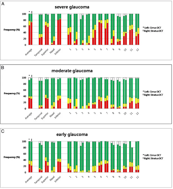
Figure 3 shows each color code frequency in Stratus and Cirrus OCT for average, quadrant, and clock-hour sectors according to the glaucoma severity. For average RNFL measurements, the
FIGURE3. Comparison of the color scale frequency for each sector between Stratus and Cirrus OCT according to the glaucoma severity. (A) Severe glaucoma group. (B) Moderate glaucoma group. (C) Early glaucoma group.
red color code was displayed more frequently by Stratus OCT than by Cirrus OCT (Stratus OCT 72.5%, and Cirrus OCT 62.5%) in the severe glaucoma group. However, there was almost no difference in the frequency of abnormal color codes (yellow or red, Stratus OCT 75.0%, and Cirrus OCT 77.5%, Fig. 3A). In the moderate and early glaucoma groups, abnormal color code frequency for average RNFL also was similar between Stratus and Cirrus OCT (Figs. 3B, 3C).
For the quadrant map, the frequency of abnormal color codes (yellow or red) was similar between Stratus and Cirrus OCT for the inferior and temporal quadrants in all glaucoma subgroups (Figs. 3A, 3B, 3C). However, for the nasal sectors, the frequency of the abnormal color code in Stratus OCT was higher than that in Cirrus OCT in all glaucoma subgroups (Stratus 27.5% and Cirrus 7.5% in the severe group, Stratus 21.1% and Cirrus 10.5% in the moderate group, and Stratus 18.2% and Cirrus 10.5% in the early group, Figs. 3A, 3B, 3C). For superior sectors, the frequency of the red color code in Cirrus OCT was higher than that in Stratus OCT in early glaucoma (Stratus OCT 21.2%, and Cirrus OCT 36.4%, Fig. 3C). A slight difference also was observed in the moderate and severe glaucoma groups (Figs. 3A, 3B).
For the 12 clock-hour map, the difference of the color code frequency was variable according to the scanned sectors and the glaucoma severity. For the 2 o’clock sectors, the frequency of the abnormal color code in Stratus OCT was much higher than that in Cirrus (Stratus 42.5% and Cirrus 12.5% in the severe group, Stratus 31.6% and Cirrus 7.9% in the moderate group, and Stratus 18.2% and Cirrus 10.5% in the early group, Figs. 3A, 3B, 3C). For the 3 and 4 o’clock sectors, Stratus OCT displayed the abnormal color code more frequently than did Cirrus OCT, especially in the severe glaucoma group (Stratus 15.0% and 30.0%, and Cirrus 0% and 7.5%, Fig. 3A).
D
ISCUSSIONThere have been several earlier studies regarding agreement of RNFL measurements between Stratus OCT and Cirrus OCT.6,9– 15However, to our knowledge there have been no reports on
agreement of RNFL color codes between the two instruments according to the glaucoma severity. In our study, RNFL color code agreement between the two OCTs was evaluated according to the glaucoma severity. Inter-device agreement of color codes between Stratus and Cirrus OCT using Cohen’s Kappa value was fair to good, except for the case of the nasal sector in normal eyes and in all glaucomatous eyes. For average inferior and temporal quadrants, color codes were consistent between the 2 OCTs and the disagreement proportion was less than 10%. However, there was considerable color code difference for the nasal and superior sectors throughout all severity groups. For the nasal sector, Stratus OCT tends to display abnormal findings, and these trends were augmented in the severe glaucoma group. For the superior sector, Cirrus OCT tends to show abnormal findings, and these trends were notable in the early glaucoma group.
Cohen’s kappa generally was fair to good; however, this value does not reflect actual agreement because each color code (red, yellow, green, and white) implies different significance with regard to the severity of glaucoma. The RNFL thicknesses are presented by a white color for those in the hypernormal range (95th to 100th percentiles), green backgrounds for those in the normal range (fifth to 95th percentiles), yellow backgrounds for those abnormal at the first to fifth percentile level, and red backgrounds for those abnormal at the first percentile level. Therefore, we grouped yellow or red colors as abnormal findings, and green or white
colors as normal findings to compare the disagreement between the 2 OCTs.
Several previous studies regarding measurement agreement have demonstrated that RNFL thicknesses measured by Cirrus OCT were thicker than those measured by Stratus OCT when the RNFL was very thin, as in severe glaucoma.6,9–12,14
Conversely, at thicker RNFL, measurements using Stratus OCT were thicker than those obtained by Cirrus OCT. In general, the absolute value of RNFL thickness difference between the two instruments increased at thicker RNFL.6,9– 12,14Our results concur with the previous results. The absolute
value of RNFL thickness difference between the two instru-ments increased when RNFL was thick. However, the proportion of color code disagreement was higher in the severe glaucoma group than it was in the early glaucoma group. The discrepancy might be due to the higher frequency of abnormal codes in the severe glaucoma group. Green color codes represent a wide range of RNFL thicknesses (fifth to 95th percentiles), while abnormal color codes represent a narrow range (less than fifth percentiles). It is reasonable that red or yellow color codes are more likely to disagree between two instruments than do green color codes.
It is known that RNFL thickness measurements obtained by Cirrus OCT usually are thicker than those measured by Stratus OCT when RNFL thickness is thin. In our study, Cirrus OCT tended to display normal color code findings compared to those of Stratus OCT for the nasal sector, and for many sectors in the clock-hour maps in the severe glaucoma group (Fig. 2A). It also is known that RNFL thickness values measured using Stratus OCT are thicker than those measured by Cirrus OCT when RNFL is very thick. In our study, Stratus OCT tended to display normal color code findings compared to those of Cirrus OCT in the early glaucoma group, especially in the superior quadrant and the 1 o’clock sector (Fig. 2C).
In our study, the RNFL color code using Stratus and Cirrus OCT showed considerable discrepancy. We suggest several possible explanations for this discrepancy. First, because these two instruments use different technologies, RNFL thickness measurements themselves obtained by Stratus and Cirrus OCT were different from each other. These instruments use a different scan registration method in locating a measurement circle, a different data-acquisition process with different scan speed, and a different segmentation algorithm; Cirrus aims to identify the bottom of the nerve fiber layer, whereas Stratus attempts to localize the top of the ganglion cell layer. RNFL color codes are derived from the RNFL measurements. Different RNFL measurements from different types of technol-ogy may have resulted in the RNFL color code disagreement between the two instruments. A second explanation for the color code disagreement is that color codes provide informa-tion on the probability of an abnormality after comparison of RNFL measurement with an internal age-matched normative database. The racial distributions in the normative databases of the two instruments are different. In the normative Stratus OCT database, very few Asian individuals (3%) were included, whereas>20% of subjects were Asian individuals in the Cirrus HD-OCT normative database (Gurses-Ozden R, et al. IOVS 2008;49:ARVO Abstract 4632). This might have resulted in the difference of the normative database, and the difference could cause color code disagreement.
There was considerable color code difference for the nasal sectors throughout all severity groups. Many studies have found that the correlation between two instruments was slightly weaker in nasal quadrant RNFL thickness.11,13,21 The
main reason might come from less reliable measurements in this sector based on Stratus OCT. Studies to determine the reproducibility of Stratus time-domain OCT have suggested
that the RNFL measurements for the nasal quadrant appeared to be the most variable.22,23
The limitations our study include the use of a small sample size in each subgroup and different test-retest variability of RNFL measurements between the two OCT instruments.21,24,25
In addition, color code agreement could not be compared directly among average, quadrant, and clock-hour sectors because agreement of average RNFL must be better than that of smaller sectors, such as quadrants or clock-hour maps.
In conclusion, there was considerable color code difference for nasal and superior sectors throughout all severity groups. For the nasal sector, Stratus OCT tends to display abnormal findings, and these trends were augmented in the severe glaucoma group. For the superior sector, Cirrus OCT tends to show abnormal findings, and these trends were notable in the early glaucoma group. RNFL color codes derived from two OCTs of different generations could not be considered interchangeable; which is similar to the case of RNFL measurement. Therefore, color code disagreements between instruments among different glaucoma severity groups must be considered when an individual undergoes longitudinal follow-up with different OCTs.
References
1. Quigley HA, Dunkelberger GR, Green WR. Retinal ganglion cell atrophy correlated with automated perimetry in human eyes with glaucoma. Am J Ophthalmol. 1989;107:453–464. 2. Quigley HA, Miller NR, George T. Clinical evaluation of nerve
fiber layer atrophy as an indicator of glaucomatous optic nerve damage. Arch Ophthalmol. 1980;98:1564–1571.
3. Sommer A, Miller NR, Pollack I, Maumenee AE, George T. The nerve fiber layer in the diagnosis of glaucoma. Arch Ophthalmol. 1977;95:2149–2156.
4. Giangiacomo A, Garway-Heath D, Caprioli J. Diagnosing glaucoma progression: current practice and promising tech-nologies. Curr Opin Ophthalmol. 2006;17:153–162.
5. Schuman JS, Hee MR, Puliafito CA, et al. Quantification of nerve fiber layer thickness in normal and glaucomatous eyes using optical coherence tomography. Arch Ophthalmol. 1995; 113:586–596.
6. Knight OJ, Chang RT, Feuer WJ, Budenz DL. Comparison of retinal nerve fiber layer measurements using time domain and spectral domain optical coherent tomography. Ophthalmolo-gy. 2009;116:1271–1277.
7. Kim NR, Lee ES, Seong GJ, Choi EH, Hong S, Kim CY. Spectral-domain optical coherence tomography for detection of localized retinal nerve fiber layer defects in patients with open-angle glaucoma. Arch Ophthalmol. 2010;128:1121– 1128.
8. Leitgeb R, Hitzenberger C, Fercher A. Performance of Fourier domain vs. time domain optical coherence tomography. Opt Express. 2003;11:889–894.
9. Huang J, Liu X, Wu Z, et al. Macular and retinal nerve fiber layer thickness measurements in normal eyes with the Stratus OCT, the Cirrus HD-OCT, and the Topcon 3D OCT-1000. J Glaucoma. 2011;20:118–125.
10. Leung CK, Cheung CY, Weinreb RN, et al. Retinal nerve fiber layer imaging with spectral-domain optical coherence tomog-raphy: a variability and diagnostic performance study. Oph-thalmology. 2009;116:1257–1263.
11. Vizzeri G, Weinreb RN, Gonzalez-Garcia AO, et al. Agreement between spectral-domain and time-domain OCT for measuring RNFL thickness. Br J Ophthalmol. 2009;93:775–781. 12. Takagishi M, Hirooka K, Baba T, Mizote M, Shiraga F.
Comparison of retinal nerve fiber layer thickness measure-ments using time domain and spectral domain optical coherence tomography, and visual field sensitivity. J Glauco-ma. 2011;20:383–387.
13. Sung KR, Kim DY, Park SB, Kook MS. Comparison of retinal nerve fiber layer thickness measured by Cirrus HD and Stratus optical coherence tomography. Ophthalmology. 2009;116: 1264–1270.
14. Seibold LK, Mandava N, Kahook MY. Comparison of retinal nerve fiber layer thickness in normal eyes using time-domain and spectral-domain optical coherence tomography. Am J Ophthalmol. 2010;150:807–814.
15. Moreno-Monta˜n´es J, Olmo N, Alvarez A, Garcia N, Zarranz-Ventura J. Cirrus high-definition optical coherence tomogra-phy compared with Stratus optical coherence tomogratomogra-phy in glaucoma diagnosis. Invest Ophthalmol Vis Sci. 2010;51:335– 343.
16. Hodapp E, Parrish RK, Anderson DR. Clinical Decisions in Glaucoma. St. Louis, MO: C.V. Mosby; 1993:84–125. 17. Budenz DL, Anderson DR, Varma R, et al. Determinants of
normal retinal nerve fiber layer thickness measured by Stratus OCT. Ophthalmology. 2007;114:1046–1052.
18. Nunnally JC. Psychometric Theory. 2nd ed. New York, NY: McGraw-Hill Book Company; 1978:50–58.
19. Bland JM, Altman DG. Statistical methods for assessing agreement between two methods of clinical measurement. Lancet. 1986;1:307–310.
20. Altman DG. Practical Statistics for Medical Research. London, UK: Chapman & Hall; 1991:407–409.
21. Hong S, Kim CY, Lee WS, Seong GJ. Reproducibility of peripapillary retinal nerve fiber layer thickness with spectral domain cirrus high-definition optical coherence tomography in normal eyes. Jpn J Ophthalmol. 2010;54:43–47.
22. Budenz DL, Chang RT, Huang X, Knighton RW, Tielsch JM. Reproducibility of retinal nerve fiber thickness measurements using the stratus OCT in normal and glaucomatous eyes. Invest Ophthalmol Vis Sci. 2005;46:2440–2443.
23. Budenz DL, Fredette MJ, Feuer WJ, Anderson DR. Reproduc-ibility of peripapillary retinal nerve fiber thickness measure-ments with stratus OCT in glaucomatous eyes. Ophthalmology. 2008;115:661–666.
24. Kim JS, Ishikawa H, Sung KR, et al. Retinal nerve fibre layer thickness measurement reproducibility improved with spec-tral domain optical coherence tomography. Br J Ophthalmol. 2009;93:1057–1063.
25. Moreno-Monta˜n´es J, Olmo N, Garcia N, Alvarez A, Garcia-Granero M. Influence of examiner experience on the reproducibility of retinal nerve fiber thickness values using Cirrus and Stratus OCTs (published online ahead of print November 1, 2011). J Glaucoma.