Acta Derm Venereol 92 399
Letters to the Editor
© 2012 The Authors. doi: 10.2340/00015555-1367
Journal Compilation © 2012 Acta Dermato-Venereologica. ISSN 0001-5555
Pityriasis rubra pilaris (PRP) is a rare papulosquamous
dermatosis characterized by keratotic follicular papules,
erythematous scaling, palmoplantar keratoderma and a
variable degree of erythroderma. Type I PRP, the most
common adult form, has a typical clinical manifestation,
and remission in these patients can be achieved within
3 years. However, the rare type II PRP presents atypical
features and has a long disease duration. We describe
here a case of type II PRP associated with rheumatoid
arthritis (RA) that achieved clinical remission with
eta-nercept therapy.
CASE REPORT
A 58-year-old woman visited our department with a 5-year history of scaly eruptions on her hands, feet, back and buttocks. She also reported arthralgia on both fingers and wrists lasting for 2 months.
Physical examination showed follicular keratotic papules on the back and buttocks, in addition to diffuse erythematous desquama-tive patches on her hands and feet (Fig. 1a and b). Swelling of her right wrist joint was also revealed. A skin biopsy on the buttock revealed alternating parakeratosis and orthokeratosis, irregular acanthosis, focal spongiosis, and lymphocytic exocytosis without Munro’s microabscess or the spongiform pustule of Kogoj. The biopsy also revealed perivascular lymphocytic infiltration on the dermis (Fig. 1e). Based on these clinicopathological findings, the patient was diagnosed with PRP.
Joint space narrowing was observed on the radiocarpal and intercarpal joints of the right wrist on X-ray examination. The blood examination showed a highly positive rheumatological factor and anti-cyclic citrullinated peptide antibody, but she was negative for human leukocyte antigen B27. The erythrocyte sedimentation rate was 22 mm/h (normal range, 0–20 mm/h) and C-reactive protein was 14.4 mg/l (0–5.3 mg/l). She fulfilled the RA criteria of 2010 American College of Rheumatology/ European League against Rheumatism classification.
The patient’s skin lesions were initially treated with oral acitretin (10 mg/day) and topical and systemic steroid (methylprednisolone 8 mg/day) for 10 months; however, she continued to report burning and itching sensation. In the rheumatological clinic, sulfasalazine (1,000 mg/day) was started for initial 9 months, but it failed to improve the arthritis. Methotrexate (12.5 mg/week) was administered for following 14 months, but the arthritis did not resolve. Finally, the patient began to receive subcutaneous in-jection of 25 mg etanercept twice a week in combination with methotrexate (12.5 mg/ week). Clinical remission of skin eruption and arthritis was achieved 2 months after etanercept therapy. This state was sustained for 9 months without any treatment mo-dification (Fig. 1c and d). However, skin lesions recurred one month after cessation of etanercept treatment.
DISCUSSION
Systemic retinoids and/or
metho-trexate have been used as first-line
therapies for PRP, but many PRP
cases are refractory to the standard
treatment. Tumour necrosis factor
alpha (TNF-α) antagonists, including
infliximab and etanercept, have been
reported as effective for patients with
recalcitrant PRP (1, 2). A total of 8
cases of PRP treated with etanercept
have been reported (Table I) (2–6).
Etanercept-induced Clinical Remission of Type II Pityriasis Rubra Pilaris With Rheumatoid
Arthritis
Jong Hoon Kim1, Min-Chan Park2 and Soo-Chan Kim1*
1Department of Dermatology and Cutaneous Biology Research Institute and 2Department of Internal Medicine, Division of Rheumatology, Yonsei University
College of Medicine, Gangnam Severance Hospital, 712 Eonjuro, Gangnam-gu, Seoul 135-720, Korea. *E-mail: [email protected] Accepted February 6, 2012.
Fig. 1. The patient showed (a) follicular hyperkeratosis on the back and buttocks, and (b) erythematous scaling lesions on the feet. After 9 months of etanercept treatment, (c) clinical remission was maintained on back, buttocks, and (d) feet. (e) Examination of the biopsy specimen revealed alternating parakeratosis and orthokeratosis, psoriasiform hyperplasia, and superficial perivascular lymphocytic infiltration (haematoxylin-eosin staining, original magnification × 200).
Included in the theme issue: INFLAMMATORY SKIN DISEASES Acta Derm Venereol 2012; 92: 339–409
400 Letters to the Editor
Three of these 8 cases were treated with 50 mg of
eta-nercept twice a week as the starting dose. The other 5
patients continuously received 50 mg once a week or
25 mg twice a week. However, there was no significant
difference in the clinical remission or maintenance dose
between the 2 groups. Our patient showed rapid clinical
remission within 2 months of treatment with 25 mg of
etanercept twice a week.
Eight cases of PRP associated with arthritis have
been reported. Only one male patient was positive for
rheumatoid factor, but his features were not sufficient
to qualify for RA diagnosis (7). As far as we know, this
is the first case of PRP associated with RA. The
concur-rent treatment of PRP and RA may be challenging (7).
This case strongly suggests that etanercept is a proper
treatment for PRP cases associated with RA. In addition,
the present case is the second example of a patient with
type II PRP who received etanercept for a long period
(2). Our patient experienced 9 months of sustained
clinical remission and relapse after drug-withdrawal,
which is similar to a previous case.
While the pathogenesis of PRP is not certain, an
im-munological response to antigenic triggers has been
proposed (8). The clinical response to etanercept in PRP
patients suggests that TNF-α-related inflammatory
reac-tions (e.g. interleukin-1, -6, and -8) are involved in the
pathogenesis of PRP. To understand the role of etanercept
in PRP treatment, further studies are needed that
investi-gate which cytokines or immune cells change in PRP skin
after anti-TNF-α treatment. It is not yet known whether
etanercept modifies the chronic course of PRP.
In conclusion, etanercept should be considered an
effective treatment option for chronic PRP, because it
can induce long-term remission.
The authors declare no conflicts of interest.
REFERENCES
Müller H, Gattringer C, Zelger B, Höpfl R, Eisendle K. 1.
Infliximab monotherapy as first-line treatment for adult-onset pityriasis rubra pilaris: case report and review of the literature on biologic therapy. J Am Acad Dermatol 2008; 59: S65–70.
Garcovich S, Di Giampetruzzi AR, Antonelli G, Garcovich 2.
A, Didona B. Treatment of refractory adult-onset pityriasis rubra pilaris with TNF-alpha antagonists: a case series. J Eur Acad Dermatol Venereol 2010; 24: 881–884.
Davis KF, Wu J, Murase JE, Rosenberg FR, Sorenson EP, 3.
Meshkinpour A. Clinical improvement of pityriasis rubra pilaris with combination etanercept and acitretin therapy. Arch Dermatol 2007; 143: 1597–1599.
Cox V, Lesesky EB, Garcia BD, O’Grady TC. Treatment of 4.
juvenile pityriasis rubra pilaris with etanercept. J Am Acad Dermatol 2008; 59: S113–114.
Seckin D, Tula E, Ergun T. Successful use of etanercept 5.
in type I pityriasis rubra pilaris. Br J Dermatol 2008; 158: 642–644.
Guedes R, Leite L. Therapeutic hotline. Treatment of pi-6.
tyriasis rubra pilaris with etanercept. Dermatol Ther 2011; 24: 285–286.
Chan H, Liu FT, Naguwa S. A review of pityriasis rubra 7.
pilaris and rheumatologic associations. Clin Dev Immunol 2004; 11: 57–60.
Mazza J, Rossi A, Weinberg JM. Innovative uses of tumor 8.
necrosis factor alpha inhibitors. Dermatol Clin 2010; 28: 559–575.
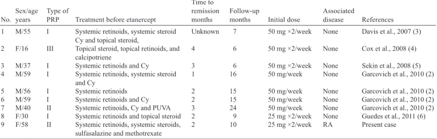
Table I. Previously published cases and the present case of pityriasis rubra pilaris (PRP) treated with etanercept
No. Sex/age years Type of PRP Treatment before etanercept
Time to remission
months Follow-up months Initial dose Associated disease References
1 M/55 I Systemic retinoids, systemic steroid
Cy and topical steroid, Unknown 7 50 mg ×2/week None Davis et al., 2007 (3)
2 F/16 III Topical steroid, topical retinoids, and
calcipotriene 4 6 50 mg ×2/week None Cox et al., 2008 (4)
3 M/37 I Systemic retinoids and Cy 3 6 50 mg ×2/week None Sekin et al., 2008 (5)
4 M/59 I Systemic retinoids, systemic steroid
and Cy 1 16 50 mg/week None Garcovich et al., 2010 (2)
5 M/56 I Systemic retinoids 2 15 50 mg/week None Garcovich et al., 2010 (2)
6 M/59 I Systemic retinoids and Cy 2 15 50 mg/week None Garcovich et al., 2010 (2)
7 M/40 II Systemic retinoids, Cy and PUVA 3 24 50 mg/week None Garcovich et al., 2010 (2)
8 F/30 I Systemic retinoids and topical steroid 2 9 25 mg ×2/week None Guedes et al., 2011 (6)
9 F/58 II Systemic retinoids, systemic steroids,
sulfasalazine and methotrexate 2 10 25 mg ×2/week RA Present case
None of the cases had complications.
Cy: Cyclosporine; PUVA: psoralen plus ultraviolet A phototherapy; RA: rheumatoid arthritis.