저작자표시-동일조건변경허락 2.0 대한민국 이용자는 아래의 조건을 따르는 경우에 한하여 자유롭게 l 이 저작물을 복제, 배포, 전송, 전시, 공연 및 방송할 수 있습니다. l 이차적 저작물을 작성할 수 있습니다. l 이 저작물을 영리 목적으로 이용할 수 있습니다. 다음과 같은 조건을 따라야 합니다: l 귀하는, 이 저작물의 재이용이나 배포의 경우, 이 저작물에 적용된 이용허락조건 을 명확하게 나타내어야 합니다. l 저작권자로부터 별도의 허가를 받으면 이러한 조건들은 적용되지 않습니다. 저작권법에 따른 이용자의 권리는 위의 내용에 의하여 영향을 받지 않습니다. 이것은 이용허락규약(Legal Code)을 이해하기 쉽게 요약한 것입니다. Disclaimer 저작자표시. 귀하는 원저작자를 표시하여야 합니다. 동일조건변경허락. 귀하가 이 저작물을 개작, 변형 또는 가공했을 경우 에는, 이 저작물과 동일한 이용허락조건하에서만 배포할 수 있습니다.
Effect of implant surface microtopography by
hydroxyapatite grit-blasting
on adhesion, proliferation and differentiation of
osteoblast-like cell line, MG-63.
by
Seong Jae Park
Major in Medicine
Department of Medical Sciences
The Graduate School, Ajou University
Effect of implant surface microtopography by
hydroxyapatite grit-blasting
on adhesion, proliferation and differentiation of
osteoblast-like cell line, MG-63.
by
Seong Jae Park
A Dissertation Submitted to The Graduate School of Ajou University
in Partial Fulfillment of the Requirements for the Degree of
Ph. D. in Medicine
Supervised by
Sung Il Song, D.D.S., M.D.S., Ph.D.
Major in Medicine
Department of Medical Sciences
The Graduate School, Ajou University
This certifies that the dissertation
of Seong Jae Park is approved.
SUPERVISORY COMMITTEE
Kyu Rhim Chung
Kwangwoo Baek
Jeong Keun Lee
Kyung Gyun Hwang
Seung Il Song
The Graduate School, Ajou University
June, 23rd, 2011
i - ABSTRACT -
Effect of Implant Surface Microtopography by Hydroxyapatite
Grit-blasting on Adhesion, Proliferation and Differentiation of
Osteoblast-Like Cell Line, MG-63.
Objective
: The purpose of this study is to evaluate the potential of the in vitro osteogenesis of microtopographically modified surfaces, RBM (resorbable blasting media) surfaces that generate a hydroxyapatite grit-blasting.Methods
: First, to produce microtopographically modified surfaces, we made the RBM surfaces using a hydroxyapatite grit-blasting and examined the surface morphology, roughness or elements. And then, to investigate the potential of the in vitro osteogenesis, we experimented the osteoblastic cell adhesion, proliferation and differentiation using the human osteoblast-like cell line, MG-63 cells. Osteoblastic cell proliferation was performed to time-course. Also osteoblastic cell differentiation was verified by four different methods of ALP activity assay, mineralization assay using alizarin red-s staining and gene expression of osteoblastic differentiation marker using RT-PCR or ELISA.Results
: Comparing with machined group, osteoblastic cell adhesion, proliferation and ALPactivity of RBM surfaces were shown higher. Also, they exhibited high level of gene expression of osteoblastic differentiation makers (osteonectin, type I collagen, Runx-2, osterix). Similar data was represented in the ELISA that RBM surface increased secretion of osteocalcin, osteopontin, TGF-beta1 and PGE2 which was known to stimulate the
ii
mineralized nodules on RBM surfaces compared with machined discs.
Conclusions
: Our results demonstrated the RBM surfaces modified with hydroxyapatite grit-blasting which stimulate the in vitro osteogenesis in MG-63 cells and raises the potential that RBM surfaces that accelerate the bone formation and finally increase bone-implant contact.Key words: Osseointegration, Osteogenesis, Microtopography, Surface roughness, RBM (resorbable blasting media), Hydroxyapatite, Biocompatibility
iii
TABLE OF CONTENTS
ABSTRACT --- i
TABLE OF CONTENTS --- iii
LIST OF FIGURES --- iv
LIST OF TABLES --- ⅴ Ⅰ. INTRODUCTION --- 1
Ⅱ. MATERIALS AND METHODS --- 4
A. MATERIALS --- 4 B. METHODS --- 4 Ⅲ. RESULTS --- 11 Ⅳ. DISCUSSION --- 21 Ⅴ. CONCLUSION --- 25 REFERENCES --- 26 국문요약 --- 33
iv
LIST OF FIGURES
Fig. 1. Surface morphologhy of titanium discs for osteoblastic cell culture --- 12
Fig. 2. Effect of surface roughness on the adhesion of MG-63 cells --- 13
Fig. 3. Results of MG-63 cell proliferation experiments --- 15
v
LIST OF TABLES
Table 1. Primer Sequences of Osteogenic Marker and Conditions for Polymerase Chain Reaction --- 10
Table 2. EDS Data of Element Contents on Machined Ti and RBM Surfaces --- 12
Table 3. Roughness Values of a Machined Ti and a RBM Surfaces Measured by
Profilometer --- 13
Table 4. Gradient of MG-63 Cell Proliferation on a Machined Ti and a RBM Surfaces During 5Days. --- 15
1
Ⅰ
. INTRODUCTION
With regards to their biological and physiochemical properties, titanium-based biomaterials have been successfully used in orthopaedic, dental and maxillo-facial surgery, especially in endosseous implants. The early osseointegration of titanium dental implants is an important factor for their clinical success(
Davies, 1998; Berglundh
et al, 2003).
The implant loading to bone is influenced by various factors, including surface chemistry, energetics and surface topography(Albrektsson and Wennerberg , 2004; Esposito
et al, 2005; Zhao
et al, 2005;Puleo and Thomas, 2006).
These surface parameters are decisive during the bone healing period in the peri-implant region. After implantation, implant surfaces contact with body fluids and interact with a number of proteins and different cell types. Cell adhesion is one of the initial stages for subsequent proliferation. Differentiation of osteoblastic cells produces bony tissue and extracellular matrix, which will ensure a high bone-implant contact. It has been shown that osteoblastic cell adhesion, growth and differentiation are related to surface chemistry, energetics and roughness(Cooper et al, 1998;
Anselme, 2000
;Zhao et al, 2005)
.Although many in vitro experiments have studied osteoblastic cell behavior on titanium surfaces, the optimal surface parameters for adhesion, proliferation and differentiation of osteoblastic cells have not yet been completely understood(
Kieswetter, 1996; Cooper et al,
1998
). But among them, surface roughness is one of the most acknowledged parameters improving the bone anchorage of titanium implants(Predecki et al, 1972; Thomas and
Cook, 1985; Carlsson et al, 1988
). A different evidence indicate that surface roughness of2
titanium implants strongly affects the behavior of osteoblastic cells. First of all, rough surfaces encourage the entrapment of fibrin protein, adhesion of osteogenic cells and mechanical stability of implants in host bone(
Lauer et al, 2001; Mustafa et al, 2001;
Ellingsen et al, 2004
). On the other hand, numerous reports indicate that increased surface roughness stimulates osteoblastic cell differentiation and bone formation(Wennerberg et al, 1996;Anselme, 2000 et al; Anselme, 2000
).To improve the clinical performance of implants and to guarantee a stable mechanical loading, there are various methods have been developed to create rough implant surfaces. Among them, the popular methods for roughening titanium implants use a biocompatible and resorbable blasting media, such as hydroxyapatite and
b
-tricalcium phosphate (b
-TCP) ceramic particles or other calcium phosphate ceramic particles(Mustafa et al, 2001; Citeau
et al, 2005; Le Guehennec et al, 2007
). These calcium phosphate biomaterials have been contemplated as a potential abrasive materials. Indeed hydroxyapatite andb
-TCP are biocompatible. They are well known to form a direct bond with surrounding tissue after bone implantation. As these calcium phosphate materials are soluble in acids, they can be easily removed from the implant surface after blasting(Sanz et al, 2001; Novaes et al, 2002
). As these treatments generate surfaces with random topography and various chemical compositions, the optimal surface properties that result in rapid osteoblastic cell adhesion, proliferation and differentiation.In view of the above-mentioned data, the main objectives of this work were (i) to develop a new technique of blasting using hydroxyapatite particles that provide a roughened surface which improves the osteoblastic cell adhesion, proliferation and differentiation and
3
(ii) to assess effects of the roughened surface with respect to osteoblastic cells. In this attempt, we examined the roughness of pure titanium discs prepared by hydroxyapatite grit-blasting technique. Two groups were investigated: polished (machined) and hydroxyapatite grit-blasted (RBM) titanium surfaces. After analyzing the properties of the implant surfaces, osteoblastic cell adhesion, proliferation and differentiation were studied.
4
Ⅱ
.
MATERIALS AND METHODS
A. Materials
Commercially pure titanium (cpTi) discs are 12mm in diameter and 1mm in thickness (Carpenter, Washington, Alabama, USA). The samples were cut from a cpTi bar and used as machined discs. Abrasive hydroxyapatite powders (40-80 mesh) (Himed, New York, NY, USA), Cell culture plastic (Falcon, Franklin Lakes, NJ, USA), Fetal bovine serum (FBS), DMEM, penicillin/streptomycin, trypsin/EDTA (HyClone, Salt Lake City, Utah, USA), Phosphate puffered saline (PBS) (Invitrogen Corporation, Inchinnan, Paisley, UK), Triton X-100, Alizarin red-s, p-nitrophenylphosphate (p-NPP), cresyl violet, MgCl2 (Sigma, St. Louis,
MO, USA), 3-(4,5-Dimethylthiazol-2-gl)-5-(3-carboxymethoxylphenyl)-2-(4-sulphophenyl-2H) tetrasolium inner salt (MTS) and hTGF-beta1 ELISA kit (Promega, Madison, WI, USA), hOsteocalcin ELISA kit (Biosource, Nivelles, Belgium), hOsteopontin and hPGE2 ELISA kit (Assay Designs, Ann Arbor, MI, USA), QuantiTechÒ Reverse Transcription kit (Qiagen, Valencia, CA, USA), PCR premix (Bioneer ,Daejeon, Korea) were prepared. All other chemicals were from standard laboratory suppliers and were of the highest purity available.
B. Methods
1.Preparation of titanium discs
CpTi discs were cleaned with acetone and ethanol. Discs were thereafter randomly divided into two groups. The polish group was obtained by polishing with silicon carbide abrasive paper (Jinan Abrasive Cloth Factory, Jinan, Shandong, China). Decreased grind
5
paper P400, P600, P1200, P2000 were successfully used to a rotative polisher at 250 rotation per minute (rpm) for 1min. The hydroxyapatite grit-blasted surface (RBM group) was prepared by blasting using hydroxyapatite powders (Himed, New York, NY, USA) of 40-80 mesh with a microhardness. The hydroxyapatite powders were blasted at air pressure of 5 bars for 10 sec. Hydroxyapatite grit-blasted (RBM) discs were then passivated by immersion for 10 min in 15% nitric acid at room temperature and rinsed deionized water. Then the samples were ultrasonically cleaned for 30min in Et-OH, washed 3 times in DW. Finally they were dried at 80°C dry oven. Before use in the cell-based assays, all discs were packaged and sterilized using autoclave machined for 20min at 120°C.
2. Surface roughness analysis
Three cpTi discs of each group were randomly selected and roughness measurements were carried out with a SV-3000 S4 digital profilometer (Mitutoyo, Tokyo, Japan) linked to an acquisition software Surfpak-SV. This profilometer system consists of a probe which scanned the surface 2.4mm in length. Results were expressed 2D parameter, as a Ra (average roughness), Rz (average maximum height of the profile) and Rt (maximum height of the profile). These parameters expressed in
m
m, gave a general description of surface roughness.3. Scanning electron microscopy (SEM) and energy-dispersive spectroscopy (EDS)
The different surface morphologies of cpTi and RBM surface were examined using a scanning electron microscopy (SEM, JEOL, Tokyo, Japan) at 20Kv. An energy-dispersive spectrophotometer (EDS) attached with the JSM-6480LV SEM has also been used to
6
characterize the elemental atomic composition of samples. Using the INCA-sight system of Oxford Instrument, elemental analysis of a defined area on the cpTi and RBM surface can easily be determined to a high degree of precision (~5 wt %).
4. Cell culture
The MG-63 cells (ATCC, Manassas, VA, USA) is a non-transformed cell line established from human osteosarcoma. This cell was routinely grown in DMEM supplemented with 10% fetal bovine serum (FBS; HyClone, Salt Lake City, Utah, USA) and 0.1 mg/ml penicillin and streptomycin (P/S) (HyClone, Salt Lake City, Utah, USA). Cells were subcultured twice a week using trypsin/EDTA and maintained at 37°C in a humidified atmosphere of air including 5% CO2. Medium was completely renewed every two days.
5. Cell adhesion
The cell adhesion assay of the MG-63 cells performed using previous report(
Ginsberg
and Che, 2004; Hutton et al, 2008
). Briefly, the cells were grown to approximately 80% confluence prior to cell adhesion assay. Mg-63 cells were cultured either onto the various titanium disc or in the culture plastic in 24 multi-well plate at a density of 1´105 cells. After1hr, culture media were removed and rinsed three times by DPBS. The cells were fixed using 4% formaldehyde solution for 1hr at 4°C and were rinsed three times by DPBS. Then the viable cells stained with cresyl violet which stained ribonucleic acid and were eluted using citric acid. Finally colorimetric measurement of cresyl violet was performed on a DTX 880 (Beckman Coulter, Fullerton, CA, USA) with optical density reading at 590nm.
7 6. Cell Proliferation
The cell proliferation assay of the MG-63cells was performed using CellTiter 96Ò Aqueous Non-Radioactive Cell Proliferation Assay kit (Promega, Madison, WI, USA) according to the manufacturer’s protocol. MG-63 cells were cultured either onto the various titanium disc or in the culture plastic in 24 multi-well plate at a density of 1´105 cells. After
indicated times, culture media was removed and 500
m
l MTS/PMS/Media mixture was added in each well for 1hr. Finally colorimetric measurement of formazan dye performed on a DTX 880 with optical density reading at 490nm. Compared to control, results were expressed as optical density and relative MTS activity.7. Alkaline phasphatase (ALP) activity
ALP activity was examined as previously described(
Engvall, 1980; Jung and
Pergande, 1980; Buxton and Murdoch, 1982; Jones et al, 1989
), in MG-63 cells cultured either onto the various titanium disc or culture plastic in 24 multi-well plates (1´105cells/well). Osteogenic medium (0.5
m
M dexamethasone, 50m
g/ml L-ascorbic acid, 10mM b-glycerolphosphate)(Chen et al, 2008; Rausch-Fan X et al, 2008
), was completely renewed every two days and after indicated times. Cells were washed twice with DPBS and collected in 0.1% ALP lysis buffer. In order to extract the dissolved proteins from the crude cell lysates, the supernatants were centrifuged at 13,000 rpm for 10 min. The protein concentrations were determined using a BCA reagent (Pierce, Rockford, IL, USA). For ALP activity measurement with isolated cell supernatants,100mg
total protein were incubated with 2mg/ml p-NPP (as a chromogenic substrate, Sigma) using ALP assay buffer. (pH 9.758
in 0.1M Glycine containing 1mM MgCl2) at 37°C for 2hr. The degree of colorimetic reaction was measured by DTX 880 for an optical density measurements at 405nm.
8. Alizarin red-s staining
The alizarin red-s staining with experimental samples was performed as reported elsewhere(
Sudo H et al, 1983; Gregory CA et al, 2004; Malladi P et al, 2006
) with a minor modifications. Briefly, MG-63 cells were cultured either onto the various titanium disc or culture plastic in 24 multi-well plates (1´105 cells/well). Osteogenic medium wascompletely renewed every two days and after indicated times. Cells were washed twice with DPBS and fixed in 10% (v/v) formaldehyde (Junsei, Chuo-ku, Tokyo, Japan) for 20min at room temperature and rinsed with triple distilled H2O. Cells were stained with 2% alizarin
red-s (Sigma-Aldrich, Oakville, ON, Canada), pH4.2, for 20min with gentle agitation. After aspiration of the unstained dye, the wells were washed five times with distilled H2O while
shaking for 5min. For quantification of staining, 500
ml
10% (v/v) acetic acid was added to each well, and the plate was heated to 70~80°C for 1hr. The cells were then scraped from the plate and centrifuged at 13000rpm for 10min. The supernatant was transferred to a new e-tube and the 0.4 volume of 10% (v/v) ammonium hydroxide added to neutralize the acid. Absorbance of extracted alizarin red-s in acetic acid solution (200ml
) was measured at 405nm. Amount of alizarin red-s was determined according to an alizarin red-s standard curve and normalized to the total protein of the cell lysates using BCA method (Pierce, Rockford, IL, USA).9 9. ELISA
MG-63 cells were cultured either onto the various titanium disc or culture plastic in 24 multi-well plates (1´105 cells/well). Osteogenic medium was completely renewed every two
days. After indicated times, supernatants were collected using centrifuge (at 13,000 rpm for 2 min) to eliminate a cell debris. The ELISA of the MG-63cells performed using each ELISA kits (hTGF-beta1 ELISA kit; Promega, hOsteocalcin ELISA kit; Biosource, hPGE2 ELISA
kit: Assay Design, hOsteopontin ELISA kit; Assay Design) according to the manufacturer’s protocol.
10. Semi-quantitative RT-PCR
Changes in osteoblast gene expression for osteonectin, Type I collagen, osteoprotegrin, BSP-2, Runx-2 and Osterix were analyzed using semi-quantitative RT-PCR. MG-63 cells cultured either onto the various titanium disc or culture plastic in 24 multi-well plates (1´105
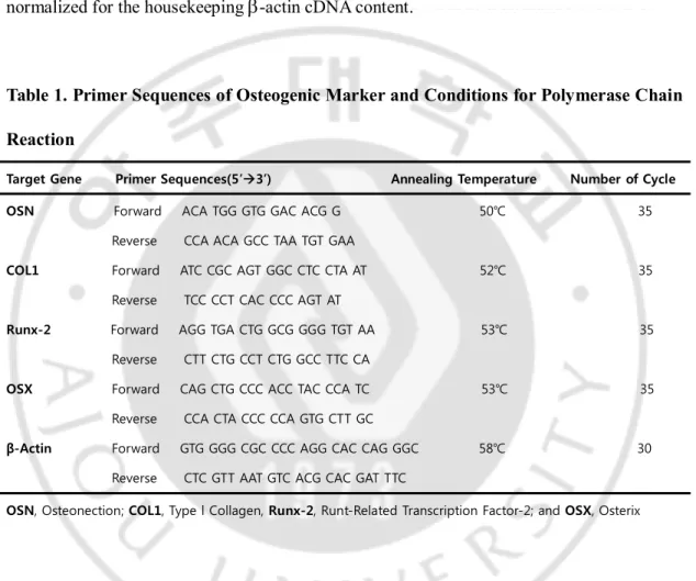
cells/well). Osteogenic medium was completely renewed every two days. After 72hr, total RNA was extracted with Trizol (Invtrogen, Carlsbad, CA, USA), according to the manufacturer’s instructions. Using the extracted RNA as a template, reverse transcription was carried out with QuantiTechÒ Reverse Transcription kit (Qiagen, Valencia, CA, USA) and RT reactions were performed in a Mastercycler (Eppendorf, Westbury, NY, USA).RT reaction mixture was incubated at 42°C for 20min heated at 94°C for 10min, and subsequentially chilled to 4°C. Then osteonectin (OSN), type I collagen (COL1), runt-related transcription factor 2 (Runx-2) and Osterix (OSX) were amplified by Mastercycler using PCR premix (Bioneer, Daejeon, Korea) (see Table 1). The captured image of amplification of
10
osteogenic marker gene expression products from 2% agarose gel stained with ethidium bromide was analyzed using a Doc-It system (UVP, Upland, CA, USA). Semi-quantitative analysis was achieved by calibration. The level of specific osteogenic marker bands was normalized for the housekeeping
b
-actin cDNA content.Table 1. Primer Sequences of Osteogenic Marker and Conditions for Polymerase Chain Reaction
Target Gene Primer Sequences(5’à3’) Annealing Temperature Number of Cycle OSN Forward ACA TGG GTG GAC ACG G 50℃ 35
Reverse CCA ACA GCC TAA TGT GAA
COL1 Forward ATC CGC AGT GGC CTC CTA AT 52℃ 35
Reverse TCC CCT CAC CCC AGT AT
Runx-2 Forward AGG TGA CTG GCG GGG TGT AA 53℃ 35
Reverse CTT CTG CCT CTG GCC TTC CA
OSX Forward CAG CTG CCC ACC TAC CCA TC 53℃ 35
Reverse CCA CTA CCC CCA GTG CTT GC
β-Actin Forward GTG GGG CGC CCC AGG CAC CAG GGC 58℃ 30
Reverse CTC GTT AAT GTC ACG CAC GAT TTC
OSN, Osteonection; COL1, Type l Collagen, Runx-2, Runt-Related Transcription Factor-2; and OSX, Osterix
11. Statistical analysis
Each experiment was repeated at least twice with similar results. Results are expressed as mean ± STDEV of triplicate determinations. For statistical analysis of experiments (N=6, three groups repeated two times), unpaired t-tests and ANOVA were performed using SPSS. (*; p<0.05, **; p<0.01, ***; p<0.001)
11
Ⅲ
.
RESULTS
A. Surface Parameter Tuning for Optimization of RBM
The purpose of this investigation was to find an optimal RBM surface that show the appropriated cell response. To decide the optimal RBM surface, we established surface roughness as a key parameter that influence to RBM surface characteristics. Briefly, cell adhesion, proliferation and osteoblastic differentiation displays tendency that increase as RBM surface roughness grows. (data not shown) Through this approach, we select the optimal RBM surface that has a 1.5
m
m range surface roughness and a following experiments used selected RBM condition.B. Surface Characterization of Optimized RBM
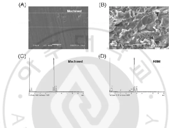
SEM indicates a quite discrepancy between each of the two surfaces. RBM surfaces exhibited a highly rugged and irregular surface (Fig. 1A, B). The roughness parameters as measured by optical profilometer are represent in Table 3. The values refer to the mean of ten measurements for each of the two types of surfaces. The surfaces scored from rougher to more smoother as follows: culture plate < machined < RBM. According to the EDS data (Fig. 1C, D), machined or RBM surface was basically similar elemental composition. The RBM surfaces possesses the phosphorous and calcium of infinitesimal quantity (Fig. 1D, Table 2.). This phenomenon is due to hydroxyapatite powder that remains behind after blasting.
12
Fig. 1. Surface morphologhy of titanium discs for osteoblastic cell culture. SEM images
of machined titanium (A) and RBM (B). Bar = 10
m
m. EDA Data of machined titanium (C) and RBM (D). Surfaces were examined using the JSM-6480LV SEM at working distance of 10m
m and accelerating voltage of 20 kV.Table 2. EDS Data of Element Contents on Machined Ti and RBM Surfaces
Element,(Weight %) Element,(Atomic %)
C p Ca Ti C P Ca Ti
Machined 1.44 0 0 98.56 5.52 0 0 94.48 RBM 1.48 0.31 0.15 98.06 5.65 0.46 0.18 93.72 C, Carbon; P, Phosphorous; Ca, Calcium and Ti, Titanium
13
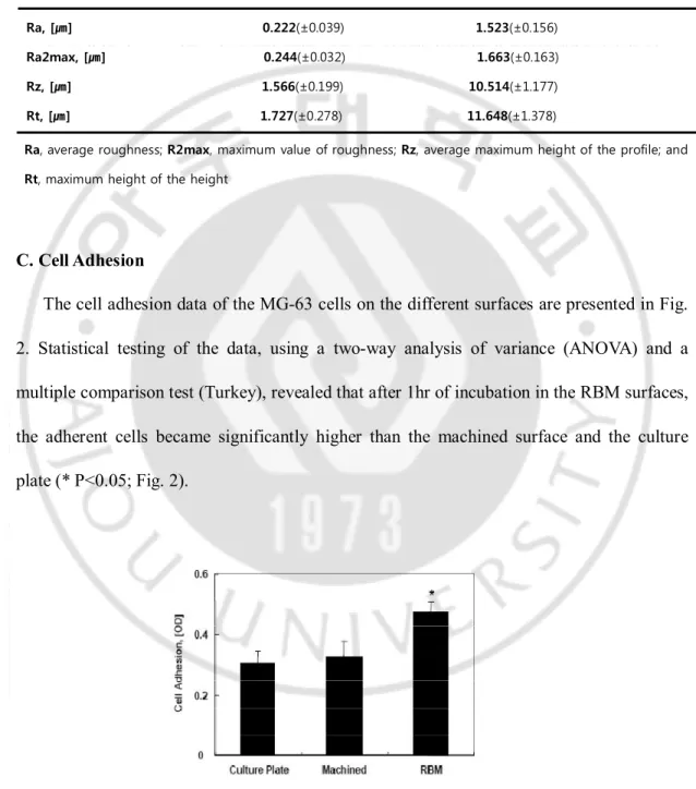
Table 3. Roughness Values of a Machined Ti and a RBM Surfaces Measured by profilometer Machined RBM Ra, [㎛] 0.222(±0.039) 1.523(±0.156) Ra2max, [㎛] 0.244(±0.032) 1.663(±0.163) Rz, [㎛] 1.566(±0.199) 10.514(±1.177) Rt, [㎛] 1.727(±0.278) 11.648(±1.378)
Ra, average roughness; R2max, maximum value of roughness; Rz, average maximum height of the profile; and Rt, maximum height of the height
C. Cell Adhesion
The cell adhesion data of the MG-63 cells on the different surfaces are presented in Fig. 2. Statistical testing of the data, using a two-way analysis of variance (ANOVA) and a multiple comparison test (Turkey), revealed that after 1hr of incubation in the RBM surfaces, the adherent cells became significantly higher than the machined surface and the culture plate (* P<0.05; Fig. 2).
14
stained with cresyl violet dye and counted using spectrophotometer on surfaces of differing roughness. Data are represented as the average ± standard deviation. RBM surface, the rough surface, were promoted the cell adhesion compared to smooth machined surface. *: Statistically significant compared with cells cultured on machined surfaces (p<0.05)
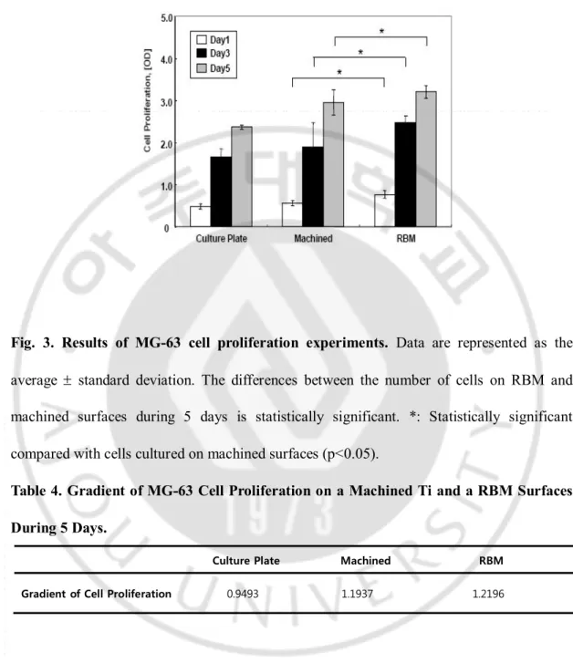
D. Cell Proliferation
To examine the proliferation of osteoblastic cells in RBM surface, we carried out a measurement of MTS activity on MG-63 cells after 1, 3, 5 days in culture. Results indicated that cell proliferation on RBM surface was increased after 1 days of culture than the machined surfaces (* P<0.05; Fig. 3). This phenomenon was observed similar at 3 days and 5 days. Also gradient of cell proliferation curve was more steep in RBM surfaces (Table 4). Therefore these results showed that proliferation of MG-63 cells was increased as early as 1 days in culture on RBM surface, compared to the machined surface.
15
Fig. 3. Results of MG-63 cell proliferation experiments. Data are represented as the
average ± standard deviation. The differences between the number of cells on RBM and machined surfaces during 5 days is statistically significant. *: Statistically significant compared with cells cultured on machined surfaces (p<0.05).
Table 4. Gradient of MG-63 Cell Proliferation on a Machined Ti and a RBM Surfaces During 5 Days.
Culture Plate Machined RBM Gradient of Cell Proliferation 0.9493 1.1937 1.2196
E. Cell Differentiation 1. ALP activity
16
osteoblastic cell differentiation was assessed by measuring the ALP activity normalized to total protein content. On RBM surfaces, we observed a significant enhancement of ALP activity (10.8±2.6%) , compared to machined surface after 7 days (*** P<0.001; Fig. 4A). These results indicate that osteoblastic cells cultured in direct contact with RBM surface increased their capability to express ALP
2. Mineralization (Quantification of alizarin red-s staining)
We used the alizarin red-s staining for quantification of mineralization induced by osteoblastic cells. As shown Fig. 4B, similar pattern in ALP activity data and MG-63 cells cultured on RBM surfaces had increased mineralization dramatically (*** P<0.001).
3. ELISA
According to the acceleration of the differentiation, various osteoblastic differentiation-related proteins, such as extracellular matrix proteins or other signaling factors, were secreted. So we checked the expression levels of osteoblastic differentiation-related proteins using ELISA methods. Osteocalcin secretion by MG-63 cells was slightly greater (** P<0.01) on RBM surface than machined surface (Fig. 4C). Moreover, osteopontin (*** P<0.001), PGE2 (*** P<0.001) and TGF-beta1 (* P<0.05) secretion were greater on RBM surface than
the machined surface (Fig. 4D, 4E, and 4F separately). As a result ELISA data showed that the RBM surface enhances the secretion of various osteoblastic differentiation-related proteins.
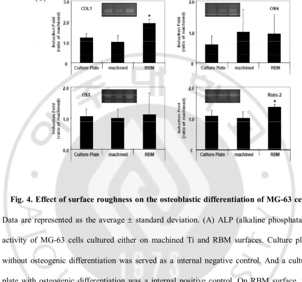
17 4. Semi-quantitative RT-PCR
Semi-quantitative RT-PCR showed significantly (* P<0.05) increased type I collagen and Runx-2 (runt-related transcription factor 2, cbfa-1, OSF-2) gene expression in MG-63 grown on RBM, relative to machined surface or culture plate (Fig. 4G). There was a slight, but insignificant, increase in OSN (osteonectin) and OSX (osterix) gene expression on the RBM surface relative to culture plate or machined surface (Fig. 4G). These results strongly suggested that RBM surface increases the expression level of osteoblastic differentiation marker gene associated with osteogenesis and osseointegration.
19
Fig. 4. Effect of surface roughness on the osteoblastic differentiation of MG-63 cells.
Data are represented as the average ± standard deviation. (A) ALP (alkaline phosphatase) activity of MG-63 cells cultured either on machined Ti and RBM surfaces. Culture plate without osteogenic differentiation was served as a internal negative control. And a culture plate with osteogenic differentiation was a internal positive control. On RBM surface, the ALP activity enhanced about 10% than machined surface (B). Mineralization properties of the machined Ti and RBM surface. MG-63 cells were cultured as before for 14 days with osteogenic differentiation medium. Total calcium deposition measured by alizarin red-s stain via extraction with acetic acid. (C-F) Effect of surface roughness on osteogenesis-related local factor levels determined by using an ELISA kit. Osteocalcin levels were measure in the conditioned media using an ELISA kit specific for human osteoclacin (C). Osteopontin
20
levels were measured in the conditioned media using an ELISA kit specific for human osteopontin (D). PGE2 levels were measured in the conditioned media using an ELISA kit
specific for humans PGE2 (E). And Active of latent TGF-
b
1 (transforming growth factor-b1
)levels were measure in the conditioned media using an ELISA kit specific for human
TGF-b
1 (F). (G) RT-PCR analysis of roughness-influenced osteogenic marker gene. Type I collagen, osteonectin, osterix and Runx-2 RNA levels in MG-63 cells exposure for 72hr on a machined Ti or RBM surface. RT-PCR products were subjected to electrophoresis on 2% agarose gel and visualized UV exposure. A representative analysis is shown inside of the each figure. And the level of specific bands was normalized for theb
-actin cDNA content. Statistically significant compared with cells cultured on machined surfaces by unpaired t-tests (* P<0.01, ** P<0.05, *** P<0.001).21
Ⅳ
.
DISCUSSION
Osteoblastic cells play a critical role in the early stages of ossteointegration. Several reports have assessed that osteoblastic cell response to on smooth surfaces more rapidly than rough surfaces while their osteoblastic differentiation was enhanced by rough topography(
Schwartz et al, 1999; Bächle and Kohal, 2004; Anselme and Bigerelle,
2005; Kim, 2006; Kim et al, 2006
). Little is known about the regulatory mechanism of the osteoblastic cell adhesion, proliferation and differentiation on the titanium discs. This study provides the evidence that the roughness and topography generated by hydroxyapatite-grit blasting (RBM) which is an important regulating factor of the osteoblastic cell adhesion, proliferation and differentiation. This was examined in six ways, using the cresyl violet staining, MTS assay, ALP activity assay, alizarin red-s staining, ELISA, semi-quantitative RT-PCR. In all six cases, the osteoblastic cell adhesion, proliferation and differentiation were dramatically enhanced in RBM surfaces. These results provided unequivocal evidence that surface roughness and topography generated by RBM surface is an important factor that enhances the osteoblastic cell adhesion, proliferation and differentiation.While the effects of surface roughness and topography remain controversial with respect to osteoblastic phenotype expression(
Wennerberg et al, 1996; Deligianni et al, 2001
), we aimed at testing whether MG-63 preserve their phenotype in direct contact with hydroxyapatite grit-blasted (RBM) surface or not. Therefore, this study further investigated the phenomenon underlying the surface roughness and topography modulating the22
osteoblastic cell behavior. Briefly, MG-63 cells cultured on very irregular surfaces, such as RBM, exhibited the increased cell adhesion, proliferation and differentiation. MTS activity was increased more steeply on RBM surfaces. ALP is a marker of osteogenic differentiation, bone formation and mineralization. Previous reports have demonstrated the influence of surface roughness on ALP activity(
Schwartz et al, 1999; Kim et al, 2006
).Like a report of Kim et al(Kim et al, 2006
), ALP activity was enhanced in parallel with the roughness parameters in our study. Moreover this osteoblastic phenotypic characterization can be based on the analysis of various osteobalstic markers including type I collagen, osteonectic, osterix and Runx-2 (cbfa-1/OSF-2). Hydroxyapatite grit-blasted (RBM) surface induced a slight increase in Type I collagen and Runx-2 expression. Also secretion of osteoblastic differentiation-related protein and deposition of mineral was enhanced in RBM surface.Nevertheless, the biological effects of residual alumina particles on titanium surfaces are controversial in the literature(
Piattelli et al, 2003; Diniz et al, 2005; Rodrigo et al,
2006; Canabarro et al, 2008
). Bone healing around residual alumina in alumina grit-blasted titanium implants is impaired. On the contrary, Sader et al.(Sader et al, 2005
) did not observe any detrimental defect of residual blasting alumina particles in vivo and in vitro experiment. Furthermore, Wennerberg et al.(Wennerberg et al, 1996
) did not find any significant differences in bone-implant contact for alumina-blasted and machined titanium implants.Calcium phosphate ceramics were used as the blasting materials in order to avoid the possible negative effects of residual alumina on the osseointegration between titanium and implant. Our results demonstrate the biocompatible and resorbable hydroxyapatite abrasive
23
particles that can be used to create titanium surface roughness. This grit-blasting process increased surface roughness of titanium implants and offered an osteoblastic cell favorable surfaces. As shown as EDS data (Table 2.), even if infitesibal calcium and phosphorous were found on the RBM surfaces after blasting and cleaning, they should not be detrimental to the osseointegration periods.
Although both alumina and hydroxyapatite grit-blasting media had comparable roughness, their surface energetics were different. The alumina-blasted surface was more hydrophobic than the hydroxyapatite grit-blasted surface. Hydrophilic surface should be more favorable to the osteoblastic cell adhesion, proliferation and differentiation(
Li et al,
2004
). These difference may be related to the presence of residual alumina particles on the surfaces. Therefore, grit-blasting titanium surfaces with a biocompatible, resorbable and osteoconductive materials like hydroxyapatite andb
-TCP ceramic particles are alternative method for avoiding the presence of residual alumina particles.In this study, osteoblastic cell adhesion, proliferation and differentiation were greater for the RBM surfaces than for the machined surfaces. This suggests that the RBM surfaces allowed more great osteoblastic cell phenotype than the machined surfaces(
Giordano et al,
2006
). Moreover we showed that rough implant surface can alter the expression of bone associated regulatory transcription factors and key osseointegration-related proteins. Maybe this occurred as a result of differences in cell adhesion, as a result of integrin-mediated adhesion and regulation of downstream signaling pathways as previously reported(Schneider et al, 1999; Schneider et al, 2001; Schneider et al, 2003;
Schneider et al, 2004
). This early osteoblastic cell differentiation in contact with roughened24
RBM surfaces may be favorable during the early phases of bone healing. Further investigations on the relationship between surface roughness, topography and chemistry should improve the understanding of the osseointegration-related osteoblastic cell behavior.
25
Ⅴ
.
CONCLUSION
In summary, this study examined the effect of microtopographically modified rough surfaces by hydroxyapatite grit-blasting on osteoblastic cell adhesion, proliferation and differentiation. Human osteoblast-like cell line, MG-63 cells, cultured on the RBM surfaces exhibited more osteoblastic cell adhesion, proliferation, differentiated osteoblastic phenotype and produced more local factors that stimulate the osteoblastic differentiation. Our results demonstrated the RBM surfaces stimulate the in vitro osteogenesis in MG-63 cells and raise the potential that RBM surfaces accelerate the bone formation and finally increase bone-implant contact.
26
REFERENCES
1. Albrektsson T, Wennerberg A. Oral implant surfaces: Part 2. Review focusing on clinical knowledge of different surfaces. International Journal of Prosthodontics 17:544-64, 2004
2. Anselme K, Bigerelle M. Topography effects of pure titanium substrates on human osteoblast long-term adhesion. Acta Biomaterilia 1:211-22,2005
3. Anselme K, Bigerelle M, Noel B, Dufresne E, Judas D, Iost A, Hardouin P. Qualitative and quantitative study of human osteoblast adhesion on materials with various surface roughnesses. Journal of Biomedical Materials Research 49:155-66, 2000
4. Anselme K. Osteoblast adhesion on biomaterials. Biomaterials 21:667-81, 2000 5. Bächle M, Kohal RJ. A systematic review of the influence of different titanium
surfaces on proliferation, differentiation and protein synthesis of osteoblast-like MG63 cells. Clinical Oral Implants Research 15:683-92,2004
6. Berglundh T, Abrahamsson I, Lang NP, Lindhe J. De novo alveolar bone formation adjacent to endosseous implants. Clinical Oral Implants Research 14:251-62, 2003 7. Buxton LE, Murdoch RN. Purification and properties of bovine dental-pulp
alkaline-phosphatase. Archives of Oral Biology 34:211-20, 1982
8. Canabarro A, Diniz MG, Paciomik S, Carvalho L, Sampaio EM, Beloti MM, Rosa AL, Fisher RG. High concentration of residual aluminum oxide on titanium surface inhibits extracellulat matrix mineralization. Journal of Biomedical Material
27
Research Part A 87A:588-97, 2008
9. Carlsson L, Rostlund T, Albrektsson B, Albrektsson T. Removal torques for polished and rough titanium implants. The International Journal of Oral & Maxillofacial
Implants 3:21-4, 1988
10. Chen KM, Ma HP, Ge BF, Liu XY, Ma LP, Bai MH, Wang Y. The influence of proepicardial cells on the osteogenic potential of marrow stromal cells in a three-dimensional tubular scaffold. Biomaterials 29:2203-16, 2008
11. Citeau A, Guicheux J, Vinatier C, Layrolle P, Pilet P, Daculsi G. In vitro biological effects of titanium rough surface obtained by calcium phosphate grit blasting.
Biomaterials 26:157-65, 2005
12. Cooper LF, Masuda T, Yliheikkila PK, Felton DA. Generalizations regarding the process and phenomenon of osseointegration: Part II. In vitro studies. The
International Journal of Oral & Maxillofacial Implants 13:163-74, 1998
13. Davies JE. Mechanisms of endosseous integration. International Journal of
Prosthodontics 11:391-401, 1998
14. Deligianni DD, Katsala ND, Koutsoukos PG, Missirlis YF. Effect of surface roughness of hydroxyapatite on human bone marrow cell adhesion, proliferation, differentiation and detachment strength. Biomaterials 22:87-96, 2001
15. Diniz MG, Pinheiro MA, Andrade Junior AC, Fischer RG. Characterization of titanium surfaces for dental implants with inorganic contaminant. Brazilian Oral
Research 19:106-11, 2005
28
bone-to implant contact with fluoride-modified titanium implants. The International
Journal of Oral & Maxillofacial Implants 19:659-66, 2004
17. Engvall E. Enzyme Immunoassay: ELISA and EMIT, Methods in Enzymology 70(A):419-39, 1980
18. Esposito M, Coulthard P, Thomsen P, Worthington HV. The role of implant surface modifications, shape and material on the success of osseointegrated dental implants: A Cochrane systematic review. European Journal of Prosthodontics & Restorative
Dentistry 13:15-31, 2005
19. Ginsberg SD, Che S. Combined histochemical staining, RNA amplification, regional, and single cell cDNA analysis within the hippocampus. Laboratory Investigation 84:952-62, 2004
20. Giordano C, Sandrini E, Busini V, Chiesa R, Fumagalli G, Giavaresi G, Fini M, Giardino R, Cigada A. A new chemical etching process to improve endosseous implant osseointegration: in vitro evaluation on human osteoblast-like cells. The
International Journal of Artificial Organs 29:772-80, 2006
21. Gregory CA, Gunn WG, Peister A, Prockop DJ. An alizarin red-based assay of mineralization by adherent cells in culture: comparison with cetylpyridinium chloride extraction. Analitical Biochemistry 329:77-84, 2004
22. Hutton LC, Castillo-Melendes M, Smythe GA, Walker DW. Microglial activation, macrophage infiltration, and evidence of cell death in the fetal brain after uteroplacental administration of lipopolysaccharide in sheep in late gestation.
29
23. Jones JV, Mansour M, James H, Sadi D, Carr RI. A substrate amplification system for enzyme-linked immunoassays: II. Demonstration of its applicability for measuring anti-DNA antibodies. Journal of Immunological Methods 118:79-84, 1989
24. Jung K, Pergande M. Influence of inorganic phosphate on the activity determination of isoenzymes of alkaline phosphatase in various buffer systems. Clinical Chimica
Acta 102:215-9, 1980
25. Kieswetter K, Schwarts Z, Dean DD, Boyan BD, The role of implant surface characteristic in the healing of bone. Critical Reviews in Oral Biology & Medicine 7:329-45, 1996
26. Kim MJ, Choi MU, Kim CW. Activation of phospholipase D1 by surface roughness of titanium in MG63 osteoblast-like cell. Biomaterials 27:5502-11, 2006
27. Kim MJ, Kim CW, Lim YJ, Heo SJ. Microrough titanium surface affects biologic response in MG63 osteoblast-like cells. Journal of Biomedical Material Research
Part A 79A:1023-32, 2006
28. Lauer G, Wiedmann-Al-Ahmad M, Otten JE, Hübner U, Schmelzeisen R, Schilli W. The titanium surface texture effects adherence and growth of human gingival keratinocytes and human maxillar osteoblast-like cells in vitro. Biomaterials 22:2799-809, 2001
29. Le Guehennec L, Lopez-Heredia MA, Enkel B, Weiss P, Amouriq Y, Layrolle P. Osteoblastic cell behaviour on different titanium implant surfaces. Acta
30
30. Li LH, Kong YM, Kim HW, Kim YW, Kim HE, Heo SJ, Koak YK. Improved biological performance of Ti implants due to surface modification by micro-arc oxidation. Biomaterials 25:2867-75, 2004
31. Malladi P, Xu Y, Chiou M, Giaccia AJ, Longaker MT. Effect of reduced oxygen tension on chondrogenesis and osteogenesis in adipose-derived mesenchymal cells.
Americal Journal of Physiology - Cell Physiology 290:C1139-C1146, 2006
32. Mustafa K, Wennerberg A, Wroblewski J, Hultenby K, Lopez BS, Arvidson K. Determining optimal surface roughness of TiO2 blasted titanium implant material
for attachment, proliferation, and differentiation of cells derived from human mandibular alveolar bone. Clinical Oral Implants Research 12:515-25, 2001
33. Novaes A, Souza S, de Oliveira P, Souza A. Histomorphometric analysis of the bone-implant contact obtained with different implant surface treatments placed side by side in the dog mandible. The International Journal of Oral & Maxillofacial
Implants 17:377-83, 2002
34. Piattelli A, Degidi M, Paolantonio M, Mangano C, Scarano A. Residual aluminum oxide on the surface of titanium implants has no effect on osseointegration.
Biomaterials 24:4081-9, 2003
35. Predecki P, Stephan JE, Auslaender BA, Mooney VL, Kirkland K. Kinetics of bone growth into cylindrical channels in aluminum oxide and titanium. Journal of
Biomedical Materials Research 6:375-400, 1972
36. Puleo DA, Thomas MV. Implant surfaces. Dental Clinics of North America 50:323-38, 2006
31
37. Rausch-Fan X, Qu Z, Qieland M, Matejka M, Schedle A. Differentiation and cytokine synthesis of human alveolar osteoblast compared to osteoblast-like cells (MG63) in response to titanium surfaces. Dental Materials 24:102-10, 2008
38. Rodrigo A, Vallés G, Saldaňa L, Rodriguez M, Martinez ME, Munueral L, Vikaboa N. Alumina particles influence the interactions of cocultured osteoblasts and macrophages. Journal of Orthopaedic Research 24:46-54, 2006
39. Sader MS, Balduino A, Soares Gde A, Borojevic R. Effect of three distinct treatments of titanium surface on osteoblast attachment, proliferation, and, differentiation. Clinical Oral Implants Research 16:667-75, 2005
40. Sanz A, Oyarzun A, Farias D, Diaz I. Experimental study of bone response to a new surface treatment of endosseous titanium implants. Implant Dentistry 10:126-31, 2001
41. Schwartz Z, Lohmann CH, Oefinger J, Bonewald LF, Dean DD, Boyan BD. Implant surface characteristics modulate differentiation behavior of cells in the osteoblastic lineage. Advances in Dental Research 13:38-48, 1999
42. Schneider GB, Perinpanayagam H, Clegg M, Zaharias R, Seabold D, Keller J, Stanford C. Implant surface roughness affects osteoblast gene expression. Journal of
Dental Research.2003;82:372–6, 2003
43. Schneider GB, Whitson SW, Cooper LF. Restricted and coordinated expression of beta3-integrin and bone sialoprotein during cultured osteoblast differentiation. Bone 24:321–7, 1999
32
preosteoblasts is affected by implant surface microtopographies. Journal of
Biomedical Material Research Part A 69A:462–8, 2004
45. Schneider GB, Zaharias R, Stanford C. Osteoblast integrin adhesion and signaling regulate mineralization. Journal of Dental Research 80:1540–4, 2001
46. Sudo H, Kodama HA, Amagai Y, Yamamoto S, Kasai S. In vitro differentiation and calcification in a new clonal osteogenic cell line derived from newborn mouse calvaria. The Journal of Cell Biology 96:191-8, 1983
47. Thomas KA, Cook SD. An evaluation of variables influencing implant fixation by direct bone apposition. Journal of Biomedical Materials Research 19:875-901, 1985 48. Wennerberg A, Albrektsson T, Andersson B. Bone tissue response to commercially
pure titanium implants blasted with fine and coarse particles of aluminum oxide.
The International Journal of Oral & Maxillofacial Implants 11:38-45, 1996
49. Wennerberg A, Albrektsson T, Andersson B. Bone tissue response to commercially pure titanium implants blasted with fine and coarse particles of aluminum oxide.
The International Journal of Oral & Maxillofacial Implants 11:38-45, 1996
50. Xavier SP, Carvalho PS, Beloti MM, Rosa AL. Response of rat bone marrow cells to commercially pure titanium submitted to different surface treatments. Journal of
Dentistry. 31:173-80, 2003
51. Zhao G, Schwartz Z, Wieland M, Rupp F, Geis-Gerstorfer J, Cochran DL, Boyan BD. High surface energy enhances cell response to titanium substrate microstructure.
33
- 국문요약 –
골모유사세포주인 MG-63에서 수산화인회석을 이용한 임플란트 표면처리가 세포 의 흡착, 성장 및 분화에 미치는 효과 아주대학교 대학원 의학과 박 성 재 (지도교수: 송 승 일) 목적: 본 연구의 목적은 흡수성 분사물질인 수산화인회석을 분사하여 표면을 미 세형태학적으로 변형시킨 RBM표면의 골형성 정도를 세포수준에서 평가 하고자 하였다. 연구방법: 우선 미세형태학적으로 변형된 표면들을 만들기 위해 수산화인회석 분 사를 이용해 RBM표면을 제작하고 표면형태, 거칠기, 구성요소들을 관찰하였다. 그런 다음 세포수준에서 골형성의 잠재성을 조사하기 위해 골모유사 세포주인 MG-63를 이용하여 골모세포의 흡착, 성장, 분화를 실험하였다. 골모세포 증식 은 시간에 따른 변화를 관찰하였고 골모세포 분화는 ALP activity assay, Alizarin red-s staing을 이용한 Mineralization assay, RT-PCR 과 ELISA를 이용한 골모세포 분화 유전자의 발현을 통하여 평가하였다.연구결과: 골모세포 흡착, 성장, ALP activity는 RBM 표면에서 Machined표면과
비교했을 때 유의성 있게 증가하였다. 그리고 RBM표면에서 높은 수준의 골모세 포 분화 Marker의 유전자 발현을 보였다. (osteonectin, type I collagen,
34
Runx-2, osterix). 비슷한 결과가 ELISA에서 관찰 되었는데, RBM 표면이 골 형성을 자극시키는 것으로 알려진 osteocalcin, osteopontin, TGF-beta1,PGE2
의 분비를 증가시키는 것을 알 수 있었다. 또한 Alizarin red-s staining은 Machined discs와 비교된 RBM표면에서 상당히 유의성 있게 미네랄화 되었음을 보여주었다.. 결론: 본 연구의 결과를 통하여 수산화인회석 분사를 이용한 RBM 표면은 MG-63 세포주에서 골 형성을 자극하는 것을 알 수 있었고 이를 통하여 골 형 성의 증가와 골과 임플란트의 접촉을 증가시킬 것으로 사료 된다. 핵심어: 골유착, 골형성, 미세형태학적, 표면 거칠기, RBM 표면처리, 수산화인회석, 생체적합성
35