저작자표시-비영리-변경금지 2.0 대한민국 이용자는 아래의 조건을 따르는 경우에 한하여 자유롭게 l 이 저작물을 복제, 배포, 전송, 전시, 공연 및 방송할 수 있습니다. 다음과 같은 조건을 따라야 합니다: l 귀하는, 이 저작물의 재이용이나 배포의 경우, 이 저작물에 적용된 이용허락조건 을 명확하게 나타내어야 합니다. l 저작권자로부터 별도의 허가를 받으면 이러한 조건들은 적용되지 않습니다. 저작권법에 따른 이용자의 권리는 위의 내용에 의하여 영향을 받지 않습니다. 이것은 이용허락규약(Legal Code)을 이해하기 쉽게 요약한 것입니다. Disclaimer 저작자표시. 귀하는 원저작자를 표시하여야 합니다. 비영리. 귀하는 이 저작물을 영리 목적으로 이용할 수 없습니다. 변경금지. 귀하는 이 저작물을 개작, 변형 또는 가공할 수 없습니다.
Surgical Outcomes for Solid Pseudopapillary Neoplasm of
Pancreas
by
Nam Hyun Baek
Major in Medicine
Surgical Outcomes for Solid Pseudopapillary Neoplasm of
Pancreas
by
Nam Hyun Baek
A Dissertation Submitted to The Graduate School of
Ajou University in Partial Fulfillment of the Requirements
for the Degree of
Master of Medicine
Supervised by
Wook Hwan Kim, MD, PhD
Major in Medicine
Department of Medical Sciences
The Graduate School, Ajou University
This certifies that the dissertation
of Nam Hyun Baek
is approved.
SUPERVISORY COMMITTEE
Wook Hwan Kim
Byung Moo Yoo
Jae Chul Hwang
The Graduate School, Ajou University
August, 2015
i
-ABSTRACT-
Surgical Outcomes for Solid Pseudopapillary Neoplasm of Pancreas
Background/Aims: Solid pseudopapillary neoplasm (SPN) is a rare exocrine tumor of the pancreas with low malignant potential. This study was designed to evaluate surgical outcome of solid pseudopapillary neoplasm (SPN).
Methodology: From Between January 1994 to November 2013, 41 patients were diagnosed with SPN of the pancreas at Ajou University Medical Center and underwent surgical resection.
Results: Of the 41 patients, 33(80.5%) were female and 8(19.5%) were male with a mean age of 34.5 years (range, 12-63 years). The most common location of SPN was the tail (43.9%). Mean diameters of SPN was 5.5 cm (range, 1.2-14.5 cm). Nineteen patients (46.3%) had non-specific abdominal symptoms that had been investigated. Surgical treatment included distal pancreatectomy in 21, pancreaticoduodenectomy in 11, segmental resection of pancreas in 4, enucleation in 2, excision in 2 and surgical biopsy in 1. Thirty-nine of the 41 patients were disease-free at a median follow-up of 59 months (range, 1-125 months).
Conclusions: Patients diagnosed as SPN should receive surgical resection because of the excellent prognosis. Closed follow-up is recommended after surgery, even in patients
without pathological malignant potential. For metastasis or recurrence, an aggressive surgical treatment is necessary because of the good possibility of long-term survival.
ii
Keywords : Solid pseudopapillary neoplasm, Recurrence, Surgical treatment
iii
TABLE OF CONTENTS
ABSTRACT ··· i TABLE OF CONTENTS ··· ii LIST OF FIGURES ··· iv LIST OF TABLES ··· v . Ⅰ INTRODUCTION ··· 1 . Ⅱ MATERIALS AND METHODS ··· 3. Ⅲ RESULTS ··· 5 A. CLINICAL FINDINGS ··· 5 B. SURGICAL OUTCOMES ··· 5 C. RECURRENT CASE ··· 6 . Ⅳ DISCUSSION ··· 11 REFERENCE ··· 14 국문요약 ··· 20
iv
LIST OF FIGURES
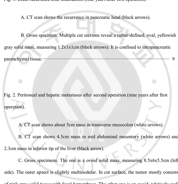
Fig. 1. Local recurrence after enucleation (four years after first operation).
A. CT scan shows the recurrence in pancreatic head (black arrows).
B. Gross specimen. Multiple cut sections reveal a rather-defined, oval, yellowish gray solid mass, measuring 1.2x1x1cm (black arrows). It is confined to intrapancreatic parenchymal tissue.··· 9
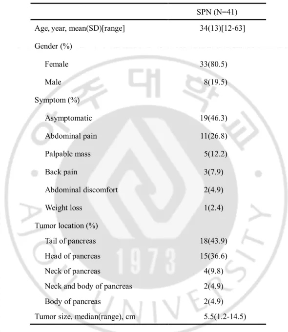
Fig. 2. Peritoneal and hepatic metastases after second operation (nine years after first operation).
A. CT scan shows about 5cm mass in transverse mesocolon (white arrows). B. CT scan shows 4.5cm mass in mid abdominal mesentery (white arrows) and 2.3cm mass in inferior tip of the liver (black arrow).
C. Gross specimens. The one is a ovoid solid mass, measuring 8.5x6x5.5cm (left side). The outer apsect is slightly multinodular. In cut surface, the tumor mostly consists of pink gray solid tissue with focal hemorrhage. The other one is an ovoid, relatively well encapsulated solid mass, measuring 5.5x4x3cm (right side)··· 10
v
LIST OF TABLES
Table 1. Clinical characteristics of patients ··· 7
1
-I. INTRODUCTION
Solid pseudopapillary neoplasm (SPN) is a relatively uncommon condition, with a reported incidence of 1% to 2% of all pancreatic tumors (Klimstra et al., 2000; Martin et al., 2002). SPN was previously known as solid and papillary epithelial neoplasm, and was first described by Frantz in 1959(Frantz, 1959). This is a tumor of the exocrine pancreas according to the World Health Organization (WHO) tumor classification system. SPN usually occurs in young women. Most patients with SPN are cured after surgical resection and have a favorable prognosis (Tipton et al., 2006).
Most reported cases of SPN are benign, but the low-grade malignant potential is 10% to 15% (Tang et al., 2005; Takahashi et al., 2006; Tipton et al., 2006; Suzuki et al., 2010). Some authors has reported that perineural invasion, vascular invasion, capsular invasion, or deep invasion into the surrounding tissue indicate malignant behavior (Tipton et al., 2006; Salla et al., 2007; Chung et al., 2009). However, none of these patients has been showed metastasis or recurrence during the follow-up period. Although SPN of the pancreas has been reported more frequently, reflecting its increased diagnosis and surgical treatment, the guidelines for this neoplasm remain unclear (Mao et al., 1995; Lam et al., 1999; Martin et al., 2002; Tipton et al., 2006). A few cases of SPN confined mainly to the liver, mesentery, and peritoneum have been reported in patients who survived for several years after surgery (Tang et al., 2005). Also, an aggressive surgical resection has been recommended for metastatic SPN (Tipton et al., 2006; de Castro et al., 2007; Machado et al., 2008). This study was designed to evaluate surgical outcome of solid pseudopapillary neoplasm (SPN) and to
2
3
-II. MATERIALS AND METHODS
Between January 1994 to November 2013, the medical records of 41 patients who were pathologically diagnosed as pancreatic SPN by surgical resection at Ajou University Medical Center were retrospectively reviewed. Clinical data included sex, age, symptoms, preoperative radiologic findings, type of surgical treatment, postoperative complication, recurrence, metastasis, and survival. This study was approved by the Ajou University Institutional Review Board.
Computed tomography (CT) had been performed for preoperative diagnosis in all patients. Two board-certified abdominal radiologists reviewed the scans, which mainly showed the presence of a heterogenously enhanced solid and cystic mass. The diagnosis of SPN of the pancreas was based on the gross and microscopic appearance, as well as immunohistochemical staining. We assumed a malignant potential if a tumor was pathologically associated with was pancreatic parenchymal invasion, capsular invasion, perineural invasion, vascular invasion, and lymph node invasion.
For the primary pancreatic disease, two experienced hepatobiliary surgeons carried out operations. According to the International Study Group of Pancreatic fistula (ISGPF) [14], pancreatic fistula (PF) is defined as any measurable drainage from an operatively or subsequently percutaneous placed drain with an amylase content greater than three times the upper limit of normal serum amylase level. Three grades of PF severity were classified as described by the ISGPF clinical criteria. If the amylase concentration was less than three times the serum level, the drain was removed on day 4~7, regardless of the volume drained. Surgical mortality was defined as any death occurring during hospitalization or within 30
4
-days postoperatively.
Patients were reviewed in the outpatient clinic 1 week after discharge from hospital. When the patient had any symptoms (abdominal discomfort, nausea, fever and chilling sensation), we recommended abdominal CT to identify intraabdominal fluid collection after surgery. Follow-up data were obtained from their outpatient clinical records. Patients were checked by abdominal CT 12 month after surgery and once per year thereafter. Survival was determined from the date of diagnosis to the date of death or to the date of last follow up. Patient survival data were obtained from the National Cancer Center in Korea. Categorical variables are expressed as frequencies (%), and continuous variables are presented as median and range.
5
-III. RESULTS
A. Clinical findings
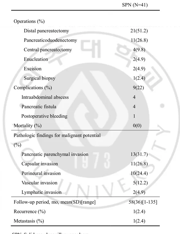
The clinical characteristics of the 41 patients with SPN are shown in Table 1. Of these patients, 33 (80.5%) were female and 8 (19.5%) were male with a mean age of 34.5±13 years (range, 12-63 years). The clinical presentation was nonspecific, comprising abdominal pain in 11 patients (26.8%), palpable mass in 5 (12.2%), back pain in 3 (7.9%), and abdominal discomfort in 2 (4.9%). 19 patients (46.3%) had non-specific abdominal symptoms found incidentally at routine examination. The mean diameter of SPN was 5.5±3.3 cm (range, 1.2-14.5 cm) and the most common location of SPN was the tail in 17 (43.9%), head in 15 (36.6%), neck in 4 (9.8%), neck and body in 2 (4.9%), and body in 2 (4.9%).
B. Surgical outcomes
40 of the 41 patients underwent complete pancreatic tumor resection by distal pancreatectomy (n=21), pancreaticoduodenectomy (n=11), segmental resection of pancreas (n=4), enucleation (n=2), and excision (n=2) according to SPN location (Table 2). Post-operative complications were evident in 9 (22%) of the 41 patients. Complications included grade A pancreatic fistula (n=4, 9.8%), intra-abdominal abscess (n=4, 9.8%; one treated by percutaneous drainage), and bleeding on the upper border of the pancreas with altered hemoglobin (n=1, 2.4%). The latter patient underwent a re-operation on postoperative day 1 to control bleeding occurring through previous incisions. Most of the patients were discharged without any symptoms for clinical observation. There was no postoperative mortality. Microscopic examination showed lymphatic invasion in 2 (4.9%), local invasion
6
-of peripancreatic tissue in 13 (31.7%), perineural invasion in 10 (24.4%), capsular invasion in 11 (26.8%), and vascular invasion in 5 (12.2%). Thirty-nine patients were disease-free at a median follow-up of 59 months (range, 1-125 months). One patient who received non-surgery showed hepatic metastasis and regression of SPN of the pancreas 7 years later.
C. Recurrent case
A 38-year-old female underwent enucleation of 8.5cm mass located in the neck of the pancreas (first operation). The tumor showed no malignant potential and the lesion was completely resected. Radiologic findings after 6 months showed no residual lesion in the abdomen. The patient was subsequently lost to follow-up for 3 years. Radiology conducted 4 years later showed local recurrence in the head of the pancreas (Figure 1A). So, she had to undergo pancreaticoduodenectomy (Figure 1B). Five years later after second operation, follow-up CT scan revealed the tumors in the liver and mesentery (Figure 2). The patient underwent resection of all local recurrences and recovered well from the third surgery. More than 6 years after third surgical procedure, the patient remains alive with no evidence of recurrence or distant metastasis.
7
-Table 1. Clinical characteristics of patients
SPN (N=41)
Age,year, mean(SD)[range] 34(13)[12-63]
Gender (%) Female 33(80.5) Male 8(19.5) Symptom (%) Asymptomatic 19(46.3) Abdominal pain 11(26.8) Palpable mass 5(12.2) Back pain 3(7.9) Abdominal discomfort 2(4.9) Weight loss 1(2.4) Tumor location (%) Tail of pancreas 18(43.9) Head of pancreas 15(36.6) Neck of pancreas 4(9.8)
Neck and body of pancreas 2(4.9)
Body of pancreas 2(4.9)
Tumor size, median(range), cm 5.5(1.2-14.5)
8
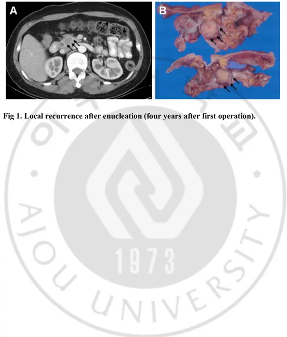
-Table 2. Surgical and pathological outcomes
SPN (N=41) Operations (%) Distal pancreatectomy 21(51.2) Pancreaticoduodenectomy 11(26.8) Central pancreatectomy 4(9.8) Enucleation 2(4.9) Excision 2(4.9) Surgical biopsy 1(2.4) Complications (%) 9(22) Intraabdominal abscess 4 Pancreatic fistula 4 Postoperative bleeding 1 Mortality (%) 0(0)
Pathologic findings for malignant potential (%)
Pancreatic parenchymal invasion 13(31.7)
Capsular invasion 11(26.8)
Perineural invasion 10(24.4)
Vascular invasion 5(12.2)
Lymphatic invasion 2(4.9)
Follow-up period, mo, mean(SD)[range] 58(36)[1-135]
Recurrence (%) 1(2.4)
Metastasis (%) 1(2.4)
9
- 10 -
Fig 2. Peritoneal and hepatic metastases after second operation (nine years after first operation).
- 11 -
IV. DISCUSSION
Solid pseudopapillary neoplasm is a relatively rare pancreatic lesion with a low-grade malignancy potential that most commonly occurs in young females in their second or third decade of life. The age of patients with SPN ranges from 2 to 72 years (average 23.9 years) (Mao et al., 1995; Panieri et al., 1998; Jung et al., 1999; Lam et al., 1999; Tipton et al., 2006). The median age at diagnosis in our study was 34.5 years, which was older than in Asian reports (Hao et al., 2006; Patil et al., 2006). SPN presents with a variety of features of SPN. Papavramidis et al. (Papavramidis and Papavramidis, 2005) summarized the clinical findings of patients presenting with 718 SPN, reporting upper abdominal pain in 46.5% and non-tender abdominal mass in 34.8%. Asymptomatic cases accidently detected after routine examination have been reported in 15.5% of cases (Duff and Greene, 1985; Martin et al., 2002; Ng et al., 2003). These tumors can be discovered by chance during diagnostic imaging or may be suspected because of the presence of an asymptomatic palpable mass in young women. In our study, 19 patients were asymptomatic symptom and 14 patients presented with dull aching abdominal pain. Tumors can be located anywhere in the pancreas, but most cases tend to occur in the body and tail.
Preoperative diagnosis of SPN has been evaluated using CT, ultrasonography (US), endoscopic US (EUS), and magnetic resonance imaging (MRI). CT scan and EUS are more sensitive and specific, and are more accurate in diagnosing SPN (Stommer et al., 1991; Trivedi et al., 1999). Procacci et al. (Procacci et al., 1996) reported a 60% accuracy of CT in the diagnosis of pancreatic tumors. MRI, US, and endoscopic retrograde cholangiopancreatography have been used in diagnosing SPN. MRI was reportedly better
- 12 -
than CT in detecting the cystic or solid components of SPN (Yu et al., 2007). Recently, EUS-Fine needle aspiration biopsy is used for definitive preoperative diagnosis (Song et al., 2012).
Surgical resection is the treatment of choice, given its curative powers in patients with SPN (Tipton et al., 2006). Patients diagnosed as SPN mainly received distal pancreatectomy with/without splenectomy because the tumor was located in body and tail of the pancreas (21/41, 51.2%). Lymphadenectomy was not recommended in any of the recent major studies because relatively strong evidence exists that incidence of lymph node metastasis is extremely rare (Yoon et al., 2001; Adamthwaite et al., 2006; Kang et al., 2006). SPN has a low malignant potential, with a reported incidence of malignant transformation of around 15%, and has relatively favorable prognosis as compared with other pancreatic neoplasm (Mao et al., 1995; Tipton et al., 2006). A few cases of SPN confined mainly to the liver, mesentery, and peritoneum have been reported in patients who survived for several years after surgery (Tang et al., 2005). Although the criteria of malignancy have not yet been clearly established, many studies has reported that perineural invasion, vascular invasion, capsular invasion, or deep invasion into the surrounding tissue indicate malignant behavior (Tipton et al., 2006; Salla et al., 2007; Chung et al., 2009). But, none of these patients has been showed metastasis or recurrence during the follow-up period.
In our study, only a single patient who underwent resection as SPN had local recurrence on follow-up. This patient had no pathologic feature suggesting malignant potential. The present data indicate that regardless of the malignant potential, all patients with SPN must be observed closely. Even if there is metastasis, aggressive surgical procedure is justified because the prognosis after surgical treatment of SPN patients even with local recurrence and
- 13 -
metastasis or invasion is good (Canzonieri et al., 2003; Parelkar et al., 2013). One patient who received non-surgery showed hepatic metastasis and regression of SPN of the pancreas 7 years later. Although there have been few reports on the natural course of SPN, the regression of SPN of the pancreas recently have been reported in several pediatric cases (Hachiya et al., 2003; Nakahara et al., 2008; Suzuki et al., 2010; Yoon and Lim, 2012) 28]. The management of metastatic SPN is poorly defined in adults. Vollmer et al. (Vollmer et al., 2003) reported on a case with liver metastasis in an adult that was aggressively managed by surgical procedure. Such management was virtually impossible in the present cases, given tumor metastasis to the liver is most cases.
In conclusion, patients diagnosed as SPN should receive adequate surgical treatment because of the excellent prognosis. Close follow-up is recommended for patients who receive surgical treatment for SPN because the malignant potential of SPN is not related to pathological features. For metastasis or recurrence, surgical treatment should be done, since long-term survival is possible.
- 14 -
REFRENCE
1. Adamthwaite JA, Verbeke CS, Stringer MD, Guillou PJ, Menon KV: Solid
pseudopapillary tumour of the pancreas: diverse presentation, outcome and histology. JOP 7: 635-642, 2006
2 Canzonieri V, Berretta M, Buonadonna A, Libra M, Vasquez E, Barbagallo E, Bearz A, Berretta S: Solid pseudopapillary tumour of the pancreas. Lancet Oncol 4: 255-256, 2003
3. Chung YE, Kim MJ, Choi JY, Lim JS, Hong HS, Kim YC, Cho HJ, Kim KA, Choi SY: Differentiation of benign and malignant solid pseudopapillary neoplasms of the
pancreas. J Comput Assist Tomogr 33: 689-694, 2009
4. de Castro SM, Singhal D, Aronson DC, Busch OR, van Gulik TM, Obertop H, Gouma DJ: Management of solid-pseudopapillary neoplasms of the pancreas: a comparison with standard pancreatic neoplasms. World J Surg 31: 1130-1135, 2007
5. Duff P, Greene VP: Pregnancy complicated by solid-papillary epithelial tumor of the pancreas, pulmonary embolism, and pulmonary embolectomy. Am J Obstet Gynecol 152: 80-81, 1985
6. Frantz VK: Tumors of the pancreas. In Atlas of tumor pathology (ed. Blumberg CW) Washington, DC, Armed Forces Institute of Pathology, pp.32-33, 1959
- 15 -
7. Hachiya M, Hachiya Y, Mitsui K, Tsukimoto I, Watanabe K, Fujisawa T: Solid, cystic and vanishing tumors of the pancreas. Clin Imaging 27: 106-108, 2003
8. Hao CY, Lu AP, Xing BC, Huang XF, Gao F, Ji JF: Solid pseudopapillary tumor of the pancreas: report of 8 cases in a single institution and review of the Chinese literature. Pancreatology 6: 291-296, 2006
9. Jung SE, Kim DY, Park KW, Lee SC, Jang JJ, Kim WK: Solid and papillary epithelial neoplasm of the pancreas in children. World J Surg 23: 233-236, 1999
10. Kang CM, Kim KS, Choi JS, Kim H, Lee WJ, Kim BR: Solid pseudopapillary tumor of the pancreas suggesting malignant potential. Pancreas 32: 276-280, 2006
11. Klimstra DS, Wenig BM, Heffess CS: Solid-pseudopapillary tumor of the pancreas: a typically cystic carcinoma of low malignant potential. Semin Diagn Pathol 17: 66-80, 2000
12. Lam KY, Lo CY, Fan ST: Pancreatic solid-cystic-papillary tumor: clinicopathologic features in eight patients from Hong Kong and review of the literature. World J Surg 23: 1045-1050, 1999
13. Machado MC, Machado MA, Bacchella T, Jukemura J, Almeida JL, Cunha JE: Solid pseudopapillary neoplasm of the pancreas: distinct patterns of onset, diagnosis, and prognosis for male versus female patients. Surgery 143: 29-34, 2008
- 16 -
14. Mao C, Guvendi M, Domenico DR, Kim K, Thomford NR, Howard JM: Papillary cystic and solid tumors of the pancreas: a pancreatic embryonic tumor? Studies of three cases and cumulative review of the world's literature. Surgery 118: 821-828, 1995
15. Martin RC, Klimstra DS, Brennan MF, Conlon KC: Solid-pseudopapillary tumor of the pancreas: a surgical enigma? Ann Surg Oncol 9: 35-40, 2002
16. Nakahara K, Kobayashi G, Fujita N, Noda Y, Ito K, Horaguchi J, Takasawa O, Obana T: Solid-pseudopapillary tumor of the pancreas showing a remarkable reduction in size over the 10-year follow-up period. Intern Med 47: 1335-1339, 2008
17. Ng KH, Tan PH, Thng CH, Ooi LL: Solid pseudopapillary tumour of the pancreas. ANZ J Surg 73: 410-415, 2003
18. Panieri E, Krige JE, Bornman PC, Graham SM, Terblanche J, Cruse JP: Operative management of papillary cystic neoplasms of the pancreas. J Am Coll Surg 186: 319-324, 1998
19. Papavramidis T, Papavramidis S: Solid pseudopapillary tumors of the pancreas: review of 718 patients reported in English literature. J Am Coll Surg 200: 965-972, 2005
20. Parelkar SV, Oak SN, Kapadnis SP, Sanghvi BV, Joshi PB, Sathe P, Mundada D, Shetty S: Solid pseudo papillary tumor of the pancreas: An unusual tumor in children. J Indian Assoc Pediatr Surg 18: 38-40, 2013
- 17 -
21. Patil TB, Shrikhande SV, Kanhere HA, Saoji RR, Ramadwar MR, Shukla PJ: Solid pseudopapillary neoplasm of the pancreas: a single institution experience of 14 cases. HPB (Oxford) 8: 148-150, 2006
22. Procacci C, Graziani R, Bicego E, Zicari M, Bergamo Andreis IA, Zamboni G, Iacono C, Mainardi P, Valdo M, Pistolesi GF: Papillary cystic neoplasm of the pancreas: radiological findings. Abdom Imaging 21: 554-558, 1996
23. Salla C, Chatzipantelis P, Konstantinou P, Karoumpalis I, Pantazopoulou A, Dappola V: Endoscopic ultrasound-guided fine-needle aspiration cytology diagnosis of solid pseudopapillary tumor of the pancreas: a case report and literature review. World J Gastroenterol 13: 5158-5163, 2007
24. Song JS, Yoo CW, Kwon Y, Hong EK: Endoscopic ultrasound-guided fine needle aspiration cytology diagnosis of solid pseudopapillary neoplasm: three case reports with review of literature. Korean J Pathol 46: 399-406, 2012
25. Stommer P, Kraus J, Stolte M, Giedl J: Solid and cystic pancreatic tumors. Clinical, histochemical, and electron microscopic features in ten cases. Cancer 67: 1635-1641, 1991
26. Suzuki M, Shimizu T, Minowa K, Ikuse T, Baba Y, Ohtsuka Y: Spontaneous
shrinkage of a solid pseudopapillary tumor of the pancreas: CT findings. Pediatr Int 52: 335-336, 2010
- 18 -
27. Takahashi Y, Hiraoka N, Onozato K, Shibata T, Kosuge T, Nimura Y, Kanai Y, Hirohashi S: Solid-pseudopapillary neoplasms of the pancreas in men and women: do they differ? Virchows Arch 448: 561-569, 2006
28. Tang LH, Aydin H, Brennan MF, Klimstra DS: Clinically aggressive solid pseudopapillary tumors of the pancreas: a report of two cases with components of undifferentiated carcinoma and a comparative clinicopathologic analysis of 34 conventional cases. Am J Surg Pathol 29: 512-519, 2005
29. Tipton SG, Smyrk TC, Sarr MG, Thompson GB: Malignant potential of solid pseudopapillary neoplasm of the pancreas. Br J Surg 93: 733-737, 2006
30. Trivedi N, Sharma U, Das PM, Mittal MK, Talib VH: FNAC of papillary and solid epithelial neoplasm of pancreas--a case report. Indian J Pathol Microbiol 42: 369-372, 1999
31. Vollmer CM, Jr., Dixon E, Grant DR: Management of a solid pseudopapillary tumor of the pancreas with liver metastases. HPB (Oxford) 5: 264-267, 2003
32. Yoon DY, Hines OJ, Bilchik AJ, Lewin K, Cortina G, Reber HA: Solid and papillary epithelial neoplasms of the pancreas: aggressive resection for cure. Am Surg 67: 1195-1199, 2001
- 19 -
33. Yoon HJ, Lim JH: Solid pseudopapillary tumor of the pancreas with hepatic
metastasis: spontaneous regression over 10-year follow-up period. Korean J Radiol 13: 648-651, 2012
34. Yu CC, Tseng JH, Yeh CN, Hwang TL, Jan YY: Clinicopathological study of solid and pseudopapillary tumor of pancreas: emphasis on magnetic resonance imaging findings. World J Gastroenterol 13: 1811-1815, 2007
- 20 - - 국문요약 -
본원에서
시행한 췌장 고형 가성유두종의 수술적 결과
아주대학교 대학원의학과 백 남 현 (지도교수 : 김 욱 환) 목적: 고형 가성 유두종은 악성 변화를 가진 비교적 드문 췌장 질환으로 알려져 있다. 이에 저자는 고형가성 유두종의 수술적 결과에 대해 알아보고자 이 연구를 진행하였다. 방법: 1994 년 1 월부터 2013 년 12 월까지, 아주대학교 병원에서 고형 가성 유두중의 완치를 목적으로 수술한 41 명을 대상으로 연구를 진행하였다. 결과: 모든 환자 군의 평균연령은 34.5 세였고, 여자 환자는 33 명 남자환자는 8 명 이였다. 고형 가성 유두종의 가장 흔한 췌장 미부였으며, 종양의 크기는 5.5cm 였다. 고형 가성 유두종의 수술적 치료로써 원위부 췌장 절제술이 21 명으로 가장 많았으며 다음으로는 췌장-십이지장 절제술, 종양 제거술 순 이였다. 41 명의 환자 중에서 1 례에서 재발을 보였으며 재발 후에도 수술적 치료가 시행되었다. 결과:고형 가성 유두종으로 진단된 환자는 수술 후 좋은 예후를 가지므로 반드시 수술적 치료가 필요하다. 그러나 고형 가성 유두종은 악성화 가능성이- 21 - 있으므로 반드시 계속적인 추적관찰이 필요하며 우리 연구에 비추어 보았을 때 추적관찰 중 재발이 있다고 하더라도 환자의 예후를 위해서라도 반드시 수술적 치료가 필요하다. 핵심어 : 고형 가성 유두종, 재발, 수술적 치료