신경초종은 Schwann 세포에서 발생하는 양성 종양이다.1) 증상은 통증이 없거나 경미한 동통을 호소하고 연결된 모신경을 따라 타 진 시 저리는 방사통을 호소하는 경우가 있다.2-4) 종괴는 주로 굴 곡근 측의 말초신경에서 발생하며,2,5) 단발성으로 느리게 성장하 며 경계가 피막으로 잘 구별되어 있다.2) 말초신경과 연결되지 않 은 고립성 신경초종은 다른 연부조직 종양과 감별이 힘들다.2-4) 몸 통에 발생하는 양성 신경초종은 그 빈도가 9% 이하로 드물다.1) 드 물게 보고되는 몸통의 신경초종 중에서도 보고된 바 없는 복직근 내에 발생한 신경초종과 흉부 지방종을 한 환자에서 동시에 경험 하여 이에 보고하고자 한다.
증례보고
63세 남자 환자로 3년 전부터 발생한 미약한 복부 통증 및 점차 커 지는 우 하복부 종물감과 비슷한 시기부터 발생한 우측 흉골 부위 종물감을 주소로 내원하였다. 환자는 특이 과거력 및 가족력은 없 었으며 다른 내과적인 질환은 없었다. 내원 시 시행한 혈액 검사 는 정상 범위였고 신체 검사상 우 하복부 종물 부위의 미약한 압 통을 호소하였다. 종물 부위의 근 위축이나 감각저하등은 없었으Case Report
J Korean Bone Joint Tumor Soc 2014; 20: 109-112 • http://dx.doi.org/10.5292/jkbjts.2014.20.2.109 www.kbjts.or.kr한 환자에서 발생한 복직근 내 신경초종과 흉부 지방종:
증례 보고
Synchronous Development of Schwannoma in the Rectus Abdominis and Lipoma in
the Chest: A Case Report
김주오 • 안기용 • 봉황세 • 이규정
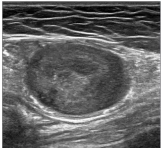
광주보훈병원 정형외과 한 환자에게 동시에 발생한 흉부 지방종과 복부 복직근 내 신경초종 1예의 경험을 보고하고자 한다. 신경초종은 드물게 보이는 양성 종양이 며 신경 조직에서 유발하는 것으로 주로 사지 굴곡부의 말초신경에서 발생한다. 저자들은 63세 남자환자에서 복부 복직근 내 발생한 신경초 종과 흉부의 지방종을 동시에 경험하여 이를 보고하고자 한다. 색인단어: 복직근, 신경초종, 지방종 며 종물은 단단하게 촉지되었고 움직임은 없었다. 단순 방사선 사 진상 특이 소견은 관찰할 수 없었으며, 시행한 초음파 검사상 경 계가 분명한 달걀 모양의 감소된 에코 음영을 확인하였으나 모신 경과 연결이 되는 소견은 확인할 수 없었다(Fig. 1). 흉복부 전산화 단층촬영 검사에서 우측 흉골 앞쪽에 종괴와 우 하복부 복직근 내의 종괴를 확인할 수 있었다(Fig. 2). 신체검사 및 영상학적 검사상 흉부 지방종과 복부 신경초종 추정진단 하에 절 제생검을 계획하였다. 우측 흉부 및 복부 종괴의 피부 부위에 위 접수일 2014년 10월 30일 심사수정일 2014년 11월 19일 게재확정일 2014년 11월 20일 교신저자 안기용 광주시 광산구 첨단 월봉로 99(산월동), 광주보훈병원 정형외과 TEL 062-602-6162, FAX 062-602-6989 E-mail [email protected]Copyrights © 2014 by The Korean Bone and Joint Tumor Society
“This is an Open Access article distributed under the terms of the Creative Commons Attribution Non-Commercial License (http://creativecommons.org/licenses/by-nc/3.0/) which permits unrestricted non-commercial use, distribution, and reproduction in any medium, provided the original work is properly cited.”
대한골관절종양학회지:제20권 제2호 2014
pISSN : 1226-4962 eISSN : 2233-9841
Figure 1. The ultrasonographic finding shows well defined heterogenous echogenic mass.
110
김주오·안기용·봉황세 외 1인 치를 표시하고 전신 마취 하에 종괴 바로 위쪽에서 수평절개를 시 행하여 우측 흉부 6번 갈비뼈 전방에서 지방조직으로 보이는 종 괴를 제거하였다. 우 하복부에서도 종괴 바로 위쪽에 수평절개를 시행하여 복직근 내에서 경계가 분명한 종괴를 확인하였다. 복직 근 내 종괴와 모신경은 연결되지 않았으며 쉽게 제거 되었다(Fig. 3). 술 후 특별한 합병증은 없었으며 12개월 추시에서 재발이나 증 세 발현 등은 보이지 않았다. 우측 흉부 종괴는 3×2×3 cm 크기의 노란색 지방조직으로 이 루어져 있었으며, 조직학적 소견상 성숙지방세포 외 다른 혈관조 직 등은 보이지 않았다. 우 하복부 종괴는 육안상 분홍빛 막에 둘 Figure 2. (A) Transverse CT image shows defined low density soft tissue mass on Rt. anterior chest wall. (B) Transverse CT image shows well-defined mass in rectus abdominis muscle.A B
Figure 4. The cross sectional finding of the intra-muscular mass demonstrates multi-septated pattern, scattered hemorrhagic clots and fibrous tissue.
Figure 5. The histologic examination of hematoxyline and eosin stained specimen shows more cellular "Antoni A" pattern (white arrow) with palisading nuclei surrounding pink areas (Verocay bodies), and "Antoni B" pattern (black arrow) with a looser stroma, fewer cells, and myxoid change (×400).
Figure 3. In the gross finding of the intra-muscular mass in rectus abdominis, a round, pink and well-capsulated mass is shown.
111
한 환자에서 발생한 복직근 내 신경초종과 흉부 지방종 러 쌓여 있었으며 크기는 4×3×3 cm이었다. 절개하여 내부를 확 인한 결과 격막으로 나뉘어져 격막 내에는 혈액 응괴와 섬유 조 직 및 석회화 소견이 관찰되었으나 괴사변형은 관찰되지 않았다 (Fig. 4). 현미경 소견상 방추형 종양세포가 중등도 정도로 세포밀 도를 보이는 부위(Antoni A)와 출혈 및 혈종이 있는 세포가 적은 점액성 부위(Antoni B)가 확인되었다(Fig. 5).고 찰
신경초종은 신경막의 신경초 조직에 존재하는 Schwann 세포에서 발생하는 양성 연부 조직 종양이다.2,6) 사지에서는 굴곡근에 주로 발생하며, 주로 상지에 많이 발생한다. 많은 경우 좌골 신경 등 큰 말초신경에서 발생하는 것으로 보고된다.2,5-8) 신경학적 증상은 거 의 없는 경우가 많고 종괴 자체의 증상을 보이나, 크기가 큰 경우 는 통증, 저림, 감각 이상, 근력 약화를 초래하기도 한다.7) 대부분 신경초종은 신경에서 발생하나 드물게 근육 내에서도 발생한다. 근육 내 신경초종은 해당 근육이 여러 개의 운동신경 섬유 지배를 받는 경우 나머지 신경이 이환 신경 증세를 보완하므로 거의 증상 을 유발하지 않는다.9) 근육 내 발생하는 경우도 드물지만 그 중 몸 통부위에서 발생하는 경우는 9% 이하로 더욱 드물게 보고 된다.1) 진단은 자기공명영상이나 초음파 검사를 통해 비교적 정확히 이루어지는데 자기공명영상에서 T1 강조 영상의 중간 신호강도 와 경미한 비균일감, T2 강조 영상의 높은 신호강도와 다양한 강 도의 균일감을 나타내는 경계가 명확한 원형의 연부 조직 음영 이 나타난다.2,5,7,8) 초음파 검사에서는 경계가 명확한 원형의 감소 된 에코 음영이 보이며 신경과의 연결이 보이는 경우 진단에 도움 이 된다. 본 환자에서도 경계가 명확한 원형의 감소된 에코 음영 을 확인하였으나 복직근 내 종괴와 신경의 연결된 것을 확인할 수 없었다. 초음파 확인 하 세침흡인 검사와 전산화 단층촬영을 통해 종괴의 위치와 신경초종을 진단할 수 있었다. 신경초종은 크기가 커지면 종괴 내에 비균질적 분엽화, 낭포, 석회화 등을 보이는데,4) 본 환자는 절개 시 여러 부위에 혈액 응괴 및 혈관 주위 유리질화 등을 확인할 수 있었다. 신경초종은 현미경 검사에서 높은 세포 밀도의 Antoni A 구역 과 낮은 세포밀도와 변성 소견을 동반하는 Antoni B 구역을 보인 다. Antoni A 구역에서는 종양 세포의 핵들이 울타리 모양으로 배 열되거나 일렬로 Verocay 체를 형성하는 특징적인 소견을 보인 다.1,6,8) 특수 면역 염색으로는 S100 면역 염색이 진단에 도움을 준 다.4) 본 환자에서도 현미경 검사에서 Antoni A 구역과 Antoni B 구역을 관찰할 수 있었다. 신경초종은 드물게 보이는 양성 종양이며 근육내에 존재하는 경우는 드물게 보고 되고 있다. 그 중 몸통의 복직근 내에 존재하 는 신경초종은 비교적 흔한 부위가 아니며 신경과의 연계가 없어 진단이 어려울 수 있다.참고문헌
1. Das Gupta TK, Brasfield RD. Tumors of peripheral nerve ori-gin: benign and malignant solitary schwannomas. CA Cancer J Clin. 1970;20:228-33.
2. Adani R, Baccarani A, Guidi E, Tarallo L. Schwannomas of the upper extremity: diagnosis and treatment. Chir Organi Mov. 2008;92:85-8.
3. Kehoe NJ, Reid RP, Semple JC. Solitary benign peripheral-nerve tumours. Review of 32 years' experience. J Bone Joint Surg Br. 1995;77:497-500.
4. Schultz E, Sapan MR, McHeffey-Atkinson B, Naidich JB, Ar-len M. Case report 872. "Ancient" schwannoma (degenerated neurilemoma). Skeletal Radiol. 1994;23:593-5.
5. Louis DS. Peripheral nerve tumors in the upper extremity. Hand Clin. 1987;3:311-8.
6. Lee SH, Jung HG, Lee HK. Neurilemoma of trunk and ex-tremities. J Korean Bone Joint Tumor Soc. 1996;4:88-93. 7. Bahk WJ RS, Kang YK, Lee AH. Schwannoma of the
extremi-ties. J Korean Bone Joint Tumor Soc. 2003;9:148-54.
8. Pyun YS, Kim SR, Joh YR. Surgical treatment of the neu-rilemoma in extremities. J Korean Bone Joint Tumor Soc. 1998;4:88-93.
9. Kwon BC, Baek GH, Chung MS, Lee SH, Kim HS, Oh JH. In-tramuscular neurilemoma. J Bone Joint Surg Br. 2003;85:723-5.
Synchronous Development of Schwannoma in the Rectus
Abdominis and Lipoma in the Chest: A Case Report
Ju-Oh Kim, Ki-Yong An, Hwang-Se Bong, and Kyu-Jung Lee
Department of Orthopedic Surgery, Gwangju Veterans Hospital, Gwangju, Korea
We experienced a case of 63 years old male patient who had synchronous rectus abdominis intramuscular schwannoma and chest wall lipoma. Schwannoma is rare benign tumor which derived from nerve sheath and mainly peripheral nerve of flexor part. The au-thors report rare synchronous schwannoma and lipoma development.
Key words: rectus abdominis muscle, schwannoma, lipoma
Received October 30, 2014 Revised November 19, 2014 Accepted November 20, 2014 Correspondence to: Ki-Yong An
Department of Orthopedic Surgery, Gwangju Veterans Hospital, 99 Chum-danwolbong-ro, Gwangsan-gu, Gwangju 506-705, Korea
TEL: +82-62-602-6162 FAX: +82-62-602-6989 E-mail: [email protected]
Case Report
J Korean Bone Joint Tumor Soc 2014; 20: 109-112 • http://dx.doi.org/10.5292/jkbjts.2014.20.2.109 www.kbjts.or.krCopyrights © 2014 by The Korean Bone and Joint Tumor Society
“This is an Open Access article distributed under the terms of the Creative Commons Attribution Non-Commercial License (http://creativecommons.org/licenses/by-nc/3.0/) which permits unrestricted non-commercial use, distribution, and reproduction in any medium, provided the original work is properly cited.”
The Journal of the Korean Bone and Joint Tumor Society Vol. 20, No. 2 (December 2014)