저작자표시-비영리-변경금지 2.0 대한민국 이용자는 아래의 조건을 따르는 경우에 한하여 자유롭게 l 이 저작물을 복제, 배포, 전송, 전시, 공연 및 방송할 수 있습니다. 다음과 같은 조건을 따라야 합니다: l 귀하는, 이 저작물의 재이용이나 배포의 경우, 이 저작물에 적용된 이용허락조건 을 명확하게 나타내어야 합니다. l 저작권자로부터 별도의 허가를 받으면 이러한 조건들은 적용되지 않습니다. 저작권법에 따른 이용자의 권리는 위의 내용에 의하여 영향을 받지 않습니다. 이것은 이용허락규약(Legal Code)을 이해하기 쉽게 요약한 것입니다. Disclaimer 저작자표시. 귀하는 원저작자를 표시하여야 합니다. 비영리. 귀하는 이 저작물을 영리 목적으로 이용할 수 없습니다. 변경금지. 귀하는 이 저작물을 개작, 변형 또는 가공할 수 없습니다.
A Thesis for the Degree of Master of Science
Studies on Transcriptional and Epigenetic Mechanisms of
NANOG
gene in Chicken Primordial Germ Cells
닭 원시생식세포에서 NANOG 유전자의
전사 및 후생 유전 조절 기전에 대한 연구
August 2017
By
HYUN GYO JUNG
Biomodulation Major
Department of Agricultural Biotechnology
Graduate School, Seoul National University
i
SUMMARY
Primordial germ cells (PGCs), the precursors of sperm and oocytes, are the only cell type which can transfer genetic information to next
generation. These distinct characteristics are largely dependent on germ cell-specific gene expression. They are controlled by coordinated actions of many key regulators such as transcription factors (TFs), RNA Binding proteins (RBPs) and specialized epigenetic modification.
Nanog, the mostly conserved protein between species, are well-known to be a core transcriptional factor in the early formation of
embryonic development. Nanog is also expressed in cells from pluripotent cells to gonads through the developmental status of PGCs, which means that the role of Nanog is to perpetually maintain germ cell characteristics
containing stemness and germness in general germ cell development. Indeed,
Nanog regulates PGCs by signaling pathways with other key transcription factors. Furthermore, induction of mouse PGC-like cells(PGCLCs) from epiblast-like cells(ESCs) is available with Nanog alone. In chicken, Nanog
expression is observed from early development. And then, the pattern of the gene is restricted to PGCs after HH3. These results suggest that Nanog in chicken is a key factor of regulation of PGC characteristics.
Recent studies find that epigenetic regulations, such as loss of 5mC in whole genome, DNA methylation, histone and chromatin
ii
chromatin modifications and also the control of epigenetic patterns in chicken are highly limited. In the previous studies, methylation and
acetylation during germ cell specification to differentiation activate germ cell specific genes and repress somatic cell genes in mouse. And also, in chicken, germ cells are also epigenetically regulated. In contrast to mammals, the H3K27me3 global level is reduced, whereas the H3K9me3 level is increased in chicken with still acetylation level in PGCs yet unknown.
In this study, we investigated the elaborate regulatory mechanisms that govern epigenetic and transcriptional programs of Nanog. Histone deacetylase (HDAC) regulates NANOG in PGCs. And also, methylation patterns of CpG islands in cNanog upstream, the regions which the
transcription factors bind to, are hypomethylated in PGCs. The result of this study using a series of experiment for functionality testing, including siRNA mediated knockdown, overexpression, immunocytochemistry, luciferase reporter assay, TFs motif analysis, and western blotting demonstrates that chicken has specific epigenetic regulation during PGC development. Intriguingly, we showed that transcriptional program of NANOG was strictly regulated by specific isotype of HDACs (HDAC1 and HDAC2) and the REST repressor complex, suggesting that avian PGCs have the different molecular regulatory mechanism from that of mammals.
In conclusion, chicken PGCs display the unique epigenetic and transcriptional program of Nanog. Our findings first time provide valuable
iii
insight on chicken PGCs to unravel important regulatory components as well as biological roles regarding the genetic and epigenetic regulation for stemness and germness of Nanog and better understanding of germ cell fate. The study of avian species can be adapted to important vertebrate model for the research of developmental biology and speciation.
Keywords: Chicken, Epigenetic Modification, NANOG, VASA, CoREST,
Primordial Germ Cells, Transcription Factors
iv
CONTENTS
SUMMARY ... i
CONTENTS ... iv
LIST OF FIGURES ... viii
LIST OF TABLES ... x
LIST OF ABBEREVIATION ... xi
CHAPTER 1.GENERAL INTRODUCTION ... 1
CHAPTER 2.LITERATURE REVIEW ... 7
1. Primordial Germ Cells ... 8
1.1 PGC specification in germ cells ... 8
1.1.1 Preformation model ... 9
1.1.2 Induction mode ... 11
1.1.3 PGC migration in chicken ... 11
1.2 Signaling pathways of PGCs ... 13
1.2.1 Transforming Growth Factor-Beta (TGF-β) signaling pathway . 14 1.2.2 Bone morphogenetic protein (BMP) signaling pathway ... 15
v
1.2.3 Stem cell factor (SCF) Signaling Pathway ... 16
1.2.4 Basic fibroblast growth factor (bFGF) signaling pathway ... 17
1.3 Germness related genes in Chicken ... 17
2. NANOG ... 19
2.1 NANOG in PGCs ... 20
2.2 NANOG in early development ... 21
2.3 NANOG in Medaka fish ... 22
2.4 NANOG in Chicken ... 22
2.5 Epigenetic regulation of NANOG ... 23
2.6 Recent study of NANOG ... 25
3. Transcriptional gene regulation ... 25
3.1 Promoter ... 26
3.1.1 Promoter Proximal elements ... 27
3.1.2 Core promoter ... 27
3.1.3 Germ cell specific promoter ... 29
3.2 Enhancer ... 30
3.3 Transcription factors... 31
4. Epigenetic modifications ... 32
4.1 Epigenetic modifications in PGCs ... 33
vi 4.2 DNA Methylation ... 36 4.2.1 DNA methylation in PGCs ... 37 4.2 Histone Acetylation ... 39 4.3 HDACs ... 40 4.3.1 HDACs in chicken ... 42
CHAPTER 3.Transcription factor confers germ cell specific transcriptional control of the chicken NANOG through CpG methylation of a proximal promoter core region ... 44
1. Introduction ... 45
2. Materials and Methods ... 47
3. Results ... 54
4. Discussion ... 66
CHAPTER 4. Epigenetically regulated moderate level of NANOG maintains PGC integrity in chicken PGCs by CoREST/HDAC Corepressor complex... 68
1. Introduction ... 69
2. Materials and methods ... 72
3. Results ... 79
vii
REFERENCES ... 102
viii
LIST OF FIGURES
CHAPTER 3
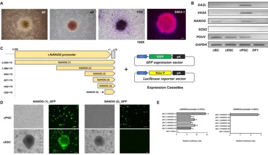
Figure 1. NANOG promoter in chicken has different regulatory mechansisms in cESCs and cPGCs respectively
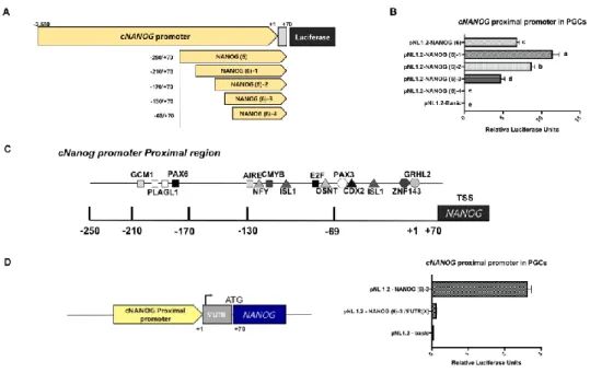
Figure 2. The proximal region of the cNANOG promoter contains a cis – regulatory elementary in the upstream of core – region
Figure 3. Selection of Transcriptional Factors in Proximal Region regulates
core-region and cis-element of cNANOG Promoter in chicken PGCs.
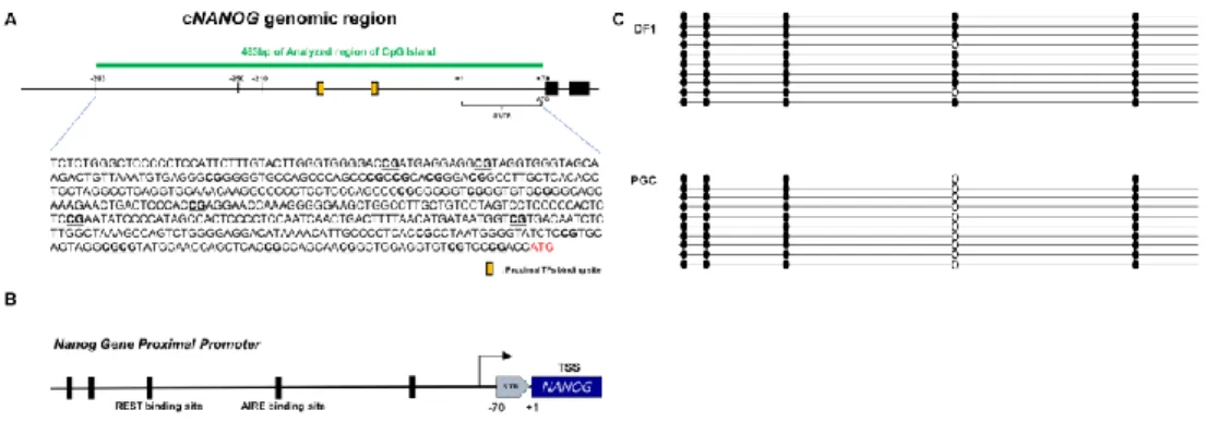
Figure 4. Methylation Analysis of proximal region shows that cNANOG
promoter is hypomethylated in chicken PGCs
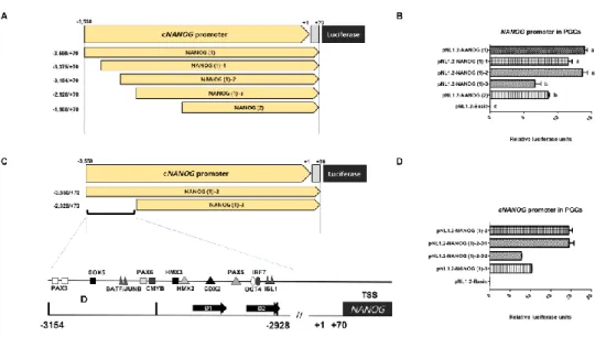
Figure 5. Selection of Transcription factor of Distal region by deletion Assay
ix
CHAPTER 4.
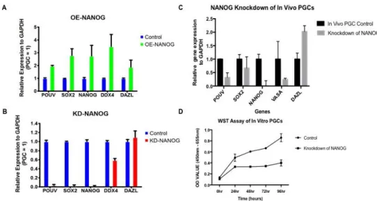
Figure 1. Relative gene expression analysis after overexpression and
knockdown of predicted transcription factors in vivo and in vitro cultured PGCs
Figure 2. NANOG maintains PGC integrity in chicken PGCs
Figure 3. Epigenetic regulation in chicken primordial germ cells.
Figure 4. REST-Corepressor Complex orchestrates Epigenetic remodeling at
the NANOG Promoter in chicken PGCs
Figure 5. Optimization of HDAC inhibitor treatment in vitro chicken PGC
Figure 6. Gene expressions of NANOG and VASA are upregulated by
inhibition of Histone deacetylase by VPA and romidepsin treatment
Figure 7. A Diagram of the role of epigenetic modifications in control of
x
LIST OF TABLES CHAPTER 3.
Table 1. List of primer sequences for cloning of the cNANOG promoter using genomic PCR
Table 2. List of primers used for quantitative real-time PCR
CHAPTER 4.
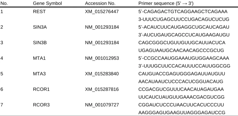
Table 1. siRNA list of HDACs and their complexes
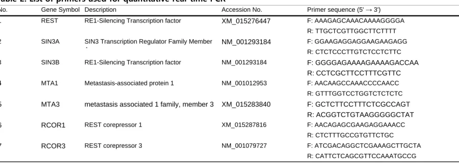
Table 2. List of primers used for quantitative real-time PCR
Table 3. List of primers used for chromatin immunoprecipitation (ChIP) - quantitative real-time PCR
Table 4. NANOG gene (Accession No. NM_001146142.1) overexpression vector and siRNA designs
xi
LIST OF ABBEREVIATION
AP2γ: Transcription factor AP-2 gamma
BLIMP1: B lymphocyte-induced maturation protein 1 BMP4: Bone morphogenetic protein 4
CHIP: Chromatin Immunoprecipitaion CVH: Chicken vasa homolog
DAZL: Deleted in azoospermia-like
DMEM: Dulbecco’s modified eagle medium eGFP: Enhanced green fluorescent protein EGK: Eyal-Giladi and Kochav
ESC: Embryonic stem cell FBS: Fetal bovine serum FGF: Fibroblast growth factors
GAPDH: Glyceraldehyde-3-phosphate dehydrogenase HDAC: Histone deacetyalse
KLF4: Kruppel-like factor 4 LIF: Leukemia inhibitory factor MEF: Mouse embryonic fibroblast
xii
PGC: Primordial germ cell
PRDM14: PR domain containing 14
qRT-PCR: Quantitative reverse transcription PCR REST: RE1 silencing transcription factor
RNAi: RNA interference siRNA: Small interacting RNA VPA: Valproic Acid
1
CHAPTER 1.
2
Primordial germ cells (PGCs), the precursors of sperm and oocytes which contains genetic information of organisms, are regulated by
coordinated actions such as Transcription factors (TFs), RNA binding proteins (RBPs), and Epigenetic modifications to acquire germ cell
specification, migration and the ability to differentiate into mature germ cells. Regulatory sequences in DNA region is a binding site of TFs to become an enhancer or repressor of the genes. In many cell types of vertebrates, TFs orchestrates gene expression by binding in characteristic motifs to procure adequate components of transcriptional machinery. And also, Chromatin epigenetic modifications, such as DNA methylation and histone post-translational modifications (PTMs) have a role in regulation of cell
characteristic such as pluripotency and germness in PGCs (Ng, Kumar et al. 2013). Despite of the diversity of the molecular mechanisms of germ cell specification in various species, major regulators to maintain germ cell characteristics during development are well conserved (Extavour and Akam 2003). During early development in many vertebrates, the patterns of DNA methylation and the associated chromatin remodeling are the crucial steps to undergo developmental steps of PGCs. )
Nanog, a core pluripotency factor, is important for acquisition and maintenance of pluripotency during embryonic development with exclusive expression of its protein in both pluripotent cells and unipotent PGCs in mice (Chambers, Colby et al. 2003, Yamaguchi, Kimura et al.
3
2005), with its role in PGCs little known (Chambers, Silva et al. 2007, Yamaguchi, Kurimoto et al. 2009, Carter, Davis-Dusenbery et al. 2014). The expression pattern of mouse NANOG appears in the inner cells of the morula of blastocyst and then be expressed in the epiblast up to early post implantation stages (Hart, Hartley et al. 2004). At this time, Nanog
expression appears in PGCs only after migration, and down regulated when the PGCs differentiate into adult germ cells (Yamaguchi, Kimura et al. 2005). Nanog in in vitro mouse model, epiblast-like cells (EpiLCs) developed from naïve pluripotent embryonic stem (ES) cells cultured in basic fibroblast growth factor (bFGF) and activin A. And also, EpiLCs acquire competence for a PGC-like fate to induce PGC-like cells (PGCLCs) with germline TFs Prdm1, Prdm14 and Tfap2c which are important for PGC
specification(Murakami, Gunesdogan et al. 2016) and bone morphogenetic protein 4 (BMP4) (Chai, Yuen et al. 1999, Hayashi, Ohta et al. 2011). In chicken, NANOG is expressed broadly in developmental stage with germ cell restricted expression pattern of protein. Chicken NANOG is restrictively expressed in scattered cells over the epiblast only in what seems to have germ cell fate throughout the epiblast at stage HH1 or HH3 (Canon, Herranz et al. 2006). In result of the studies, Nanog is revealed as an important regulator both in pluripotent cells and PGCs. Thus, many studies have been tried to identify the transcriptional regulators to enhance or repress of Nanog gene to deeply understand the molecular mechanisms how cell-specific Nanog expression is regulated.
4
Recent studies find that epigenetic regulations like loss of 5mC in whole genome, DNA methylation, histone and chromatin modifications have a crucial role in PGCs. Recent studies find that epigenetic regulations like loss of 5mC in whole genome, DNA methylation, histone and
chromatin modifications have a crucial role in PGCs to eliminate paternal imprinting in mammals (Kawasaki, Lee et al. 2014). However, studies of histone and chromatin modifications and also the control of epigenetic patterns in chicken are highly limited. In the previous studies, methylation and acetylation during germ cell specification to differentiation activate germ cell specific genes and repress somatic cell genes in mouse. And also, in chicken, the regulation is regulated epigenetically. In contrast to mammals, the H3K27me3 global level is reduced, whereas the H3K9me3 level is increased in chicken. However, other epigenetic modifications such as the level of acetylation/deacetylation level in PGCs still yet unknown in vertebrates.
Acetylation/deacetylation regulates gene expressions by relaxation or condensation of chromatin structure by Histone
acetyltransferase (HAT) and Histone deacetylase (HDAC), respectively (Grozinger and Schreiber 2002). CoREST, NURD, and SIN3A complexes, the three major Class I HDAC complexes are abundantly existed in cells which have pluripotency. The CoREST complex in both mouse and human has been shown to establish and maintain pluripotency in the formation of
5
iPS cells (Yang, Wang et al. 2011), and the SIN3A/HDAC complex in mouse ESCs increase transcriptional functions with NANOG and also reprogramming efficiency. This means that HDAC can co-localize and co-activate with NANOG to efficiently regulated transcriptional programs at the chromatin level (Saunders, Huang et al. 2017). The CoREST
([co]repressor for element-1 silencing transcription factor) complex was firstly identified in mammals to be associated with the repressor for element-1 silencing transcription factor (REST)/neuronal restrictive silencing factor to have a crucial role in regulating neuronal gene
expression and neuronal stem cell fate (Su, Kameoka et al. 2004). CoREST proteins are conserved chromatin modifying complexes composed of integral subunits. REST is broadly expressed in mouse early development (Chen, Paquette et al. 1998, Grimes, Nielsen et al. 2000). Mouse CoREST mRNA at E8.5 strongly expressed in head mesenchyme and becomes to have an omnipresent expression by E11.5 (Grimes, Nielsen et al. 2000). Using Knockout model of REST in mouse, REST was validated to regulate
retarded cell growth and also the death of widespread apoptotic cells in E9.5 to E11.5 (Chen, Paquette et al. 1998) These complexes combine histone demethylase and deacetylase and have been identified in many species, such as Drosophila (Dallman, Allopenna et al. 2004), C. elegans (Smialowska and Baumeister 2006) and mammals (Cowger, Zhao et al. 2007). CoREST complex is also associated with lincRNA HOTAIR in breast cancer (Gupta, Shah et al. 2010, Tsai, Manor et al. 2010). These results suggest that
6
CoREST might have a role in germ cells. NANOG also has a crucial role in embryonic and germline development, and also establish naïve
pluripotency in the final reprogramming stage (Saunders, Faiola et al. 2013).
In summary, evolutionally well-conserved NANOG has many roles in pluripotency cells and also, unipotent PGCs in many vertebrates. However, TFs and Epigenetic regulation of NANOG, especially histone acetylation in the chicken PGCs are mostly unknown. Therefore, it will be valuable to investigate the molecular and epigenetic mechanisms that maintain cell characteristics in the chicken PGCs, thus providing information about the germ cell development.
7
CHAPTER 2.
8
1. Primordial Germ Cells
The first germline cell population is called primordial germ cell(PGC)s in various species (Saitou and Yamaji 2010). Primordial germ cell(PGC)s, the precursors of both the spermatogonia and oocytes, have totipotent state that undergo meiosis to generate gametes and only can deliver genetic information to next generation. Because of their unique functions, PGCs have different regulatory system from other early developmental cells, and are regulated by transcription factor, signaling pathway and epigenetic regulation. Primordial germ cells in many species have different mode of development during embryonic development.
1.1 PGC specification in germ cells
PGC development commonly undergoes early and late germ cell development – PGC specification, migration into genital ridges, and germ cell differentiation. In mammals, PGCs are specified during early germ cell development. Fertilization of the oocyte by the sperm is the first step of PGC specification. This results in a totipotent zygote that give rise to all cell lineages of an organisms including germ cell lineage itself. Thus, PGC
9
D. melanogaster, zebrafish and X. laevis, germ cell specification occurs by preformation and identity of germline is continuously passed via the oocyte to the PGCs during early embryogenesis and the PGCs inherit maternally supplied germ plasm (preformation mode) Germ plasm complex is composed of RNA and conserved proteins in many species and these
proteins have a role as RNA binding factors and regulate mRNA translation. Otherwise, specification of germ cell fate in mice occurs by induction and the germline must be induced from a subset of embryonic cells and are induced by cell signaling (induction mode) (Extavour and Akam 2003).
1.1.1 Preformation model
In Drosophila melanogaster, oocyte has localized mRNA and protein before fertilization. Polar granule is assembled in the posterior region of the oocyte (Illmensee and Mahowald 1974), and oskar, a component of polar granule, forms pole cells in posterior region and to have the maternal pore plasm proteins like vasa, tudor, valois after celluarization. (Mahowald 2001).
In Caenorhabditis elegans, electron dense mRNA-protein complex called P granules are scattered throughout cytoplasm in one-cell embryo (P0), and then move to the posterior cytoplasm of the embryo
10
during zygote formation after decision of embryonic polarity(Hird, Paulsen et al. 1996). During embryo development, P granules are firstly distributed to germline blastomere P1, and repeat division asymmetrically for 3 times so that the P4 blastomere of 16 to 24 cell stage become the PGC (Deppe, Schierenberg et al. 1978, Strome and Wood 1982, Sarmah, Muralidharan et al. 2013). The P4 cell always becomes the PGC overall the nematodes. VASA
homolog (one of the components of P granule) and Sm protein (spliceosome component) have a crucial role in the distribution of PGCs with maternal inheritance of the germ plasm (Arkov and Ramos 2010). In this species, RNA-binding proteins like VASA-related RNA helicases, the Tudor-domain proteins, NANOS, the Arg methyltransferse PRMT5 and Argonaute proteins are the conserved major germ plasm elements to determine and maintain germ cell fate (Strome and Updike 2015).
In several vertebrate species like Xenopus laevis, PGCs are specified by preformation mode. As in flies, germ plasm, in which RNAs, proteins and mitochondria are accumulated, is assembled during oogenesis and localized to the vegetable pole (Tada, Mochii et al. 2012). During cleavage stages, maternally loaded germ granules, segregated in vegetal plasm, finally accumulate to the PGCs has also been documented in these vertebrates. The transmission of maternal germ granules evolved
independently with absence of maternal germ granules in early embryos (Chang, Torres et al. 2004).
11
1.1.2 Induction mode
Induction mode is conserved in most mammalian species. PGC specification to acquire germ cell fate occurs by transcriptional factors and extrinsic signals because of no maternal germ plasm in these species. Bone morphogenetic proteins (BMP) signaling pathway and WNT signaling pathways are two common pathways in human and mice (Tang, Kobayashi et al. 2016). In this signaling pathways, small set of signaling molecules and also the zygotic transcription factors critically regulate instruction of a small number of proximal epiblast cells becoming PGCs (de Sousa Lopes, Hayashi et al. 2007).
1.1.3 PGC migration in chicken
The migration of PGCs has been studied in numerous species in vertebrates, for example, mouse, zebrafish and Drosophila. In zebrafish,
Dnd1 regulates the migration and polarization of PGCs for the initial onset of germ cell migration (Weidinger, Stebler et al. 2003). In Drosophila, endoderm – 1 (Tre1) traps germ cells to regulate initiation of migration (Kunwar, Sano et al. 2008).
12
Especially, CXC motif receptor (CXCR4)/Stromal derived factor (SDF1) system is used to make it possible. SDF1 and its receptor, CXCR4 are also essential in both mouse and zebrafish (Ara, Nakamura et al. 2003, Molyneaux, Zinszner et al. 2003). In chicken, SDF1 and CXCR4 were determined to be expressed in the habitat where PGCs reside in after the later stages of their migration and also during migration (Stebler, Spieler et al. 2004).
Chicken primordial germ cells were first identified in the germinal crescent region and arise from epiblast and migrate from anterior of the primitive streak to the germinal ridge. In this region, PGCs find their way to the gonad using blood vessels.
In stage X (EG&K), avian PGCs or their precursors are localized in the central zone of the area pellucida on the ventral surface of the epiblast. During stage XI to XIV, these cells progressively translocate into the
hypoblast. PGCs arisen from the epiblast are carried to the germinal crescent region at stage 4 to 8, and then they migrate into the genital ridges via blood stream, the circulatory system, at stage 9 to 10 (Swift 1914). PGCs have a distinct genetic differentiation mechanism in male and female embryos. In genetically female embryos, PGCs differentiate into oogonia after 8d of incubation, whereas PGCs in genetically male embryos differentiate into spermatogonia after 13d of incubation.
13
The unique migration patterns of PGCs through the circulatory system are the characteristics of avian species and these characteristics can be used for the tools for the study of early PGC development, a laborious technique in other species.
1.2 Signaling pathways of PGCs
Based on the several researches of maintenance of PGCs in vitro, such as BMP4’s function of activation of germ cell specific genes, Prdm1 and
Prdm14 (Ohinata, Ohta et al. 2009), extrinsic factors to stimulate signal transduction have crucial roles in PGC development as the key
transcriptional regulators (Saitou and Yamaji 2010).
The growth factors, BMP4, LIF, SCF, retinoic acid and FGF, which are contained in each signal, are required for PGC survival and proliferation (Whyte, Glover et al. 2015). In mammal, using a cocktail of growth factors, KLF4, LIF, BMP4, SDF-1, bFGF and compounds (N-acetyl-l-cysteine, forskolin, retinoic acid) enable the survival and self-renewal of PGCs in the absence of somatic cells in culture (Farini, Scaldaferri et al. 2005).
14
Leukemia inhibitory factor (LIF), basic fibroblast growth factor (bFGF), bone morphogenetic protein 4 (BMP4) and kit ligand are the well-known signaling pathways of primordial germ cells related to proliferation and survival of primordial germ cells (De Felici and Barrios 2013).
Like-wise other species, chicken PGCs also proliferate and the proliferation of PGC during embryogenesis, specification, differentiation and also PGC survival is controlled by a cell signal.
1.2.1 Transforming Growth Factor-Beta (TGF-β) signaling pathway
Transforming Growth Factor-Beta (TGF-β) super family is composed of a large and diverse polypeptide morphogen groups: TGF –β themselves, Bone Morphogenetic Proteins (BMPs) and the Growth and Differentiation Factors (GDFs) (Ripamonti, Ferretti et al. 2009). TGF-β is activated from a complex before binding to its receptors. Ligand binding to receptors by highly conserved juxta-membrane region, known as the GS domain. TGF-β family members have distinct temporal and tissue specific expression patterns so that they play crucial roles in the development and also repair of tissues in organisms like immune cell lineages, dendritic cells, etc (Yan, Liu et al. 2009)
15
In the result of screening signaling pathway and genes related to male PGC production, TGF-β is related to the regulation of male germ cell differentiation from chicken ESCs. During induction to PGCs, Expression of Nanog and Sox2 in ESCs are decreased. In contrast, Stra8, Dazl, Integrin-α6 and c-kit, the germ cell specific genes, are increased. In the result of study, TGF- β regulates germ cell differentiation (Zhang, Wang et al. 2016).
1.2.2 Bone morphogenetic protein (BMP) signaling pathway
In mice, PGCs require Bmp2 from the visceral endoderm and Bmp4 and Bmp8b from extra-embryonic ectoderm. Expression of Bmp4
and Bmp8b from the extra-embryonic ectoderm for generation of sufficient cell numbers to induct for germ cell (Chai, Yuen et al. 1999, Ying, Liu et al. 2000, Ying and Zhao 2001). Also, key transcriptional regulators Blimp1 (Prdm1), Prdm14 and AP2γ in competent epiblast cells express to respond BMP signals to have a crucial role in PGC specification. A tripartite
transcription factor network represses somatic mesodermal program, activates germ cell genes, and also resets epigenome as a basal state. (Magnusdottir, Dietmann et al. 2013).
16
In human, SOX17 from endoderm is the key regulator of hPGCLCs acting in the upstream of Blimp1 to promote germ cell
development (Irie, Weinberger et al. 2015). In contrast, only the proximal epiblast cells receiving BMP signaling can induce PGCs expressing Blimp1 from somatic tissues (Kanai-Azuma, Kanai et al. 2002).
1.2.3 Stem cell factor (SCF) Signaling Pathway
The ligand stem cell factor (SCF), and its receptor c-kit, are essential for PGC survival and migration in the mouse (Matsui, Zsebo et al. 1990, Dolci, Williams et al. 1991, Gu, Runyan et al. 2009), and also, this signaling pathway, which is important for germ cell survival and migration in mice has also been identified in the chicken. Chicken c-KIT is expressed in both stage 19HH embryos in the presumptive genital ridge and cultured chicken PGCs (Tang and Zhang 2007). Similar to many other genes, the in
ovo studies of SCF in avian germ cells has not been determined yet. SCF without addition of hSCF or mSCF for PGC culture derivation has been used in cultured PGC medium (van de Lavoir, Diamond et al. 2006, Macdonald, Glover et al. 2010, Rengaraj, Zheng et al. 2010). SCF is
requires as an PI3K inhibitor, a down-stream target of c-kit. This results in a loss of PGC proliferation in culture (Macdonald, Glover et al. 2010).
17
1.2.4 Basic fibroblast growth factor (bFGF) signaling pathway
Basic fibroblast growth factor (bFGF) ligand activates the Map kinase signaling pathway. Mitogen – Activated Protein Kinases (MAPKs) are activated by many different cell signals. MAPKs deliver extracellular signals from activated receptors. Especially, in nucleus, MAPKs regulates appropriate gene regulatory by activation of gene transcription, protein synthesis, cell cycle regulation, apoptosis and differentiation. bFGF signaling in PGCs inhibition of mitogen – activated protein kinase (MEK) results in reducing of proliferation of cultured PGCs (Macdonald, Glover et al. 2010, Rengaraj, Zheng et al. 2010).
1.3 Germness related genes in Chicken
Specification and maintenance of PGC characteristics are
determined by major factors and germ line specific RNA-binding proteins such as VASA, DAZL and DND1. These proteins may have a role in repressing translation to prevent their differentiation into somatic cells and to maintain PGC characteristics (Houston and King 2000).
18
The conserved and universal molecular determinants in germ line maintenance are called vasa and vasa-like DEAD box RNA helicase genes. Vasa is a member of the DEAD box protein family contains nine conserved sequence motifs to unwind duplex RNA in a discontinuous and manner to have a role in pre-mRNA splicing, ribosome biogenesis, nuclear export, translational regulation and degradation. The vasa gene firstly isolated in
Drosophila (Lasko and Ashburner 1988), and vasa homologous genes have been characterized in germ cells across diverse organisms. In some organisms like mouse, vasa only found in late germ cell development stage because of no maternal inheritance in germ plasm (Toyooka, Tsunekawa et al. 2000).
In chicken, PGC specification is yet unknown problem. However, studies of germness genes are continuously studied in views of their
characteristics and expressions. Chicken vasa homologue (CVH) is one of the germ plasm components detected in initial developmental stages and is also localized to cytoplasm of germ cells and co-localized to spherical mitochondrial cloud in growing oocytes (Gustafson and Wessel 2010). Early developmental expression of CVH in chicken embryos supports that chicken PGC specification may follow the predetermination model determined by maternally inherited factors and is restricted to chicken germ cells during early embryogenesis (Tsunekawa, Naito et al. 2000). Tracking cells using Cvh promoter - GFP expression vector shows the clue of Cvh expression’s crucial role in differentiation into the germ cell fate and germline
19
germline competency in newly derived pluripotent cells (Lavial, Acloque et al. 2009).
Dazl (Deleted in Azoospermia-Like) is a member of the DAZ
gene family. Dazl regulates translation in various organisms as a germ cell specific RNA-binding protein (Collier, Gorgoni et al. 2005). DAZL
regulates spermatogenesis and oogenesis (Ruggiu, Speed et al. 1997). Dazl
gene also protect germ cells in the from apoptosis (Lin and Page 2005). In chicken, DAZL is detected in germ cells until the adult germ cell (Rengaraj, Zheng et al. 2010), and cDAZL (chicken DAZL) is expressed specifically in PGCs during their migration from EGK stage X to HH stage11. This is the difference from mammalian species. In mammalian species, DAZL is
expressed in late PGCs and regulate pre-meiotic and meiotic genes to make fertile germ cells in both sexes, for example, fetal gonocytes, spermatogonia and spermatocytes in adult testis (Fu, Cheng et al. 2015). Therefore, study of
cDAZL expression pattern suggests origin and central formation of PGCs in chicken and the difference between other species (Lee, Choi et al. 2016). Above experiments indicate that germ cell-specific RNA-binding proteins are required for germ cell development and survival.
20
Nanog is a multi-domain protein of which contains two
transactivation domains, N-terminal transactivation domain (ND) and C-terminal transactivation domain (CD) and Tryptophan-rich domain (WR) (Saunders, Faiola et al. 2013).
To understand the specific gene expression, confirmation of gene expression induced by deletion or mutation of targeting site predicted by specific gene promoter analysis. In the research of Nanog promoter, there are identification in mice and human. In mice, 180bp before transcription start site (TSS), and, in human, 118bp before TSS has OCT4/SOX2 binding site and has a crucial role of Nanog gene expression (Kuroda, Tada et al. 2005, Rodda, Chew et al. 2005). This region is highly-conserved between species. Nanog promoter in mice is activated by binding with protein factors; STAT3, T and FOXD3, and is repressed by factors like p53 and TCF3 (Pan and Thomson 2007). And also, OCT4 and SOX2 interact with KLF4 and PBX1 protein to regulate Nanog in human ESCs (Chan, Zhang et al. 2009).
2.1 NANOG in PGCs
Induction of PGCs is regulated by transcription factors and epigenetic changes to upregulate its characteristics. In the previous researches, there were few factors to have a key role in this mechanism.
21
Nanog, a key pluripotency factor in Inner Cell Mass (ICM) of Blastocyst, also expresses in PGCs (Yamaguchi, Kimura et al. 2005). Epiblast-like cell(EpiLC)s are induced from naïve state embryonic stem cell(ESC)s by Nanog with basic fibroblast growth factor (bFGF) and activin A in vitro and acquire PGC-like characteristics (Hayashi, Ohta et al. 2011). In recent study, in contrast to the result that bone morphogenetic protein 4 (BMP4), PR domain zinc finger protein 1 (Prdm1; Also known as Blimp1), PR domain zinc finger protein 14 (Prdm14) and Transcription factor AP-2 gamma (Tfap2c) are necessary to the induction of Primordial germ cell-like cell(PGCLC)s from EpiLCs, Nanog can induce PGCLCs by induction of established 4 PGC specific transcription factors (Murakami, Gunesdogan et al. 2016).
2.2 NANOG in early development
Nanog protein is a transcription factor that binds to consensus sequence ((C/G)(G/A)(C/G)C(G/C)ATTAN(G/C)) (Mitsui, Tokuzawa et al. 2003). Nanog have some roles in different states. Nanog interacts with the main self-renewal factors, Oct4, Sox2, Rex-1 synergistically and cyclically to contribute to self-renewal (Shi, Wang et al. 2006, Schulz and Hoffmann 2007). And also, Nanog binds to the promoter of Gata6 and represses Gata6 gene expression (Singh, Hamazaki et al. 2007). Nanog directly binds to
NF-22
kB, so that Nanog represses transcriptional activation of NF-kB, the gene induces differentiation, and maintains the characteristics of stem cells with Stat3 (Torres and Watt 2008).
2.3 NANOG in Medaka fish
In Medaka fish, Nanog has a different regulation with mammal and it is required for proliferation and S-phase transition in embryonic development. Thus, zNanog, which has a homology with mammalian Nanog, generates protein in nucleus and express mRNA in cytoplasm to repress PGCs’ proliferation in embryonic development (Wang, Liu et al. 2016).
2.4 NANOG in Chicken
In chicken, the pattern of Nanog is differ from mice. Nanog in chicken is not shown in hypoblast in HH1 to HH3, and is shown as scattering shape in cells in which have a fate to become PGCs in the
germinal crescent between Area pellucida and Area opaca at HH5 to HH8. After that, Nanog expression cells have same patterns with chicken PGC
23
specific genes, Vasa and Dead. Continuously, Nanog is also expressed in PGCs from HH12 to HH17 – HH20, when PGCs migrate into gonad (Canon, Herranz et al. 2006).
2.5 Epigenetic regulation of NANOG
In the epigenetic side, Nanog has two main roles: methylation and chromatin condensation. In methylation, Nanog has an interaction with Prdm14 correlated to PRC2, H3K27me complex, to regulate genes like fibroblast growth factor receptor 1/2 (Fgfr1/2), which determine system. This results in repression of differentiation. And also, it helps broad hypo-methylation using De novo DNA methyltransferase, Dnmt3A/3B/3L (Nakaki and Saitou 2014). Activators of Key enhancers marked by
H3K4me1/2, and these also marked by H3K27ac. MLL4/KMT2D is a main enhancer to p300, which is an acyltransferase and is required for H3K27ac. Nanog has an interaction with MLL4 and this results in relation of Nanog and H3K27ac (Wang, Lee et al. 2016).
In chicken, Nanog expression has a role in PGC proliferation and survival similar to mammals and is observed in HH12 to HH17-HH20, and this means Nanog regulates PGC migration from vascular system to genital ridge (Canon, Herranz et al. 2006). Methylation pattern of Nanog promoter
24
is hypo-methylated in sperm and strongly hyper-methylated in other somatic cells. This pattern is also same in Ddx4, Dnd1 and Dazl, which are related to chicken PGC proliferation and survival with Transcription factors unknown (TANAKA, Tomoki et al. 2014).
In Chromatin level, Nanog also have some different roles. Formation of opened and uncondensed chromatin defines the quantity of pluripotency. Nanog in mouse ESCs regulates pluripotency related to the formation of heterochromatin. In time, Sall1, heterochromatin related protein, maintains opened chromatin state with low level of H3K9me3 by direct co-factor (Novo, Tang et al. 2016).
Nucleosomes are basic units of chromatin that which is the histone compaction of DNAs. Nucleosomes are known to have a crucial role in packing of genes. Nucleosome itself works as an epigenetic regulator with the most crucial transcription factor, Oct4, competitively. This
regulation forms nucleosome depleted region (NDR) in DNA regulatory region of Nanog to start the transcription, and nucleosome forms NDR before the regulation of Oct4 to regulate de novo DNA methylation
(Hammachi, Morrison et al. 2012). As the result, Nanog has a possibility to have interactions with other transcription factors to regulate DNA and histone modification in chicken PGCs.
25
2.6 Recent study of NANOG
In recent studies, Nanog–RNA binding proteins, YBX1 and ILF3 upregulate pluripotency related genes in mouse ESCs (Guo, Xue et al. 2016). Prmt7 regulates micro RNA, miR 24-2, which represses Oct4, Nanog, Klf4 and c-Myc, to regulate stem-ness (Lee, Chen et al. 2016). And also, Cbfa2t2 combine with mouse Prdm14 and Oct4 to increase pluripotency related genes and lineage-specific genes. This gene also increases repressed chromatin modification genes to regulate PGC expression from ESCs (Tu, Narendra et al. 2016). Induction of germ cells also are related to Nanog
gene, known as a core pluripotency factor. NANOG can induces germ cells in primed epiblast in vitro by interacting enhancers of Prdm1 and Prdm14 in EpiLCs in mouse (Murakami, Gunesdogan et al. 2016).
3. Transcriptional gene regulation
Gene expression regulated by Promoter is spatially and temporally controlled. Transcriptional regulation by promoter results in signaling pathways and this transcriptional regulation is managed by the interactions between transcriptional factors including enhancers, silencers, insulators in cis-regulatory region of promoters (Novina and Roy 1996, Goodrich and Tjian 2010). Specific gene expressions are the result of one or more
26
transcriptional factors on promoters called enhancers and repressors. Thus, studies on cis elements and different algorithms of specific gene promoters and also transcriptional start points and transcription factor binding sites is important for exploring the modulation of gene expression regulatory network. (Bucher, Fickett et al. 1996, Fickett and Hatzigeorgiou 1997).
3.1 Promoter
Promoter is a direct tool of gene regulation and exists in the upstream of gene. In the upstream of gene, mRNA synthesis in eukaryotic cells initiates by complex and highly regulated process. This process is the result of general transcription factors and RNA polymerase II which assemble into a pre-initiation complex at the binding motif of the core promoter. (Struhl 1989, Da Silva and Srikrishnan 2012).
Regulatory elements that map near genes are cis-acting DNA sequences. Cis-acting DNA sequences contains two elements: Core
promoter and Promoter Proximal Elements. Core promoter has a basal level expression which contains binding site for TATA-binding protein and associated factors. Promoter Proximal Elements has a level for
transcriptional regulation and also translational regulation. These elements contain binding sites for transcription factors. Eukaryotic Promoter elements
27
are so various that their combinations of core and proximal elements are found near each gene.
3.1.1 Promoter Proximal elements
Promoter proximal elements has key roles in gene expression in many developmental stages and also in many different type of cells, surprisingly, also in ubiquitously expressed genes. Activators which are important in transcription regulation are recognized by promoter proximal elements.
Housekeeping genes such as β-actin are ubiquitous in all cell types for basic cellular functions. These genes have common promoter proximal elements because of their ubiquitous characteristics. Also, housekeeping genes are recognized by activator proteins found in all cells. Otherwise, Genes expressed only in some specific cell types or at singular times have promoter proximal elements recognized by activator proteins found only in specific types or times.
28
The definition of core promoter is as the minimal DNA region sufficient to direct low levels of activator-independent (basal) transcription by RNAP II in vitro (Butler and Kadonaga 2002). Core promoter typically extends approximately 40bp up-and down- stream relative to the
transcription start site and can contain several distinct core promoter sequence elements recognized by the general transcription factor TFIID which promotes the process of forming pre-initiation complexes (Goodrich and Tjian 2010).
Core promoters in higher eukaryotes are highly diverse in
structure, and, so far, only TATA box which contain an A/T-rich sequence, with the consensus TATAAA, and initiator element are shown to be capable of directing accurate RNAP II transcription initiation independent of other core promoter elements. TATA-mediated transcription initiation is well known. Nevertheless, initiator and other core promoter elements are known very little(Kadonaga 2012).
In the past, the core promoter was regarded as being found in every cellular gene. Nowadays however, not only distal promoters and enhancers but also core promoter and initiators are involved in
transcriptional regulation by interacting cell-specific trans-acting factors and mechanisms(Juven-Gershon and Kadonaga 2010). Also, germ cells are observed to express core promoter-associated regulatory factors with various core promoter architecture contribute to regulate germ-cell-specific
29
transcriptional signals of appropriate gene expression related to somatic gene repression and germ-ness upregulation genes(Lenhard, Sandelin et al. 2012). These studies can be adapted to germ cell transcription by specialized core promoters in application like embryonic study and production of transgenic animal using germ cell-specific promoter vectors (Bhullar, Schmidt et al. 2001, Song, Lai et al. 2016).
3.1.3 Germ cell specific promoter
Germ cell specific promoter is a unique set of core promoter-associated transcription factors not found in somatic cells. These promoters can used in producing transgenic animals including small size (~100bp) and relatively high GC content (Xiao, Kim et al. 2006).
Cell-type-specific Transcription activation factors (TAFs) and TBF related factors (TRFs) have unique roles during development, differentiation and cell proliferation. Proliferation and differentiation regulated by specific factors and promoter such as positive and negative cell cycle regulators, differentiation promoting transcription factors and epigenetic regulation, for example, chromatin remodeling promoting proliferation and differentiation (Ruijtenberg and van den Heuvel 2016).
Recent studies showed an analysis of endogenous genes and regulators observed in the context of nearly homogeneous population of single, specific, differentiated cell type and in distinct cell cycle stages.
‘Non-30
prototypic’ core promoter recognition factors, including cell-type specific TAFs and TRFs have a critical role in driving cell specific programs of transcription to regulate specific sets of genes such as germ cell specification or maintenance of gene expression states during embryonic development (Goodrich and Tjian 2010). By using GFP reporters contain ALF promoter, which has a regulatory role in male and female gametogenesis, CCCTC-binding factor (CTCF) binds to ALF promoter to regulate germ cell gene expression. (Kim, Li et al. 2006). By research germ cell specific promoters, on-off germ cell gene promoter regulation has an insight in application for transgenic animal and developmental studies.
3.2 Enhancer
Eukaryotic enhancers are one of the cis-regulatory elements. Enhancers are required for maximal transcription of genes involved in tissue-specific gene expression. Enhancers can exist upstream or downstream of the transcription initiation site. Enhancers are DNA
sequences that could modulate from a far distance base pairs away from the initiation site. Enhancers could regulate transcriptional ability by interaction with transcription factors independent of their location, distance or
orientation. (Banerji, Rusconi et al. 1981). Ability of enhancer by interaction with transcription factors independent of their location, distance or
31
together (Matharu and Ahituv 2015). The distinct chromatin feature of enhancers to regulate the cell-specific gene expression mechanisms. OCT4 expression in mouse is regulated by stage/tissue specific enhancer in mouse that proximal and distal enhancer are respectively active in the epiblast of mouse embryos and in the germline cells. (Yeom, Fuhrmann et al. 1996).
3.3 Transcription factors
Transcription factors are also called activator proteins and silencer proteins by their role in gene expression. These bind to promoter, enhancer and silencer DNA by interactions with other proteins. These interactions activate/repress and increase/decrease transcription as much as 100-fold above basal levels. Transcriptional activators bind to specific promoters and enhancers at specific times to increase transcriptional levels by two
structural domains: DNA-binding domain and Transcription-activator domain.
Transcription factors regulates central mechanism of promoter regulation by site-specific binding. Germ cell specific factors have their roles in germ cell specific gene regulation in many species. The gene expression program which specialized transcription in male germ cells of Drosophila
and MousePrimary Spermatocytes were discovered. A Testis-Specific DREAM Complex (tMAC) regulated meiotic arrest of Drosophila and
32
Cyclic AMP response element (CRE) binding protein (CREB) and cyclic AMP response element modulator (CREM) have regulatory pathway with ACT and KIF17 in haploid cells. And also, Two TBP – related factors, TRF2 and TRF3, maintains self-renewal of spermatogonia in male and restrictedly expresses in the ovary, respectively (White-Cooper and Davidson 2011).
4. Epigenetic modifications
Epigenetics including remodeling of nucleosomes, DNA methylation, posttranslational modifications of histone proteins, and chromatin reorganization modulates chromatin structure to regulate gene expression allows cell specific characteristics. Integration of TSS location with related functions, such as histone methylation (Yan and Boyd 2006) and acetylation sites (Pokholok, Harbison et al. 2005), the position of nucleosomes (Wiren, Silverstein et al. 2005), and the position of
transcription factor binding sites are the clues of transcriptional regulation of proximal to genes.
Chromatin has two broad classes: Euchromatin and Heterochromatin. Euchromatin is the majority chromatin in its
de-condensed state during interphase, and it only condenses during mitosis This contains most genes and this region is an active region. Heterochromatin is
33
highly condensed even in interphase. When the chromosome packaged, this influences gene activity. Nucleosomes in the de-compacted area unwind to make transcription initiate. Transcription factors are non-histone proteins which unwind nucleosomes and remove histones at 5’ end of genes and open to interaction with RNA polymerase so that recognize promoter and initiate gene expression (Butler and Kadonaga 2002). In the result of Epigenetic regulation, DNA are modified in both DNA and Histone level (Rothbart and Strahl 2014).
4.1 Epigenetic modifications in PGCs
Epigenetics must be reset during germ cell specification and establish new epigenetic states for renewal of acquirement of totipotency (Smallwood and Kelsey 2012). Therefore, epigenetic modifiers have key roles in germ cell development in germ cell specific fate such as meiosis and maintaining genomic integrity through stage and cell specific gene regulation. In the researches of germ cell studies, germ cells acquire unique cell types by a series of epigenetic events (Sasaki and Matsui 2008).
Epigenetic regulation of germ cell development has an extensive spectrum. Epigenetic reprogramming has a crucial role in PGC mechanism to reacquire totipotency in pre-migratory and pluripotency in migratory
34
germ cells (Seki, Hayashi et al. 2005). It occurs from PGC specification to colonization and migration into developmental gonads of the early germ cell development, so that germ cells mature to acquire the capacity of germ cell characteristics after fertilization (Jang, Seo et al. 2013).
Epigenetic Modification has a critical role to regulate germ cell specific gene and establish the characteristics of PGCs. During migration of PGCs into gonads, DNA methylation and H3K9me2 is decreased, and H3K27me3 is increased. After migration into gonads, H3K27me3 is hypo-methylated to regulated germ cell specific genes (Tollervey and Lunyak 2012). Due to the features that PGCs have both activation and repression mechanism, H3K4me3 and H3K9Ac is temporarily increased during PGC development. For these reasons, Epigenetic regulation is highly important for PGCs to acquire totipotency during embryonic development. (Khromov, Pantakani et al. 2011).
4.1.1 Epigenetic modifications in Chicken PGCs
In Chicken, transcriptome analysis of primordial germ cell – specific genes including genes related to cell cycle was reported, and also the result that chicken’s embryonic stem cell is similar to mouse’s primordial germ cell was reported (Rengaraj, Lee et al. 2012, Jean, Oliveira et al. 2015).
35
Nevertheless, some of the germ-cell specific regulator genes are differently expressed differ from mouse.
In case of the genes, SOX2/SOX3, SOX2 expresses mostly in cultured cESCs. In opposition to SOX2, SOX3 expresses in cES, and also PGCs and Hypoblast (Zhang and Klymkowsky 2007, Acloque, Ocana et al. 2011). DAZL, DND1, DDX4, PIWIL1 and GTSF1, CALR3, GPR149 is involved in differentiation of mammalian germ cells, (Edson, Lin et al. 2010, Ikawa, Tokuhiro et al. 2011, Jean, Oliveira et al. 2015) and they also have a role of germ cell specific genes in avian species (Lee, Choi et al. 2016, Sekinaka, Hayashi et al. 2016). In spite of the fact that OCT4/POUV, NANOG, ENS/ERNI, CDX2’s expression in both cESCs and PGCs, TRIM71, KLF1, SOX3, CFC1B, OTX2 and EOMES expresses only in cESCs, and KLF2, DAZL, DDX4 express only in PGCs.
Epigenetic regulation of chicken PGCs was not unknown until recently. In Chicken, Upregulation of the gene, which transcribes
SUV39H2/KMT1B, Histone Methyltransferase, results in elevation of H3K9me3, the most heterochromatin model, in cPGCs related to cESCs. In contrast, the expression of H3K27me3 is lower in chicken’s PGCs. DNA methylation in PGCs regulates the expression of DNMT1, DNMT3B and HELLS/SMARCA6 to maintain its pattern and have an important role. In result, epigenetic regulation of chicken PGCs differs from mouse PGCs in
36
that H3K9me3-abundant heterochromatin and limitative accumulation of H3K27me3 (Zhang, Elsayed et al. 2015, Kress, Montillet et al. 2016).
4.2 DNA Methylation
DNA methylation regulates gene expression usually by inhibition of transcriptions. In vertebrates and plants, many genes contain CpG islands near their promoters (Lister, Mukamel et al. 2013). CpG islands are DNA sequences in which contains lots of CG repeats (Gardiner-Garden and Frommer 1987). CpG has different pattern in housekeeping genes and tissue-specific genes. In housekeeping genes, The CpG islands are un-methylated (Schug, Schuller et al. 2005). Otherwise, Methylation pattern of tissue – specific genes are methylated in CpG islands. Transcriptional silencing via methylation blocks transcription factor binding to induce heterochromatin. Only a fraction of CpG associated promoters have TATA-like elements. Control of germ cell-specific promoters are broadly similar in several sequences like a TTCAAA element, a GC-rich region with a number of CpG island and un upstream TC-rich region (Han, Xie et al. 2004). This result means that germ cell – specific promoters could be regulated by epigenetic regulation including DNA methylation. Though the sequences are broadly similar, Factors related to their regulation has
37
unknown selective activation during germ cell differentiation and somatic silencing using epigenetic regulation.
4.2.1 DNA methylation in PGCs
DNA methylation leads to proper epigenetic programming of PGCs from pre-implantation development (Durcova-Hills, Hajkova et al. 2006). During germ cell development, nearly complete demethylation and re-methylation (Seisenberger, Peat et al. 2013).
DNA methylation contributed by the paternal gamete is de-methylated through a hydroxyl-de-methylated intermediate catalyzed by the Tet Methyl-cytosine Dioxygenase 3 (TET3) member, one of the TET family (Gu, Guo et al. 2011, Wossidlo, Nakamura et al. 2011, Smith, Chan et al. 2012). After fertilization, loss of global DNA methylation levels continues until the blastocyst stage (Guibert, Forne et al. 2012) After specification of the ICM, global re-methylation of the genome occurs (Borgel, Guibert et al. 2010). Global resetting of DNA methylation patterns occurs at germ cell development and at early in embryogenesis (Sasaki and Matsui 2008).
Generally accurate heritability of DNA methylation and its stability makes erasure of DNA methylation might only be possible either by breaking DNA or replicating DNA in the absence of DNMT1 and such
38
enzymes like DNMT3A, DNMT3B and also 5hmC (Reik 2007, Kangaspeska, Stride et al. 2008, Stadler, Murr et al. 2011)
During implantation of the blastocyst, the first wave of epigenetic reprogramming occurs. Existed DNA methylation patterns at CpG islands are erased to become a barrier of undergoing somatic cell fate. Only some regions related to evolutionarily young and potentially hazardous
retrotransposons remain highly methylated (Tang, Dietmann et al. 2015). When PGCs migrate to the genital ridge epigenetic remodeling observed only in paternal or maternal imprinted loci of germ cells has started. Transcriptional repression and DNA methylation decreases in migrating PGCs are regulated by H3K9 di-methylation (H3K9me2). Subsequently, proper repressive chromatin state of the PGC genome is maintained by the H3K27 tri-methylation (H3K27me3), another repressive mark. Theses repressions by methylation marker is related to repression of somatic genes and contribution of acquiring totipotency in migrating PGCs (Sasaki and Matsui 2008).
After germ cell differentiation, distinct sex-specific DNA re-methylation patterns of mature oocytes and sperm during germ cell development occur. (Durcova-Hills, Hajkova et al. 2006).
39
4.2 Histone Acetylation
The N-terminal tails of the histone H3 and H4 contain many Lysine residues, and can be modified by acetylation, methylation,
phosphorylation, ubiquitination and SUMOylation to regulate the interactions both between histone-DNA and histone-histone (Ridsdale, Hendzel et al. 1990, Wang, Zang et al. 2009), and these post-translational modifications regulate gene transcription.
Acetylation and deacetylation of lysine residues on histone tails are related to initiation and elongation. In transcription start sites (TSSs), Chromatin remodeling in promoter region is regulated by acetylation of nucleosomes. And also, Acetylation destabilizes nucleosome structure to facilitate RNA polymerase II binding to activate mRNA synthesis result in transcription activation (Wang, Zang et al. 2009). Histone acetylation is commonly associated with transcriptional activation to plays crucial roles in modulating chromatin structure and functions (Shahbazian and Grunstein 2007). The acetylation state of a chromatin locus is controlled by two
classes of antagonizing histone modifying enzymes, histone acetyltransferases (HATs) and histone deacetylases (HDACs). These enzymes add to or
40
4.3 HDACs
Histon deacetylation is associated with transcriptional
corepressors (Kadosh and Struhl 1997). These enzymes are highly conserved from yeast to human. The 18 HDACs in humans are grouped into four classes based on their homology with yeast orthologs and other phylogenetic analyses: class I (HDAC1,2,3, and 8), class II (HDAC4,5,6,7,9 and 10), class III (Sirt1,2,3,4,5,6, and 7) and class IV (HDAC 11) (de Ruijter, van Gennip et al. 2003, Yang and Gregoire 2005, Yang and Seto 2008). Among the four classes, HDAC1 and HDAC2 in class I mostly regulate gene expression in nucleus and the others regulate in both nucleus and cytoplasm according to the mechanism of the cells. The functions of these enzymes are various and substrate specificity between these are little. Though these enzymes have small substrate specificity, these enzymes can have a various function by their co-factors (Shi and Whetstine 2007). Among these, HDAC 1,2,3 and 8 in class I mostly regulate gene expressions using co-repressor complexes: Sin3, NuRD (nucleosome remodeling and deacetylation), CoREST (co-repressor for element-1-silencing transcription factor) and SMRT (silencing mediator of retinoid and thyroid receptors)/NCoR (nuclear receptor co-repressor) (Kelly and Cowley 2013). HDAC1 and HDAC2 are the main components of all four complexes. The most well-known HDAC1/2 complexes are Sin3A, NuRD and CoREST complex. These complexes use transcription factor and histone recognition site in complexes to have roles
41
in repression of genes, regulation of cell cycle and DNA repair, etc. according to the cell types.
HDAC mostly have researched in the study of cancer regulatory mechanism than embryology. For example, AML1 – ETO fusion protein, discovered in t(8;21) AML patients, represses the regulation of AML1 targeting genes attracting HDAC1/2/3 by ETO elements. And, this protein result in inhibition of bone marrow differentiation and induction of cell transformation. In the mechanism of the protein, HDAC1/2/3, DNMT1, DNMT3A and EZH2, which also regulate in mechanism of stem cells and PGCs are contained and are being researched (Gong, Li et al. 2016). And also, well-known HDAC inhibitors being used in the researches have various activities and combination ability in inhibition of specific HDACs and their complexes. For example, Romidepsin and VPA, which inhibit HDAC1/2 and broad HDACs respectively, have the highest combination ability with CoREST complexes (Robertson, Hurley et al. 2013). In mice, HDACs also repress p21 and p57 to regulate G1-to-S-phase transition with no negative effect in B-cell to improve cell proliferation (Yamaguchi, Cubizolles et al. 2010).
In terms of embryological study of HDACs, most studies in mice are divided into two developmental stages; early embryo before fertilization to Blastocyst and oocyte, the functional germ cell after migration into genital ridge of PGCs. In mice oocyte, HDACs regulate acetylation in meiosis (Kim,
42
Liu et al. 2003), and HDAC2 regulates de novo methylation of DNMT3A2 in oocyte, late of PGC development, to forms imprints and repress repetitive element result in protecting genomic integrity (Ma, de Waal et al. 2015). In case of HDAC1, HDAC1 repression in ESCs regulates HDAC1/2 complex to acquire specification of differentiated cell by regulating H3K56Ac (Dovey, Foster et al. 2010). In the development of mice in pre-implantation stage, SIN3A, one of the HDAC1/2 complexes, maternally regulates gene
expression related to reprogramming until middle of 2-cell stage (Jimenez, Melo et al. 2015).
4.3.1 HDACs in chicken
In chicken, according to Pubmed, HDAC1,2,3,8 and 9 has Phylogenetic similarity between other species between HDACs, which translate proteins. In comparison, chicken’s HDAC 1,2 and 3 have 480,488 and 428 amino acids and resemble 93.8, 97.1 and 97.0%, respectively (Takami, Kikuchi et al. 1999).
In the embryonic study of HDACs in chicken, HDAC3, Dnmt3a and Dnmt3b are highly expressed in ESCs. On the other hand, the
expressions of Dnmt1, HAT and POUV are decreased to regulate the differentiation to embryonic body (EB) cells. At that time, methylation
43
increases and H3 acetylation decreases, so that they regulate consistent gene expression of POUV to maintain pluripotency and prevent differentiation (Jiao, Wang et al. 2013). In the result of co-treatment of trichostatin A (TSA), HDAC inhibitor, and 5-azadC, DNMT inhibitor to regulate transcription of genes, they synergistically upregulate NANOG expression and decrease overall methylation in chicken ESCs (Wang, Wang et al. 2016). And also, treatment of tamibarotene (AM80), RARα activator, and TSA promotes STRA8 gene’s promoter in chicken ESCs to induce spermatogonial stem cell (SSC)s which express Integrinβ1 (Zhang, Zuo et al. 2015).
44
CHAPTER 3.
Transcription factor confers germ cell specific
transcriptional control of the chicken
NANOG
through CpG methylation of a
45
1. Introduction
Primordial germ cells (PGCs) are the precursors of differentiated germ cells, sperm and egg, has two main distinguishable properties: stemness to maintain pluripotency and self-renewal, germness to undergo PGC development from PGC specification to mature germ cell
differentiation to be able to deliver genetic information to next generations. Despite of the diversity of the molecular mechanisms of germ cell
specification in various species, major regulators to maintain germ cell characteristics during development are well conserved (Extavour and Akam 2003).
For examples, germ cell related genes, Vasa and Dazl , are expressed in germ line cells in various species to support the germ cell development. (Takeda, Mishima et al. 2009, Gustafson and Wessel 2010, Lesch and Page 2012). Pluripotency genes such as Nanog, Oct4 and Sox2 are also express in PGCs in many organisms. The importance of these genes in PGCs are revealed by the studies that the mis-regulation of these genes cause apoptosis in PGCs, still their functions yet unknown (Saito, Takeda et al. 2003, Kehler, Tolkunova et al. 2004, Chambers, Silva et al. 2007). The recent studies of pluripotency factor for the control of epigenetic status in germ cells assume the relationship between germ cells and the roles of pluripotency n the cells. (Leitch and Smith 2013).
Nanog, a core pluripotency factor, is important for acquisition and maintenance of pluripotency during embryonic development with
46
exclusive expression of its protein in both pluripotent cells and unipotent PGCs in mice (Chambers, Colby et al. 2003, Yamaguchi, Kimura et al. 2005), with its role in PGCs little known (Chambers, Silva et al. 2007, Yamaguchi, Kurimoto et al. 2009, Carter, Davis-Dusenbery et al. 2014). The expression pattern of mouse NANOG appears in the inner cells of the morula of blastocyst and then be expressed in the epiblast up to early post implantation stages (Hart, Hartley et al. 2004). At this time, Nanog
expression appears in PGCs only after migration, and down regulated when the PGCs differentiate into adult germ cells (Yamaguchi, Kimura et al. 2005).
According to the evolutionary conservation between homologues in many vertebrates (Jauch, Ng et al. 2008), Nanog in human ES cells has been shown to have the same role in regulation of pluripotency as its mouse counterpart (Hyslop, Stojkovic et al. 2005).
Regulatory sequences in DNA region is a binding site of TFs to become an enhancer or repressor of the genes. In many cell types of vertebrates, TFs orchestrates gene expression by binding in characteristic motifs to procure adequate components of transcriptional machinery.
Nanog in in vitro mouse model, epiblast-like cells (EpiLCs) developed from naïve pluripotent embryonic stem (ES) cells cultured in basic fibroblast growth factor (bFGF) and activin A. And also, EpiLCs acquire competence for a PGC-like fate to induce PGC-like cells (PGCLCs) with germline TFs Prdm1, Prdm14 and Tfap2c which are important for PGC
47
specification(Murakami, Gunesdogan et al. 2016) and bone morphogenetic protein 4 (BMP4) (Chai, Yuen et al. 1999, Hayashi, Ohta et al. 2011). In chicken, NANOG is expressed broadly in developmental stage with germ cell restricted expression pattern of protein. Chicken NANOG is restrictively expressed in scattered cells over the epiblast only in what seems to have germ cell fate throughout the epiblast at stage HH1 or HH3 (Canon, Herranz et al. 2006).
In result of the studies, Nanog is revealed as an important
regulator both in pluripotent cells and PGCs. Thus, many studies have been tried to identify the transcriptional regulators to enhance or repress of
Nanog gene to deeply understand the molecular mechanisms how cell-specific Nanog expression is regulated. Thus, in this study, we tried to validate cell specific regions of cells and TFs, and Also, to confirm that epigenetic modification regulates cell specific Nanog promoter activities by methylation analysis.
2. Materials and Methods
Experimental Animals and Animal Care
The care and experimental use of chickens were approved by the Institute of Laboratory Animal Resources, Seoul National University (SNU- 150827-1). The chickens were maintained according to a standard
48
management program at the University Animal Farm, Seoul National University, Korea. The procedures for animal management, reproduction, and embryo manipulation adhered to the standard operating protocols of our laboratory.
Collection of chick stage X embryos from eggs
Chick stage X embryos were separated from the egg using sterilized paper (Chapman, Collignon et al. 2001) and the shell membrane and albumen were detached from the yolk. A piece of square-type filter paper (Whatman, Maidstone, Kent, UK) with the hole at the center was placed over the germinal disc. After cutting around the paper containing the stage X embryos, it was gently turned over and transferred to saline buffer to further remove the yolk and the vitelline membrane for embryo collection (Pannett and Compton 1924), and were classified according to the cleavage stages proposed by Eyal-Giladi and Kochav (Eyalgiladi and Kochav 1976). Unfertilized and abnormal embryos were identified by the morphological criteria of blastoderms.
49
Chicken blastodermal cells from White Leghorn chicken stage X – stage XII blastodermal cells were used (Pain, Clark et al. 1996). Chicken embryonic stem cell (ESC) line was maintained and subcultured with
knockout Dulbecco’s Modified Eagle’s Medium (DMEM, Gibco, CA, USA) supplemented with 10% (vol/vol) Fetal Bovine Serum (FBS, Hyclone, Utah, USA), 2% (vol/vol) chicken serum (Sigma-Aldrich, St.Louise, MO, USA), 2 mM L-glutamine (Gibco), 1× non-essential amino acids (Gibco), 1×β-mercaptoethanol (Gibco), and 1×Antibiotic–Antimycotic (Gibco, CA, USA). rch LIF (20 ng/mL, Sigma) was used. Cultured cells were serially subcultivated every second or third day to inhibit differentiation by gentle pipetting without any enzyme. The cells were separated gently using the tip of a micropipette and disseminated onto a new plate. Half or all of the incubation medium was replaced daily with fresh medium. Chicken ES cells were cultured in an incubator at 37℃ with an atmosphere of 5% CO2 and
60–70% relative humidity.
Culture of DF-1 cell line
Chicken DF-1 fibroblast cell line was maintained and subcultured with Dulbecco’s modified Eagle’s medium (DMEM) (Hyclone, Utah, USA) supplemented with 10% (vol/vol) Fetal Bovine Serum (FBS) (Hyclone, Utah, USA), and 1X Antibiotic-Antimycotic (Gibco, CA, USA). DF-1 cells were