Over-expression of thymosin
β4 promotes abnormal
tooth development and stimulation of hair growth
HEE-JAE CHA*
,1, DEBORAH PHILP
2, SOO-HYUN LEE
3, HYE-SUNG MOON
4,
HYNDA K. KLEINMAN
2and TAKASHI NAKAMURA
21Department of Parasitology and Genetics, College of Medicine, Kosin University, Busan, S. Korea, 2Craniofacial Developmental Biology and Regeneration Branch, National Institute of Dental and Craniofa-cial Research, National Institutes of Health, Bethesda, MD, USA, 3Department of Pharmaceutics, College of
Pharmacy, Seoul National University, Seoul, S. Korea and 4Department of Obstetrics and Gynecology, School of Medicine, EWHA Womans University, Seoul, S. Korea
ABSTRACT Thymosin βββββ4 has multi-functional roles in cell physiology. It accelerates wound healing, hair growth and angiogenesis, and increases laminin-5 expression in corneal epithelium. Furthermore, thymosin βββββ4 stimulates tumor growth and metastasis by induction of cell migration and vascular endothelial growth factor-mediated angiogenesis. Using a construct on the skin-specific keratin-5 promoter, we have developed thymosin βββββ4 over-expressing transgenic mice to further study its functional roles. Thymosin βββββ4 in adult skin and in embryonic stages of the transgenic mouse was analyzed by both Western blot and immunohistochemistry. The over-expression of thymosin βββββ4 was observed especially around hair follicles and in the teeth in the transgenic mice. We examined the phenotype of the thymosin βββββ4 over-expressing mice. Hair growth was accelerated. In addition, the transgenic mice had abnormally-shaped white teeth and dull incisors. We found that the expression of laminin-5 was up-regulated in the skin of the transgenic mice. We conclude that thymosin βββββ4 has an important physiological role in hair growth and in tooth development.
KEY WORDS: thymosin β4, transgenic mice, hair growth, tooth development, laminin-5
Introduction
Thymosin β4, a 4.9 kDa protein containing 43 amino acids, is an actin-sequestering peptide. It binds with monomeric globular actin and inhibits the polymerization of actin (Safer et al., 1991). Thymosin β4 plays an important role in cell motility due to its participation in the rapid polymerization/depolymerization of actin (Pantaloni and Carlier, 1993; Kang et al., 1999). Thymosin β4 has multiple biological activities, including promotion of angiogenesis (Grant wt al., 1995; Grant et al., 1999) and wound healing (Philp et al., 2004). Previously, we also showed that thymosin β4 stimulates tumor growth and metastasis by induction of cell migration and vascular endothelial growth factor (VEGF)-medi-ated angiogenesis (Cha et al., 2003). Its role in malignancy has been reported by several labs using transfection approaches, and its level is also elevated in many metastatic lesions (Cha et al., 2003; Wang et al., 2004).
Functional studies using exogenously delivered thymosin β4
BIOLOGY
www.intjdevbiol.com*Address correspondence to: Hee-Jae Cha. Department of Parasitology and Genetics, College of Medicine, Kosin University, Pusan, S. Korea, 34, Annam-dong, Seo-gu, Busan, 602-703, S. Korea. Fax: +82-51-990-3081. e-mail: hcha@kosin.ac.kr
Accepted: 24 November 2008. Final author corrected PDF published online: 25 November 2009.
ISSN: Online 1696-3547, Print 0214-6282
© 2009 UBC Press Printed in Spain
Abbreviations used in this paper: CMV, cytomegalo virus; K5, keratin 5 promoter;
LM-5, laminin-5; VEGF, vascular endothelial growth factor.
demonstrated its role in wound healing and in hair growth in both normal rats and mice (Philp et al., 2004). When examining the distribution of endogenous thymosin β4 through sequential phases of depilation-induced hair growth, we found that a significant number of thymosin β4-expressing cells are present in the devel-oping hair follicle. In addition, isolated clonogenic hair follicle keratinocytes, which are related to hair follicle stem cells, produce thymosin β4 when cultured in vitro for 7–10 days. The presence of exogenous thymosin β4 causes a dose-dependent decrease in the expression of the stem cell marker K15 by these cells, suggesting that thymosin β4 may promote initial stem cell fate determination (Philp et al., 2004). A critical step in the hair growth cycle is the movement of some of the stem cells downward from bulge region, which is located at the middle of hair follicles and is
enriched in stem cells. Their differentiated progeny contribute to complete re-growth or regeneration of the lower, cycling portion of the follicle. Thymosin β4 promotes the migration of these stem cells and their immediate progeny and, thus, exerts its promoting effect on hair growth.
Laminin-5 (LM-5) is a unique sub-epithelial basement mem-brane glycoprotein composed of α3/ β3/γ2 chains (Burgeson et al., 1994). LM-5 is a component of anchoring filaments localized in the basement membrane region of the skin, cornea, conjunc-tiva, and other tissues. It interacts with laminin-6 and -7 to form a stable association with the basement membrane structure (Carter et al., 1991; Kallunki et al., 1992; Ljubimov et al., 1995; Champliaud et al., 1996). LM-5 is induced by thymosin β4 in the cornea (Sosne et al., 2004). LM-5 is expressed by migrating keratinocytes and is expressed in hair follicles (Chuang et al., 2003). Furthermore, integrin beta-1, the major receptor component of laminin-5, has a significant role in hair follicle development (Brakebusch et al., 2000; Raghavan et al., 2000) suggesting that thymosin β4 may stimulate hair growth, in part, by increasing laminin-5 expression. The expression of thymosin β4 is closely related to the devel-opment of certain organs. Thymosin β4 has been studied in chick embryogenesis, including the development of the nervous sys-tem, cardiovascular syssys-tem, and feather buds (Dathe et al., 2004). It has been detected in the nervous system of the develop-ing rat brain, and durdevelop-ing differentiation of embryonic cells into cardiac cells, thus, suggesting that the expression of thymosin β4 may be involved in the development of the nervous system and the heart, (Gomez-Marquez et al., 1993; Gomez-Marquez et al., 1996; Anadon et al., 2001). Thymosin β4 is also expressed early in the developing tooth and may be important in dentinogenesis (Akhter et al., 2005). Thus, thymosin β4 is likely important in the early development of several organs, but this has not been directly demonstrated. Here, we used over-expressing mice to determine the physiological role of this protein in development.
Results
Generation of transgenic mice expressing thymosin βββββ4 under the control of the keratin 5 (K5) promoter and char-acterization of its expression
First, we expressed the thymosin β4 transgene under con-trol of the cytomegalovirus (CMV) promoter. However, we did not get any positive transgenic mice among hundreds of new born pups. We concluded that mice expressing thymosin β4 on the CMV promoter died at an early embryonic stage. As thymosin β4 is a key protein regulating cell migration, we hypothesize that over-expressing thymosin β4 may affect cell migration of early stage embryogenesis resulting in embryonic lethality. Next, we focused on the analysis of thymosin β4 functions in ectodermal development. We successfully created an epithelial-specific thymosin β4 transgenic mouse model a using keratin 5 promoter. We used this promoter because many functions of thymosin β4 occur in the skin, including wound healing and hair growth. The K5-thymosin β4 transgene was made by sub-cloning the mouse thymosin β4 cDNA into a vector containing a 5.2 kb fragment of the bovine K5 promoter, the rabbit β-globin intron 2, and the SV40 polyadenylation signal (Fig. 1A). Two founder transgenic mice were identified by PCR analysis of tail DNA using primers specific for the
thymosin β4 and β-globin intron (Fig. 1A). All of the transgenic mice grew to normal size and were fertile.
Immunohistochemistry on adult skin sections revealed that both lines of founder mice over-expressed thymosin β4 in the skin especially in the hair follicles (Fig. 1B). The over-expres-sion was confirmed by Western blot analysis with total protein from the skins of the transgenic and wild type mice (Fig. 1C).
Fig. 1. Generation of thymosin βββββ4 over-expressing mice. Construction of the K5 thymosin β4 transgene (A). The fragment containing the full-length mouse thymosin β4 cDNA (0.2 Kb) was cut using SnaBI and Nhe I and inserted into A bluescript KS plasmid containing a 5.2 kb fragment from the bovine K5 promoter, a 0.6 kb fragment containing the rabbit b-globin intron, and a 1.1 kb fragment containing two 3' poly A signal sequences. The transgene was isolated by cutting with Acc65I and injected into the pronuclei of fertilized oocytes of FVB/N mice to create transgenic founder mice. The primer sequences for the detection of the transgene were based on rabbit β-globin intron and thymosin β4 as indicated. Immunohistochemical analysis of thymosin β4 expression in the skin of transgenic mice (B). Skins of 6 week old mice were immunostained with a rabbit polyclonal antibody to thymosin β4 (1:500 dilution). Antibody binding was detected using a DAKO EnVision-peroxi-dase system. Western blot analysis thymosin β4 expression in the skin of transgenic mice (C). Total lysates from 6 week old mouse skin were separated by electrophoresis and transferred to a polyvinylidene difluoride membrane. The membrane was incubated in rabbit polyclonal thymosin β4 (1: 5000 dilution) and GAPDH (1:2000 dilution) and detected with an enhanced chemiluminescence detection kit.
B
C
A
Expression of thymosin β4 during development of transgenic mice embryos was also analyzed by immunohistochemistry from E-7 to P-3 stage. There was no significant difference in thymosin β4 expression until E-13 stage mice and over-ex-pressed thymosin β4 was detected after E-13 stages (Data not shown). Thymosin β4 was highly expressed in the forming anal and nasal cavities (Fig. 2 D,E) and around developing teeth (Fig. 2F). Thymosin β4 was also strongly expressed in the outer root sheath of the follicles of the whiskers and in the hair follicles of the transgenic mice (Fig. 2E).
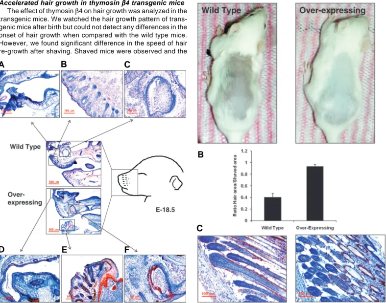
Accelerated hair growth in thymosin βββββ4 transgenic mice
The effect of thymosin β4 on hair growth was analyzed in the transgenic mice. We watched the hair growth pattern of trans-genic mice after birth but could not detect any differences in the onset of hair growth when compared with the wild type mice. However, we found significant difference in the speed of hair re-growth after shaving. Shaved mice were observed and the
speed of hair re-growth was compared by measuring the hair area on the shaved spot at 2 weeks using an image analysis program. The speed of hair growth on the skin of the transgenic mice was accelerated as shown in Fig. 3. These data suggest that the increased expression of thymosin β4 in the hair follicles (Fig. 1B) promoted hair re-growth in the transgenic mice. To analyze the expression pattern of thymosin β4 on the hair follicle, we stained for thymosin β4 in the skin of transgenic mice. As shown in Fig. 3B, thymosin β4 was expressed on
Fig. 2 (Left). Expression of thymosin βββββ4 in the developing anal and nasal cavities of transgenic mice. Thymosin β4 was highly expressed in the forming anal and nasal cavities (D,E) and around developing teeth (F). Thymosin β4 was also strongly expressed in the whiskers and hair of the transgenic mice (E). Immunohistochemical analysis of thymosin β4 expression was conducted with 18.5 day mouse embryos. Whole embryos were immunostained with a rabbit polyclonal antibody to thymosin β4 (1:500 dilution). The antibody binding was detected with the use of the DAKO EnVision-peroxidase system.
Fig. 3 (Right). Analysis of hair re-growth. The effect of thymosin β4 on hair growth was analyzed in cycling wild type and transgenic mice. Shaved mice were observed for 2 weeks at which time the photos were taken (A). To compare the speed of hair growth, white colored hair area was measured and divided by whole shaved area using image analysis software, Image J. Six mice were used for each group and the experiment was repeated three times (B). Expression of thymosin β4 in the hair of transgenic mice. Thymosin β4 was highly expressed on the surface of the hair follicles especially on the external root sheath (C).
B
C
D
E
F
A
B
C
A
surface of the hair follicles especially in the external root sheath.
Induced expression of laminin-5 in thymosin βββββ4 transgenic mice
We examined the expression of laminin-5 in the thymosin β4 over-expressing mice since laminin-5 was one of the candidate genes associated with hair growth among the proteins up-regulated by thymosin β4. Western blot analysis and immuno-histochemistry were used with the skin of wild type and trans-genic mice to characterize the expression of laminin-5. As shown in Fig. 4A, Western blot shows that the expression of laminin-5 was increased approximately 5-fold in the transgenic mice relative to the wild type mice. Immunohistochemistry showed that the expression of lamin5 was specifically in-creased in the root of the hair follicles and this staining pattern is different from that of thymosin β4 (Fig. 4B). The expression of laminin-5 around the root of the hair follicle suggests that laminin-5 is induced by the over-expression of thymosin β4 and that this induced laminin-5 expression may stimulate hair
follicle stem cells and play a role in the thymosin β 4-stimulated hair growth.
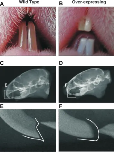
Unusual tooth morphology in thymosin βββββ4 transgenic mice
Thymosin β4 was highly expressed around developing teeth in the transgenic mice. In addition, thymosin β4 was reported to be elevated in tooth development at embryonic day 13 (E-13) of tooth development and decreased after birth (Akhter et al., 2005). These data suggest that thy-mosin β4 plays an important role in tooth development. Tooth morphology was further analyzed in the transgenic mice. As shown in Fig. 5, the morphology of teeth of the transgenic mice was significantly different from that of the wild type mice. The teeth are fragile and show a chalky color, indicating enamel hypoplasia similar to the teeth from the ameloblastin or amelogenin knockout mouse (Fukumoto et al., 2004) (Fig. 5A). As shown in x-ray photography, the teeth have dull incisors compared to the teeth of wild type mice (Fig. 5B). We have examined more than 20 mice and 100% of the transgenic mice have this phenotype. These data suggest that over-expression of thymosin β4 causes abnormal tooth development, and indicates that thymosin β4 plays an important role in enamel matrix deposition and/or production.
Discussion
Thymosin β4 is the most abundant β-thymosin in mam-mals. It forms a 1:1 complex with ATP-G-actin (monomeric actin) and inhibits the polymerization of actin (Safer et al., 1991). Thymosin β4 plays an important role in cell motility due to its participation in the rapid polymerization/depoly-merization of actin (Pantaloni and Carlier, 1993; Kang et al., 1999). Recently, diverse functional roles of thymosin β4 have also been reported, including angiogenesis (Grant et al., 1995; Grant et al., 1999), wound healing (Philp et al., 2004), and tumorigenesis (Wang et al., 2004). To investi-gate the role of thymosin β4 in vivo, we constructed Over-expressing Wild Type Over -express ing Wild Type Lamininγ2 100 mm 100 mm 50 mm 20 mm
B
A
Fig. 4. Expression and localization of laminin-5 in thymosin βββββ4 over-expressing mice. Western blot analy-sis of laminin-5 in the skin of trans-genic mice (A). Total lysates from 6 week old mice skin were separated by electrophoresis and transferred to a polyvinylidene difluoride membrane. The membrane was incubated with
rabbit polyclonal laminin γ2 (1: 1000 dilution) and detected with an enhanced chemiluminescence detection kit. Immunohistochemical analysis of laminin-5 in the skin of transgenic mice (B). Skins of 6 week old mice were immunostained with a rabbit polyclonal antibody to laminin γ2 (1: 100 dilution). The antibody binding was detected using a DAKO EnVision peroxidase system.
transgenic mice over-expressing thymosin β4. Highly expressed thymosin β4 in the skin accelerated hair growth and induced abnormally-shaped white teeth and dull incisors. These results suggest that the keratin 5 promoter stimulated high expression of thymosin β4 in hair follicles and in germ line stem cells regulating tooth development.
A recent study showed that thymosin β4 promotes laminin-5 (LM-5) expression (Sosne et al., 2004). Using gene expression analysis with human corneal epithelial cells treated with thymosin
β4, the laminin-5 γ2 chain was increased by more than 2-fold over untreated cells. LM-5 is one of the proteins involved in hair growth. The expression of LM-5 is highly increased in hair follicles (Chuang et al., 2003). In addition, hair follicle integrity is depen-dent on keratinocyte expression of integrin β1, the major receptor component of laminin-5 (Brakebusch et al., 2000). Furthermore, conditional ablation of integrin β1 results in severe defects in hair follicle invagination (Raghavan et al., 2000). These studies as well as ours suggest that thymosin β4 increased LM-5 expression and that this induction of LM-5 stimulated hair growth. As shown in Fig. 5, LM-5 expression in the skin was increased in thymosin
β4 over-expressing mice and this induction of LM-5 was concen-trated in hair follicles.
The involvement of thymosin β4 in tooth development was previously reported by Akhter et al. (Akhter et al., 2005) who identified thymosin β4 expression in early tooth development. We found (Fig. 5) that over-expression of thymosin β4 affected tooth development resulting in fragile teeth that have a chalky color, indicating enamel hypoplasia similar to the ameloblastin knockout mouse teeth (Fukumoto et al., 2004). These phenotype is also similar to the urokinase-type plasminogen (u-PA) activator over-expressing mice controlled by kertain 5 promoter (Zhou et al., 1999). These results suggested that keratin 5 promoter was expressed at the site of teeth development and that thymosin β4 also plays an important role in tooth development like u-PA. In ectodermal organs, including hair follicles, skin, and ectodermal derivatives of teeth, the morphology of the initial developmental stage is similar and shares many genes, which are involved in cell proliferation and in differentiation (Nakamura et al., 2004). We suggest that over-expressing thymosin β4 may affect teeth germ cell migration since thymosin β4 has been found to promote the migration of many cell types (Akhter et al., 2005). It is also possible that high amounts of thymosin β4 disturb dental epithelial
E F Over-expressing Wild Type
B
C
D
E
F
A
Fig. 5. Tooth morphology of thymosin βββββ4 over-expressing mice. The teeth of the transgenic mice were blunter due to significant fragility and they appeared chalky in color, indicating enamel hypoplasia (A,B). X-ray photography shows that the teeth from the transgenic mice have dull incisors compared to the teeth of wild type mice (C,D,E,F).
cell polarization by inhibiting actin polymerization in preameloblast and/or integrin expression, because dental epithelial polarization is important for secretion of enamel matrix and migration of ameloblasts.
Materials and Methods
Construction of the K5 thymosin βββββ4 transgene and generation of transgenic mice
A bluescript KS plasmid containing a 5.2 kb fragment from the bovine K5 promoter, a 0.6 kb fragment containing the rabbit β-globin intron, and a 1.1 kb fragment containing two 3' poly A signal sequences (Angela et al., 1998) were obtained from Dr. Yoshi Yamada (NIH, MD, USA). This plasmid was derived from constructs originally obtained from Dr. Jose Jorcano (CIEMAT, Madrid, Spain). A 0.2 kb fragment containing the full-length mouse thymosin β4 cDNA was cut using SnaBI and Nhe I and inserted into the K5 vector. The transgene was isolated by cutting with Acc65I followed by sucrose gradient centrifu-gation. The purified fragment was injected into the pronuclei of fertil-ized oocytes of FVB/N mice to create transgenic founder mice (Sreenath
et al., 1999). The founder mice were back-crossed to the wild type
FVB/N mice and analyzed. Integration of the transgene in the founder mice was detected by either Southern blot or PCR analysis. DNA was isolated from tail biopsies using classical proteinase K digestion. For Southern blot analysis, 10 ug of genomic DNA was digested overnight by KpnI and hybridized with a 0.2 kb fragment containing thymosin β4. For PCR analysis, 0.5 ug of genomic DNA was amplified for 35 cycles using Takara ExTaq DNA polymerase. The primer sequence for detection of the transgene was designed based on the rabbit β-globin intron and thymosin β4 as indicated at Fig. 1. For all experiments, a minimum of 5 animals were used for each data point and each experiment was repeated at least three times.
Western blot analysis and immunostaining
Western blot analysis was conducted as described (Champliaud et
al., 1996). Briefly, 100 μg of lysate from 6- week old mice skin was separated by electrophoresis on a Novex 4-20% Tris–glycine gel (Invitrogen, CA, USA). The protein concentrations of the lysates were determined by the bicinchoninic acid protein assay system (Pierce, Rockford, IL), and equal protein amounts of each sample were sepa-rated by electrophoresis on Novex 4-20% Tris–glycine gels. Equal protein loading was confirmed by Coomassie blue staining of duplicate gels after electrophoresis. The gels were incubated for 1 hour in phosphate-buffered saline (PBS) containing 10% glutaraldehyde (Sigma-Aldrich, St. Louis, MO), washed three times for 20 minutes in PBS, and further incubated in a blotting buffer containing Novex Tris– glycine transfer buffer (Invitrogen) and 20% methanol for 30 minutes at room temperature. Proteins were transferred to a polyvinylidene difluoride membrane (Invitrogen) by electrotransfer. The membrane was pre-incubated for 2 hours in Tris Buffered Saline (TBS) containing 5% skim milk and 0.05% Tween 20 (TBS-T). The membrane was incubated for 1 hour at room temperature in TBS-T plus antibodies (rabbit polyclonal thymosin β4, 1: 5000 dilution; ALPCO Diagnostics, Windham, NH) or with rabbit polyclonal laminin γ2 (1: 1000 dilution). The membranes were washed five times with PBS-T and then incu-bated with a species-appropriate horseradish peroxidase–conjugated secondary antibody (Santa Cruz Biotechnology) for 1 hour at room temperature. The membranes were washed four times with TBS-T, and bound antibody was detected with an enhanced chemilumines-cence detection kit (Amersham Biosciences, Buckinghamshire, UK). For immunohistochemistry, the skin of 6 week old mice or whole embryos at various stages were fixed with 4% formaldehyde (Sigma-Aldrich) and paraffinized. The sections were deparaffinized and immunostained with a rabbit polyclonal antibody to thymosin β4 (1:
500 dilution) or laminin γ2 (1: 100 dilution). The antibody binding was detected with the use of an EnVision peroxidase system (DAKO, Carpinteria, CA).
Analysis of hair growth
The effect of thymosin β4 on hair growth was analyzed in 28 day old (cycling) wild type and transgenic mice. Shaved mice were observed for 2 weeks at which time photos were taken. To compare the speed of hair growth, white colored hair area was measured and divided by whole shaved area using image analysis software (Image J, NIH, Bethesda, MD). Six mice were used for each group and the experiment was repeated three times.
Radiographic analysis
After euthanization with CO2, 6-week-old mouse heads were dis-sected out and sliced sagittally into two symmetrical halves. Tooth mineral density was analyzed by a microradiographic technique using X-ray imaging with a standard setting of 110 sec x 20 kV (Model MX20, Faxitron x-ray Corporation, Wheeling, IL). Six mice were used for each group and the experiment was repeated three times.
Acknowledgements
This work was supported by the National Research Foundation of Korea (NRF) grant funded by the Korea government (MEST) (KRF-20090066740) and a grant from the Kosin University College of Medicine (2007).
References
AKHTER, M., KOBAYASHI, I., KIYOSHIMA, T., MATSUO, K., YAMAZA, H., WADA, H., HONDA, J.Y., MING, X., and SAKAI, H. (2005). Possible functional involve-ment of thymosin beta 4 in developing tooth germ of mouse lower first molar.
Histochem. Cell. Biol. 124: 207-213.
ANADON, R., RODRIGUEZ, MOLDES I., CARPINTERO, P., EVANGELATOS, G., LIVIANOU, E., LEONDIADIS, L., QUINTELA, I., CERVINO, M.C., and GOMEZ-MARQUEZ, J. (2001). Differential expression of thymosins beta(4) and beta(10) during rat cerebellum postnatal development. Brain Res. 894: 255-265. ANGELA, M.P., SUSAN, M.F., CLAUDIO, J.C., and DAVID, G.J. (1998).
Deregu-lated expression of E2F1 induces hyperplasia and cooperates with ras in skin tumor development. Oncogene 16: 1267-1276.
BRAKEBUSCH, C., GROSE, R., QUONDAMATTEO, F., RAMIREZ, A., JORCANO, J.L., PIRRO, A., SVENSSON, M., HERKEN, R., SASAKI, T., TIMPL, R., WERNER, S., and FASSLER, R. (2000). Skin and hair follicle integrity is crucially dependent on beta 1 integrin expression on keratinocytes. EMBO J. 19: 3990-4003.
BURGESON, R.E., CHIQUET, M., DEUTZMANN, R., EKBLOM, P., ENGEL, J., KLEINMAN, H., MARTIN, G.R., MENEGUZZI, G., PAULSSON, M., SANES, J.,
et al.,1994). A new nomenclature for the laminins. Matrix Biol. 14: 209–211.
CARTER, W.G., RYAN, M.C., and GAHR, P.J. (1991). Epiligrin, a new cell adhesion ligand for integrin alpha 3 beta 1 in epithelial basement membranes. Cell 65: 599–610.
CHA, H.J., JEONG, M.J., and KLEINMAN, H.K. (2003). Role of Thymosin beta-4 in tumor metastasis and angiogenesis. J. Natl. Cancer Inst. 95: 1674-1680. CHAMPLIAUD, M.F., LUNSTRUM, G.P., ROUSSELLE, P., NISHIYAMA, T., KEENE,
D.R., and BURGESON, R.E. (1996). Human amnion contains a novel laminin variant, laminin 7, which like laminin 6, covalently associates with laminin 5 to promote stable epithelial– stromal attachment. J. Cell Biol. 132: 1189–1198. CHUANG, Y.H., DEAN, D., ALLEN, J., DAWBER, R. and WOJNAROWSKA, D.F.
(2003). Comparison between the expression of basement membrane zone antigens of human interfollicular epidermis and anagen hair follicle using indirect immunofluorescence. Br. J. Dermatol. 149: 274-278.
DATHE, V., and BRAND-SABERI, B.(2004). Expression of thymosin beta-4 during chick development. Anat Embryol (Berl) 208: 27-32.
FUKUMOTO, S., KIBA, T., HALL, B., IEHARA, N., NAKAMURA, T., LONGENECKER,
G., KREBSBACH, P.H., NANCI, A., KULKARNI, A.B., and YAMADA,Y. (2004). Ameloblastin is a cell adhesion molecule required for maintaining the differen-tiation state of ameloblasts. J. Cell. Biol. 167: 973-983.
GOMEZ-MARQUEZ, J., FRANCO DEL AMO, F., CARPINTERO, P., and ANADON, R. (1996). High levels of mouse thymosin beta-4 mRNA in differentiating P19 embryonic cells and during development of cardiovascular tissues. Biochim.
Biophys. Acta. 1306: 187-193.
GOMEZ-MARQUEZ, J., PEDRARES, J.I., OTERO, A., and ANADON, R. (1993). Prominent expression of the actin-sequestering peptide Fx gene in the hippoc-ampal region of rat brain. Neurosci. Lett. 152: 41-44.
GRANT, D.S., KINSELLA, J.L., KIBBEY, M.C., LAFLAMME, S., BURBELO, P.D., GOLDSTEIN, A.L., and KLEINMAN, H.K. (1995). Matrigel induces thymosin beta 4 gene in differentiating endothelial cells. J. Cell Sci. 108: 3685–3694. GRANT, D.S., ROSE, W., YAEN, C., GOLDSTEIN, A., MARTINEZ, J., AND
KLEINMAN, H.K. (1999). Thymosin beta-4 enhances endothelial cell differen-tiation and angiogenesis. Angiogenesis 3: 125–135.
KALLUNKI, P., SAINIO, K., EDDY, R., BYERS, M., KALLUNKI, T., SARIOLA, H., BECK, K., HIRVONEN H., SHOWS, T.B., and TRYGGVASON, K. (1992). A truncated laminin chain homologous to the B2 chain: structure, spatial expres-sion, and chromosomal assignment. J. Cell Biol. 119: 679–693.
KANG, F., PURICH, D.L., and SOUTHWICK, F.S. (1999). Profilin promotes barbed-end actin filament assembly without lowering the critical concentration. J. Biol
Chem 274: 36963–36972.
LJUBIMOV V., BURGESON R.E., BUTKOWSKI R.J., MICHAEL A.F., SUN T.T., and KENNEY M.C. (1995). Human corneal basement membrane heterogene-ity: topographical differences in the expression of type IV collagen and laminin isoforms. Lab. Invest. 72: 461–473.
NAKAMURA T., UNDA F., DE-VEGA S., VILAXA A., FUKUMOTO S., YAMADA K.M., and YAMADA Y. (2004). The Krüppel-like factor epiprofin is expressed by epithelium of developing teeth, hair follicles, and limb buds and promotes cell proliferation. J. Biol. Chem. 279: 626-634.
PANTALONI, D., and CARLIER, M.F. (1993). How profiling promotes actin filament assembly in the presence of thymosin β4. Cell 75: 1007–1014.
PHILP, D., GOLDSTEIN, A.L., and KLEINMAN, H.K. (2004). Thymosin β4 pro-motes angiogenesis, wound healing, and hair follicle development. Mech.
Ageing Dev. 125: 113–115.
PHILP, D., NGUYEN, M., SCHEREMETA, B., ST.-SURIN, S., VILLA, A.M., ORGEL, A., KLEINMAN, H.K., and ELKIN, M. (2004). Thymosin beta-4 increases hair growth by activation of hair follicle stem cell. FASEB J. 18: 385–387. RAGHAVAN, S., BAUER, C., MUNDSCHAU, G., Li, Q., and FUCHS, E. (2000).
Conditional ablation of beta1 integrin in skin. Severe defects in epidermal proliferation, basement membrane formation, and hair follicle invagination. J.
Cell Biol. 150: 1149-1160.
SAFER, D., ELZINGA, M., and NACHMIAS, V.T. (1991). Thymosin beta 4 and Fx, an actin-sequestering peptide, are indistinguishable. J. Biol Chem 266: 4029– 4032.
SOSNE, G., XU, L., PRACH, L., MROCK, L.K., KLEINMAN, H.K., LETTERIO, J.J., HAZLETT, L.D., and KURPAKUS-WHEATER, M. (2004). Thymosin beta 4 stimulates laminin-5 production independent of TGF-beta. Experimental Cell
Research 293: 175–183.
SREENATH, T.L., CHO, A., MACDOUGALL, M., and KULKARNI, A.B. (1999). Spatial and temporal activity of the dentin sialophosphoprotein gene promoter: differential regulation in odontoblasts and ameloblasts. Int. J. Dev. Biol. 43: 509–516.
VERMA, A. K. and BOUTWELL, R.K., (1980). Effects of dose and duration of treatment with the tumor-promoting agent, 12-O-tetradecanoylphorbol-13-ac-etate on mouse skin carcinogenesis. Carcinogenesis 1: 271-276.
WANG, W.S., CHEN, P.M., HSIAO, H.L., WANG, H.S., LIANG, W.Y., and SU, Y. (2004). Overexpression of the thymosin β-4 gene is associated with increased invasion of SW480 colon carcinoma cells and the distant metastasis of human colorectal carcinoma. Oncogene 23: 6666–6671.
ZHOU H.M., NICHOLS, A., WOHLWEND, A., BOLON, I., and VASSALLI, J.D. (1999). Extracellular proteolysis alters tooth development in transgenic mice expressing urokinase-type plasminogen activator in the enamel organ.
Further Related Reading, published previously in the Int. J. Dev. Biol.
See our Special Issue Epigenetics and Development edited by Saadi Khochbin and Stefan Nonchev at: http://www.ijdb.ehu.es/web/contents.php?vol=53&issue=2-3
See our Special Issue Skin Development edited by Danielle Dhouailly at: http://www.ijdb.ehu.es/web/contents.php?vol=48&issue=2-3
Hair cell regeneration in the avian auditory epithelium
Jennifer S. Stone and Douglas A. Cotanche Int. J. Dev. Biol. (2007) 51: 633-647
BMP signalling in craniofacial development
Xuguang Nie, keijo Luukko and Paivi Kettunen Int. J. Dev. Biol. (2006) 50: 511-521
Detection of differentially expressed genes in the early developmental stage of the mouse mandible.
H Yamaza, K Matsuo, T Kiyoshima, N Shigemura, I Kobayashi, H Wada, A Akamime and H Sakai Int. J. Dev. Biol. (2001) 45: 675-68
Differential expression of laminin-5 subunits during incisor and molar development in the mouse.
K Yoshiba, N Yoshiba, D Aberdam, G Meneguzzi, F Perrin-Schmitt, C Stoetzel, J V Ruch and H Lesot Int. J. Dev. Biol. (2000) 44: 337-340
Timing of the expression of enamel gene products during mouse tooth development.
M Zeichner-David, H Vo, H Tan, T Diekwisch, B Berman, F Thiemann, M D Alcocer, P Hsu, T Wang, J Eyna, J Caton, H C Slavkin and M MacDougall
Int. J. Dev. Biol. (1997) 41: 27-38
Integrin regulatory switching in development: oscillation of beta 5 integrin mRNA expression during epithelial-mesenchymal interactions in tooth development.
S Yamada, K M Yamada and K E Brown Int. J. Dev. Biol. (1994) 38: 553-556