Jung-Sun Lee
1*, Jonghoe Byun
2*,
Jung-Min Kim
1, Chae-Young Kim
3,
Byong-Moon Kim
3, Ji Hyung Chung
4,
Yangsoo Jang
4and Duk-Kyung Kim
1,5 1Department of MedicineSamsung Medical Center
Samsung Biomedical Research Institute Sungkyunkwan University School of Medicine Seoul 135-710, Korea
2Neuroscience Research Institute Gachon Medical School Incheon 405-760, Korea 3Research Laboratories Dong-A Pharm. Co. Ltd.
Yongin-si, Kyunggi-do 449-900, Korea 4Yonsei Cardiovascular Research Institute Yonsei University
Seoul 120-749, Korea
5Corresponding author: Tel, 82-2-3410-3413;
Fax, 82-2-3410-3849; E-mail, [email protected] *These authors contributed equally to this work. Accepted 20 July 2005
Abbreviations: VEGF, vascular endothelial growth factor; bFGF, basic fibroblast growth factor; vWF, von Willebrand factor; α-SMA, smooth muscle α-actin
Abstract
We investigated expression profiles and biological effects of the naked DNA vectors in the heart. To this end, naked DNA vector was injected into the apex of the beating rat heart after thorocotomy. When the expression of LacZ reporter was examined by reverse transcription-PCR and histochemical staining for β-galactosidase, LacZ expression was
detected only in the heart, suggesting limited dissemination of the injected vector in vivo. Even within the heart, LacZ expression was limited to the injection area (apex). Similar observations were made with other transgenes such as VEGF and basic fibroblast growth factor (bFGF), where 77% and 69% of the total transgene exprssion were detected in the heart segments containing the apex.
Although VEGF and bFGF expressions were de -tected until 2 weeks after DNA injection, the highest levels of VEGF and bFGF were observed on day 5 and day 1, respectively. The optimal doses of the vectors were 10 µg and 25 µg for the VEGF and bFGF
vectors, respectively. Interestingly, injection of bFGF vector led to 50% increase in the level of endogenous murine VEGF expression. Consistent with this finding, the number of vessels that stained positive for α-smooth muscle actin was increased
in the bFGF vector-injected heart. These results suggest that simple injection of naked DNA vector may be sufficient to induce significant angio -genesis in the myocardium and that naked DNA gene therapy may be a feasible approach for the treatment of ischemic heart disease.
Keywords: angiogenesis; basic fibroblast growth
fac-tor; gene therapy; heart; myocardium; vascular endo-thelial growth factor
Introduction
The number of patients suffering from ischemic heart disease (IHD) is growing rapidly these days (Jones
et al., 1983; Mukherjee et al., 1999). Despite
conti-nued advances in the treatment of IHD, up to 15% of patients are not amenable to conventional therapies or receive incomplete revascularization (Wu et al., 2004). For the treatment of such no-option patients, development of a novel therapeutic modality is urgently needed (Sellke et al., 2003; Ye et al., 2003). Recently, therapeutic angiogenesis that is based on exogenous administration of angiogenic growth fac-tors has emerged as a promising strategy for the treatment of tissue ischemia (Lee et al., 2000b; Kim
et al., 2004). The potential of therapeutic
angiogene-sis has been validated in various animal models of limb or myocardial ischemia and recently in patients treated with VEGF and bFGF (Banai et al., 1994; Harada et al., 1994; Kim et al., 1994; Unger et al., 1994; Lazarous et al., 1996; Koh et al., 2002; Yamamoto et al., 2003; Hughes et al., 2004).
Gene transfer can be an effective means to provide localized delivery of a high concentration of angio-genic growth factors to the ischemic tissue. Among several gene delivery vectors that are currently used
Cardiac expression profiles of the naked DNA vectors encoding
vascular endothelial growth factor and basic fibroblast growth
factor
to induce angiogenesis, naked DNA vectors provide the simplest means of expressing target genes (Ver-ma et al., 1997; Lee et al., 2000b). When used alo-ne, it avoids infectious agents and cumbersome production procedures that are often associated with viral or other gene transfer vectors (Sellke et al., 2003). Importantly, the injected DNA vectors mostly remain episomal with greatly reduced risk for the integration into host chromosome. Also the naked DNA vectors essentially have no limit for the size of inserts and thus can accommodate larger inserts that can hardly be used effectively in other gene delivery systems (Heilmann et al., 2003).
The two dominant families of angiogenic factors that have been widely studied are FGF and VEGF (Rutanen et al., 2004; Wu et al., 2004). The thera-peutic potential of these growth factors, either as protein or gene, has been confirmed in many pre- clinical studies (Carmeliet, 2000a; Isner et al., 2002; Simons et al., 2002; Kim et al., 2003; Wu et al., 2004). VEGF gene therapy has been shown to in-crease myocardial perfusion and function in coro-nary artery diseases (Lee et al., 2000a; Isner et al., 2002). Also beneficial long-term effects of FGF were reported recently (Carmeliet, 2000a; Lee et al., 2000a; Sellke et al., 2003). However, despite these diverse applications of VEGF and bFGF for ischemic heart diseases, comparative expression studies on the naked DNA vectors encoding VEGF and bFGF have been limited.
In the present study, we evaluated cardiac expres-sion profile of the naked DNA vectors encoding VEGF and bFGF for their therapeutic potential in ischemic heart disease. The time course of expre-ssion, dose response, and spatial distributions were examined in a comparative manner. Also the in-crease in vessel count was examined for their bio-logical efficacy. Considering the fact that little atten-tion has been paid to the behavior of the naked DNA vectors injected into the heart, our study provides important information pertinent to the development of novel angiogenic gene therapy for the no-option patients who are not treated by conventional meth-ods (Ruel et al., 2003; Sellke et al., 2003).
Materials and Methods
Naked DNA vectorThe pGT2-VEGF plasmid DNA vector was con-structed by cloning the human VEGF165 cDNA into the eukaryotic expression vector, pGT2 whose back-bone carries a prokaryotic replication origin (ColE1), a bacterial kanamycin resistance gene, and a eukaryotic expression cassette consisting of HCMV promoter, an adenovirus tripartite leader sequence,
SV40 late poly A, and SV40 enhancer. The plasmid DNA was produced to clinical grade according to the proprietary process established at the Dong-A Pha-rm. Co. (Korea). Briefly, E. coli (DH5 ) cells carrying the plasmid were grown in a kanamycin-containing medium in a 20 L fermentor. The fermentation broth was subjected to a series of purification steps in-cluding alkaline lysis, PEG precipitation, anion ex-change chromatography, and gel filtration chroma-tography. The purified plasmid was dialyzed against a formulation buffer (Saline, pH 7.0).
Animal experiments
All animal experiments conform to the Guide for the Care and Use of Laboratory Animals published by the US National Institute of Health (NIH Publications No. 85-23, revised 1996). Male Sprague-Dawley (SD) rats weighing 300 grams were used for naked DNA injection. The rats were anesthetized with intra-peritoneal injection of ketamine-xylazine (50 mg/kg and 2 mg/kg, respectively), heated in warm pad (37oC), intubated, and mechanically ventilated with room air. Under aseptic conditions, a thoracotomy was performed through the left fifth intercostal spa-ce, and the heart was exposed. Cardiac injections were made directly into the apex of the left ventricle. Fifty microliter of DNA solution containing each plasmid was injected into the apical portion of the beating left ventricle, under direct visualization, using a 29-gauge needle. The DNA injection caused im-mediate sub-epicardial edema, with the tissue color changing from red to pink. The chest was then closed and the animals allowed to recover for specific periods of time before sacrifice. Five to ten rats were used per each group.
Measurement of VEGF and bFGF
Rats were killed at the indicated time points following cardiac injection. After trimming away the atria, one fourth segment of the heart containing apex region (approximately 200 mg around the injection site) was excised, frozen by immersing in liquid nitrogen, and stored at -75oC until needed. The harvested heart was homogenized in 1 ml of ice-cold homo-genization buffer (0.1 M Tris-HCl, pH 7.8 containing 2 mM EDTA and 0.1% Triton X-100). Homogenates were centrifuged at 14,000 rpm for 10 min at 4oC. Supernatants were collected to measure the level of human VEGF, bFGF and mouse endogenous VEGF using Quantikine immunoassay kits (R&D systems, MN) according to the manufacturer’s protocols. Total protein concentration of the supernatant was also determined by Bradford assay (Bio-Rad, CA). The results were expressed as picograms of cytokine per milligram protein.
RT-PCR
Total RNA was isolated from the heart tissues ac-cording to the protocol of RNeasy minikit (Qiagen, CA) and the cDNA synthesized using the Super-script First-Standard Synthesis system for RT-PCR (Invitrogen, CA, USA). The LacZ cDNA was ampli-fied using the forward primer (5'-CTGCATAAACC-GACTACACA-3') and the reverse primer (5'-TTTCA-ATATTGGCTTCATCC-3'). The forward and reverse primers for GAPDH were 5'-CGTGGAAGGACTCAT-GAC-3' and 5'-CAAATTCGTTGTCATACCAG-3', re-spectively.
Histological analysis
For general histological analysis, the heart sections were stained with hematoxylin and eosin (H & E). For immunostaining, the hearts were collected im-mediately after sacrifice and embedded in OCT medium. The frozen sections (5 m thickness) were stained with von Willebrand factor (vWF) antibody (Dako, Carpinteria, CA) to count the number of capillaries. In addition, the frozen sections were stained with the monoclonal antibody against
smoo-th muscle -actin ( -SMA; clone 1A4, Dako, Carpin-teria, CA) to count the number of mature vessels. Anti- -SMA antibodies were detected by VECTASTAIN Universal Quick Kit (Vector Laboratories, Burlin-game, CA) according to the manufacturer’s instruc-tions. Three sections having equal separation dis-tance were examined per animal. The capillaries were counted and averaged from 10 representative fields at 400 × magnification.
LacZ staining
The heart tissue was rinsed three times in cold phos-phate-buffered saline (PBS), embedded in OCT med-ium, and quick frozen in liquid nitrogen. Cryostat sec-tions (7 m) were air dried, rehydrated in PBS (pH 7.4) for 10min, and developed in a substrate solution [5 mM K3Fe(CN)6, 5 mM K4Fe(CN)6, 2 mM MgCl2, 0.01% sodium deoxycholate, 0.02% NP-40, 1 mg/ml X-gal] overnight at 37oC.
Statistical analysis
The data are expressed as mean ± SEM. One-way
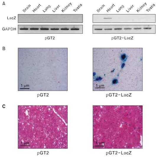
Figure 1. Analysis of biodistribution and safety of injected DNA. (A) Assessment of LacZ mRNA ex-pression in different tissues after pGT2 naked DNA injection. Total RNA (1 g) isolated from the in-dicated tissue at Day 5 was used for RT-PCR analysis. (B) Histoche-mical staining for -galactosidase expression in the injected area (apex) of the heart. Ten microgram of pGT2-LacZ DNA was injected in-to the rat heart followed by X-Gal staining at Day 5. -galactosidase expression was only shown in the apex of the heart. No acute organ toxicities were observed in the in-jected rat. Original magnification, × 400. (C) The hearts from each group were sectioned at 7 m thick-ness and stained with H & E (× 200 magnification). Salient patho-logical findings were not observed in the heart.
analysis of variance followed by Bonferroni’s post hoc multiple comparison tests were used to evaluate statistical differences between the groups. A P value of less than 0.05 was considered statistically signi-ficant.
Results
LacZ transgene expression following cardiac injection
To investigate transgene expression from the naked DNA vector in the heart, we injected 10 g of pGT2- LacZ vector into the rat myocardium and analyzed the LacZ expression by RT-PCR and LacZ staining (Figure 1) at day 5. The empty vector pGT2 served as a negative control for LacZ transgene. Following cardiac injection, other organs were also examined for the presence of unintended LacZ expression which would indicate dissemination of the vector. In
RT-PCR analysis (Figure 1A), the band correspon-ding to the LacZ mRNA was easily detected in the heart sample, whereas none were detected in sam-ples from other organs or the empty vector-injected heart. This implies that LacZ expression from the pGT2-LacZ vector was restricted to the injected organ. When the heart tissues were subjected to immunohistochemical staining for -galactosidase expression (Figure 1B), LacZ-positive cells were identified as blue staining-cells near the injection area. The intense deep blue staining also suggested that the pGT2-LacZ vector drove high level expres-sion of LacZ. No adverse effects were found along the needle track as determined by the H & E staining of the heart tissue (Figure 1C).
Angiogenic gene expression from the naked DNA vector injected into the myocardium
Next, we examined angiogenic gene expression
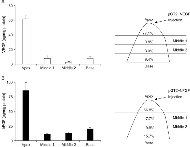
Figure 2. Spatial distribution of VEGF and bFGF expression. Five days after injection of the naked DNA into the apex of heart, the rats were sacrificed and the excised hearts were divided into four segments ranging from apex to base. Using ELISA, each segment was assayed for the expression of ei-ther VEGF (A) or bFGF (B). The ELISA data was normalized against the amount of protein in the given tissue. The data are mean ± SEM (n = 5). The percentage of expression is also shown schematically on the right panel.
from the naked DNA vector in the myocardium. To this end, cDNAs of the most widely studied growth factors, VEGF and bFGF, were inserted into the pGT2 vector and injected into the rat heart. Since localized expression pattern was observed in the case of LacZ transgene (Figure 1B), we again examined spatial distribution of each cytokine gene expression by quantifying the expression level of VEGF and bFGF along the heart axis from the base to the apex (Figure 2). At day 5, the isolated heart was cut into four pieces and each segment was homogenized and measured for the level of trans-gene expression using ELISA. The amount of pro-teins extracted from each segment was similar between the VEGF and bFGF. Consistent with the LacZ study, most of the VEGF and bFGF expression (77.1% and 66.8%, respectively) was restricted to the apex segment containing the injected region. These findings suggest that cardiac injection of the naked DNA vector can bring about significant local expression of therapeutic gene in the injected area and thus can be a feasible approach for the treat-ment of heart diseases having local pathologic regions such as ischemic heart disease.
Dose response and time course of transgene expression
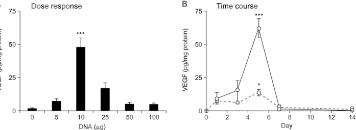
To optimize gene expression from the cardiac-inject-ed nakcardiac-inject-ed DNA vector, we examincardiac-inject-ed dose response and time course of VEGF and bFGF gene ex-pression. As shown in Figure 3A, the optimal dose for pGT2-VEGF was estimated at 10 g. Doses
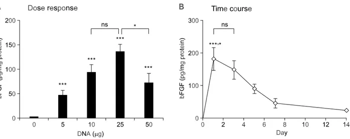
higher than 10 g did not lead to further increase, but rather resulted in a decrease of expression (Figure 3A). We have also examined the time course of VEGF gene expression after direct intramyo-cardial injection of pGT2-VEGF (Figure 3B). The maximum level of VEGF expression was observed at day 5 with two different doses (10 g and 25 g) of pGT2-VEGF. Compared with the gradual increase in gene expression until day 5, rapid drop in VEGF expression was observed after day 5. This is in contrast to the bFGF case, where rapid increase in gene expression was followed by gradual decrease in bFGF expression (Figure 4B). Also, the optimal dose for bFGF expression was estimated to be 25 g, which is higher than the optimal dose of VEGF. Overall, the tissue expression level of bFGF was approximately 3-fold higher than that achieved with VEGF. Also, compared with the short duration of VEGF expression, bFGF expression persisted even after day 14. These differences in expression profile between VEGF and bFGF may be explained in part by the short-half and secretory nature of VEGF (Ferrara et al., 1992).
Vascularity following angiogenic gene expression Next, we examined angiogenic response in the myocardium following cardiac injection of pGT2- LacZ, pGT2-VEGF, and pGT2-bFGF. The number of capillaries was counted from the heart sections stained with vWF, which was then normalized by the number of myocytes. As shown in Figure 5A, VEGF or bFGF group did not result in a significant increase
Figure 3. Dose response and time course of the VEGF expression from the naked DNA vector in the rat heart. (A) Dose response of VEGF expression. Different amounts of pGT2-VEGF DNA were injected into the apex region of the rat heart. Five days after DNA injection, the heart segment containing injected area (apex) was excised and the level of VEGF protein determined. Normal saline solution was used as a negative control. Values are given as mean ± SEM (n=20). *** P < 0.001 vs. other groups. (B) Time course of VEGF expression. Following injection of 10 g (circle) or 25 g (square) of pGT2-VEGF DNA, the heart segment containing injected area (apex) was excised and assayed for VEGF using ELISA at each time point. Values are given as mean ± SEM (n = 10 for each dose). *** P < 0.001, * P < 0.05 compared to Day 1.
in capillary density compared with control (LacZ) group. However, when the sections were stained for the presence of smooth muscle -actin ( -SMA) marker that is expressed in both pericytes and smooth muscle cells of mature blood vessels, the
FGF and VEGF group had significantly increased the number of -SMA-positive vessels than the control (LacZ) group (Figure 5B). Statistically sig-nificant differences were not found between the VEGF and bFGF groups.
Figure 5. Increased vessel formation following the naked DNA vector injection. (A) Capillary density of the rat myocardium evaluated in frozen section stained with vWF. Capillary/muscle fiber ratio refers to the number of capillaries divided by the number of myofibers. Original magnification, × 400. (B) Arteriole density evaluated in longitudinal section of the myocardium stained with the antibody against -SMA. Values are given as mean±SEM (n = 5). *** P < 0.001, ** P < 0.01 compared to LacZ group. ns, not significant.
Figure 4. Dose response and time course of the bFGF expression from the naked DNA vector in the rat heart. (A) Different amounts of pGT2-bFGF DNA were injected into the rat heart. Five days after DNA injection, the heart segment containing injected area (apex) was excised and the level of bFGF protein determined. Normal saline was used as a negative control (0 g). Values are given as mean ± SEM (n = 8-9). *** P < 0.001 compared to control (0 g). * P < 0.05. ns; not significant. (B) Following injection of DNA (25 g) into the rat heart, the heart segment containing injected area (apex) was excised and assayed for the level of bFGF using ELISA at each time point. Values are given as mean ± SEM (n=8-9). *** P < 0.001 com-pared to Day 7 and Day 14. * P < 0.05 comcom-pared to Day 5. ns, not significant.
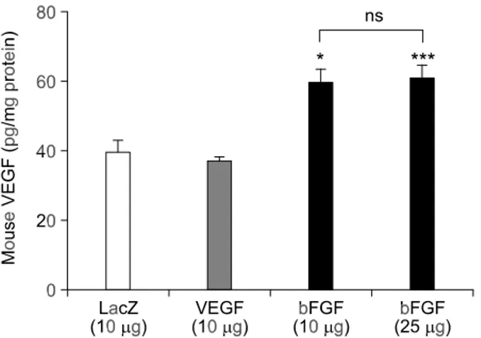
bFGF induces endogenous VEGF expression Since it was previously reported that bFGF can induce expression of VEGF (Stavri et al., 1995; Ra-bie et al., 2004), we investigated whether the similar effect can be found in the rat myocardium. When the rat hearts were injected with pGT2-LacZ, pGT2- VEGF, and pGT2-bFGF and analyzed for the level of endogenous murine VEGF level, significant increase in the level of murine VEGF was observed in the group injected with pGT2-bFGF (Figure 6). This indicates that overexpression of human bFGF can upregulate the expression of endogenous murine VEGF, whereas overexpression of VEGF or control LacZ gene has little effect. There was no further increase in murine VEGF level with higher dose of pGT2-bFGF (25 g).
Discussion
This study was carried out as part of the pre-clinical evaluation of the naked DNA vectors encoding two typical angiogenic genes, VEGF and bFGF. The pGT2 DNA vectors were developed for high level expression of therapeutic gene and our preliminary study indicated that it drove high level expression of target gene in the muscle tissues both in vitro and in
vivo (unpublished data). Considering the fact that
little is known about the behavior of naked DNA vectors injected into the myocardium, our study provides much-needed information with regard to the spatial and temporal characteristics of transgene expression in the heart. Also, the optimal dose and
time point of angiogenic gene expression deter-mined in myocardial tissue as well as the evidence of increased vascularity following VEGF and bFGF vector injection should prove very useful for the researchers in the field. Since less invasive per-cutaneous catheterization can be easily applied for intramyocardial injection of naked DNA, the results of our study has an implication for the development of novel angiogenic gene therapy for ischemic heart diseases.
Naked DNA vector may be an effective strategy for therapeutic angiogenesis in ischemic myocardium (Tsurumi et al., 1996; Tio et al., 1999; Vale et al., 2001; Rutanen et al., 2004). Despite the instability and short duration of the injected DNA, the increa-sed vascularity following pGT2-VEGF and pGT2- bFGF implies that local injection of naked DNA vector is sufficient for effective cellular uptake and biological effects in the myocardium. This result is supported by other studies where administration of the VEGF DNA vector facilitated local angiogenesis and the reperfusion of the ischemic tissues (Mack CA et al., 1998; Rutanen J et al., 2004). Regarding the utility of naked DNA vector, the use of non-viral vectors is disproportionately high in cardiovascular field, which is in contrast to the widespread appli-cation of viral vectors in other areas. Approximately half of the gene therapy trials in cardiovascular field used naked DNA vectors (Isner, 2002). This can be explained in part by the relatively high transfection efficiency of naked DNA in the myocardium (Lee et
al., 2000b; Isner et al., 2001; Heilmann et al., 2003).
Moreover, the simplicity of DNA vector may have been an important factor in the choice of vector. Nevertheless, the use of viral vectors is still a good option to increase the magnitude and duration of gene expression at the expense of decreased safety. One of the concerns associated with myocardial injection of naked DNA vector is its expression in unintended organs. Since complete retention of the vector at the injection area is unlikely to occur even under the most carefully controlled conditions, dis-semination of injected DNA and its expression in various organs need to be monitored closely. In the present study, however, expression of the injected DNA was not detected in other organs by RT-PCR assays (Figure 1). Also inadvertent dissemination of DNA into other tissues via the bloodstream was not detected at 2 hr after the intramyocardial admini-stration in most organs (data not shown). This may be in part due to a rapid degradation of the plasmid DNA by the plasma deoxyribonuclease (DNase) and the enzymes in other body compartments such as the liver (Acharya et al., 2004). Considering the high degradation rate of the plasmid DNA in vivo and the fact that the dose (25 g DNA) used in this study is
Figure 6. bFGF upregulates endogenous expression of mouse VEGF. pGT2-VEGF(10 g)or pGT2-bFGF (10 g or 25 g) DNA was injected into the apex region of the rat heart. Five days after DNA injection, the injected area was excised and the level of mouse VEGF protein determined. Induction of endogenous VEGF is noted in bFGF group. Values are given as mean ± SEM (n = 8-9 for each dose). * P < 0.05, *** P < 0.001 compared to LacZ group. ns; not significant.
merely several-fold higher (per bodyweight) than that expected in clinical applications, it is highly unlikely that significant amount of plasmid DNA will reach the non-target organs via blood circulation and persist in these organs in actual clinical situations. The risk of insertional mutagenesis or germ line transmission also appears to be negligible as the genomic inte-gration in the injected tissue or the mRNA production in the testis was not detected by PCR or RT-PCR (data not shown). These observations all together suggest that local injection of naked DNA vector may be a safe procedure with little concern for the adverse effects.
With regard to the safety issues related to forced upregulation of angiogenic factors, systemic admini-stration of VEGF can induce hypotension and car-ries theoretic risk of inducing occult tumors and inappropriate angiogenesis in the retina and the synovium. However, the relatively low expression level that is achieved using naked DNA vector is expected to compromise such effects. Compared with our previous study using pCN-VEGF121 (Jeong
et al., 2002), the present study reveals different
VEGF expression pattern in terms of dose response and time course of expression, which could be explained in part by the different vector, VEGF isoform (VEGF165), and the experimental methods used. In addition, more animals were used in the present study to provide extensive comparisons. As such, the results of the present study would help to increase our understanding of the behavior of naked DNA vectors encoding angiogenic genes in the myocardium.
One of the reasons for the use of nonischemic heart in this study was that collateral growth begins in normoxic areas. On the other hand, we can minimize confounding effects of ischemic tissues, in which there is an upregulation of various endo-genous growth factors and their receptors (Rutanen
et al., 2004). When this latter point is acknowledged,
upregulation of endogenous VEGF following bFGF vector injection in the normoxic heart (Figure 6) implies that there may be even stronger induction of VEGF in the ischemic heart. This upregulated ex-pression of endogenous VEGFs may act in concert with the delivered bFGF, creating enhanced respon-se in the myocardium. Indeed, the number of mature vessels was larger in bFGF-treated group than in VEGF-treated group (Figure 5), consistent with our speculation. In other aspect, hemangioma formation and vascular leakage that may be seen with VEGF gene therapy were not observed with FGF-encoding vectors at doses that allow for biological response (Lopez et al., 1997; Carmeliet, 2000b; Lee et al., 2000a). Therefore, it appears that administration of bFGF may be advantageous over that of VEGF in
terms of safety as well as efficacy. However, more study would be required to elaborate on this point. In conclusion, we provide evidence that naked DNA vectors are capable of delivering angiogenic genes safely to myocardium and that the delivered VEGF and bFGF genes are capable of inducing significant angiogenesis in vivo in nonischemic heart. Since it is reasonable to expect that this naked DNA strategy can be used clinically as an adjunct or alternative to conventional revascularization thera-pies to promote collateral vessel formation, our re-sults should have implications for many current and future angiogenic gene therapy trials.
Acknowledgement
This work was supported by Ministry of Commerce, Industry, and Energy, Republic of Korea (project #: 10007404), the Korea Science and Engineering Foundation (SRC, Molecular Therapy Research Center), and the National Research Laboratory Grant from the Korea Institute of Science and Technology Evaluation and Planning (M1-0203-00-0048) to DK Kim.
References
Acharya MM, Khamesra SH, Katyare SS.Effect of repeated intraperitoneal exposure to picrotoxin on rat liver lysosomal function.Indian J Exp Biol 2004; 42:808-11
Banai S, Jaklitsch MT, Shou M, Lazarous DF, Scheinowitz M, Biro S, Epstein SE, Unger EF. Angiogenic-induced enhance-ment of collateral blood flow to ischemic myocardium by vascular endothelial growth factor in dogs. Circulation 1994; 89:2183-9
Carmeliet P. Mechanisms of angiogenesis and arteriogene-sis. Nat Med 2000a;6:389-95
Carmeliet P. VEGF gene therapy: stimulating angiogenesis or angioma-genesis? Nat Med 2000b;6:1102-3
Harada K, Grossman W, Friedman M, Edelman ER, Prasad PV, Keighley CS, Manning WJ, Sellke FW, Simons M. Basic fibroblast growth factor improves myocardial function in chronically ischemic porcine hearts. J Clin Invest 1994; 94:623-30
Heilmann CA, Attmann T, Thiem A, Haffner E, Beyersdorf F, Lutter G. Gene therapy in cardiac surgery: intramyocardial injection of naked plasmid DNA for chronic myocardial ischemia. Eur J Cardiothorac Surg 2003;24:785-93
Henry TD, Annex BH, McKendall GR, Azrin MA, Lopez JJ, Giordano FJ, Shah PK, Willerson JT, Benza RL, Berman DS, Gibson CM, Bajamonde A, Rundle AC, Fine J, McCluskey ER; VIVA Investigators. The VIVA trial: Vascular endo-thelial growth factor in Ischemia for Vascular Angioge-nesis. Circulation 2003;107:1359-65
Landolfo CK, Lowe JE, Annex BH, Landolfo KP. Therapeutic angiogenesis in chronically ischemic porcine myocardium: comparative effects of bFGF and VEGF. Ann Thorac Surg 2004;77:812-8
Isner JM. Myocardial gene therapy. Nature 2002;415:234-9 Isner JM, Vale PR, Symes JF, Losordo DW. Assessment of risks associated with cardiovascular gene therapy in human subjects. Circ Res 2001;89:389-400
Jeong JO, Byun J, Jeon ES, Gwon HC, Lim YS, Park J, Yeo SJ, Lee YJ, Kim S, Kim DK. Improved expression by cytomegalovirus promoter/enhancer and behavior of vas-cular endothelial growth factor gene after myocardial injec-tion of naked DNA. Exp Mol Med 2002;34:278-4
Jones EL, Craver JM, Guyton RA, Bone DK, Hatcher CR Jr, Riechwald N. Importance of complete revascularization in performance of the coronary bypass operation. Am J Cardiol 1983;51:7-12
Kim HJ, Jang SY, Park JI, Byun J, Kim DI, Do YS, Kim JM, Kim S, Kim BM, Kim WB, Kim DK. Vascular endothelial growth factor-induced angiogenic gene therapy in patients with peripheral artery disease. Exp Mol Med 2004;36: 336-44
Kim JH, Kim JC, Shin SH, Chang SI, Lee HS, Chung SI. The inhibitory effects of recombinant plasminogen kringle 1-3 on the neovascularization of rabbit cornea induced by angio-genin, bFGF, and VEGF. Exp Mol Med 1999;31:203-9 Kim KS, Hong YK, Lee Y, Shin JY, Chang SI, Chung SI, Joe YA. Differential inhibition of endothelial cell proliferation and migration by urokinase subdomains: amino-terminal frag-ment and kringle domain. Exp Mol Med 2003;35:578-85 Koh GY, Kim I, Kwak HJ, Yun MJ, Leem JC. Biomedical significance of endothelial cell specific growth factor, angio-poietin. Exp Mol Med 2002;34:1-11
Lazarous DF, Shou M, Scheinowitz M, Hodge E, Thirumurti V, Kitsiou AN, Stiber JA, Lobo AD, Hunsberger S, Guetta E, Epstein SE, Unger EF. Comparative effects of basic fibro-blast growth factor and vascular endothelial growth factor on coronary collateral development and the arterial response to injury. Circulation 1996;94:1074-82
Lee RJ, Springer ML, Blanco-Bose WE, Shaw R, Ursell PC, Blau HM. VEGF gene delivery to myocardium: deleterious effects of unregulated expression. Circulation 2000a;102: 898-901
Lee Y, Park EJ, Yu SS, Kim DK, Kim S. Improved expression of vascular endothelial growth factor by naked DNA in mouse skeletal muscles: implication for gene therapy of ischemic diseases. Biochem Biophys Res Commun 2000b; 272:230-5
Lopez JJ, Laham RJ, Carrozza JP, Tofukuji M, Sellke FW, Bunting S, Simons M. Hemodynamic effects of intracoronary VEGF delivery: evidence of tachyphylaxis and NO depen-dence of response. Am J Physiol 1997;273:1317-23
Mack CA, Patel SR, Schwarz EA, Zanzonico P, Hahn RT, Ilercil A, Devereux RB, Goldsmith SJ, Christian TF, Sanborn TA, Kovesdi I, Hackett N, Isom OW, Crystal RG, Rosengart TK. Biologic bypass with the use of adenovirus-mediated
gene transfer of the complementary deoxyribonucleic acid for vascular endothelial growth factor 121 improves myo-cardial perfusion and function in the ischemic porcine heart. J Thorac Cardiovasc Surg 1998;115:168-76
Mukherjee D, Bhatt DL, Roe MT, Patel V, Ellis SG. Direct myocardial revascularization and angiogenesis-how many patients might be eligible? Am J Cardiol 1999;84:598-600 Rabie AB, Lu M. Basic fibroblast growth factor up-regulates the expression of vascular endothelial growth factor during healing of allogeneic bone graft. Archives of Oral Biol 2004; 49:1025-33
Ruel M, Sellke FW. Angiogenic protein therapy. Semin Tho-rac Cardiovasc Surg 2003;15:222-35
Rutanen J, Rissanen TT, Markkanen JE, Gruchala M, Sil-vennoinen P, Kivela A, Hedman A, Hedman M, Heikura T, Orden MR, Stacker SA, Achen MG, Hartikainen J, Yla- Herttuala S. Adenoviral catheter-mediated intramyocardial gene transfer using the mature form of vascular endothelial growth factor-D induces transmural angiogenesis in porcine heart. Circulation 2004;109:1029-35
Sellke FW, Ruel M. Vascular growth factors and angio-genesis in cardiac surgery. Ann Thorac Surg 2003;75:685-90 Simons M, Annex BH, Laham RJ, Kleiman N, Henry T, Dauerman H, Udelson JE, Gervino EV, Pike M, Whitehouse MJ, Moon T, Chronos NA. Pharmacological treatment of coronary artery disease with recombinant fibroblast growth factor-2: double-blind, randomized, controlled clinical tri-al. Circulation 2002;105:788-93
Stavri GT, Zachary IC, Baskerville PA, Martin JF, Eru-salimsky JD. Basic fibroblast growth factor upregulates the expression of vascular endothelial growth factor in vascular smooth muscle cells. Circulation 1995;92:11-4
Tio RA, Tkebuchava T, Scheuermann TH, Lebherz C, Mag-ner M, Kearny M, Esakof DD, IsMag-ner JM, Symes JF. Intra-myocardial gene therapy with naked DNA encoding vascular endothelial growth factor improves collateral flow to ischemic myocardium. Hum Gene Ther 1999;10:2953-60
Tsurumi Y, Takeshita S, Chen D, Kearney M, Rossow ST, Passeri J, Horowitz JR, Symes JF, Isner JM. Direct intra-muscular gene transfer of naked DNA encoding vascular endothelial growth factor augments collateral development and tissue perfusion. Circulation 1996;94:3281-90
Unger EF, Banai S, Shou M, Lazarous DF, Jaklitsch MT, Scheinowitz M, Correa R, Klingbeil C, Epstein SE. Basic fibroblast growth factor enhances myocardial collateral flow in a canine model. Am J Physiol 1994;266:588-95
Vale PR, Losordo DW, Milliken CE, McDonald MC, Gravelin LM, Curry CM, Esakof DD, Maysky M, Symes JF, Isner JM. Randomized, single-blind, placebo-controlled pilot study of catheter-based myocardial gene transfer for therapeutic angiogenesis using left ventricular electromechanical map-ping in patients with chronic myocardial ischemia. Circulation 2001;103:2138-43
Verma IM, Somia N. Gene therapy -- promises, problems and prospects. Nature 1997;389:239-42
Min JJ, Fishbein MC, Crystal R, Gambhir SS. Molecular imaging of the kinetics of vascular endothelial growth factor gene expression in ischemic myocardium. Circulation 2004; 6:685-91
Yamamoto M, Sakakibara Y, Nishimura K, Komeda M, Tabata Y. Improved therapeutic efficacy in cardiomyocyte
transplantation for myocardial infarction with release system of basic fibroblast growth factor. Artif Organs 2003;27:181-4 Ye L, Haider HKh, Jiang S, Ge R, Law PK, Sim EK. High efficiency transduction of human VEGF165 into human ske-letal myoblasts: in vitro studies. Exp Mol Med 2003;35: 412-20