Clinicopathologic characteristics of
second primary cancers
in patients with gastric cancer
Chan Kim
Department of Medicine
Clinicopathologic characteristics of
second primary cancers
in patients with gastric cancer
Directed by Professor Sun Young Rha
The Master's Thesis submitted to the Department of
Medicine, the Graduate School of Yonsei University in
partial fulfillment of the requirements for the degree of
Master of Medicine
Chan Kim
This certifies that the Master's Thesis of
Chan Kim is approved.
---
Thesis Supervisor : Sun Young Rha
---
Committee Member : Hei-Cheul Jeung
---
Committee Member : Joo Hee Kim
The Graduate School
Yonsei University
ACKNOWLEDGEMENTS
I would especially like to thank Prof. Dr. Sun Young Rha for her
persistent support and inspiration. Also, I learned more analysis
and theoretical statistics than I thought I could ever understand.
Her advice and support will always be treasured and never
forgotten. Sincere gratitude goes out to my reviewers, Prof. Dr.
Hei-Cheul Jeung and Prof. Dr. Joo Hee Kim who had the patience
and fortitude to read my thesis and provided constructive criticism.
Their guidance not only improved my dissertation but also will
benefit my future work.
I also sincerely thank to Hyunkyung Lee
for her help in collecting database. In addition, I equally thanks to
my family for their love, sacrifice and encouragements.
<TABLE OF CONTENTS>
ABSTRACT···1
I. INTRODUCTION···2
II. MATERIALS AND METHODS···4
III. RESULTS···6
1. Clinicopathologic features of the patients···6
2. Site distribution of SPC···7
3. Time interval ···10
4. Comparison of synchronous and metachronous cancer···11
5. Survival outcome and causes of death···13
IV. DISCUSSION···16
V. CONCLUSION···23
REFERENCES···24
LIST OF FIGURES
Figure 1. Site distribution of SPCs in gastric cancer patients···7
Figure 2. Site distribution of SPCs according to gender ···8
Figure 3. Time interval between diagnosis of the gastric cancer and
SPC···10
Figure 4. The Kaplan-Meier survival curve according to stage of
gastric cancer ···14
Figure 5. The Kaplan-Meier survival curve for gastric cnacer with
metachronous SPC···15
LIST OF TABLES
Table 1. Clinicopathologic features of gastric cancer with SPC···6
Table 2. Site distribution of SPC in gastric cancer patients···7
Table 3. Clinicopathologic features of gastric cancer with
synchronous and metachronous SPCs···12
Table 4. Site distribution of the SPC and causes of death ···15
Table 5. Treatment modalities of gastric cancer with SPC
1
<Abstract>
Clinicopathologic characteristics of second primary cancers
in patients with gastric cancer
Chan Kim
Department of Medicine
The Graduate School, Yonsei University
(Directed by Professor Sun Young Rha)
Due to improved survival rate, gastric cancer patients have an increased risk of developing second primary cancers (SPC). The purpose of this study is to evaluate the clinicopathologic features and outcomes of SPC in gastric cancer patients.
Between January 1995 and December 2004, 8,839 patients diagnosed with gastric cancer were reviewed retrospectively based on the clinical information from health records and the survival database from the Korea National Statistical Office.
Of 8,839 patients, 253 (2.9%) had a SPC. Of these, 123 (48.6%) had a synchronous cancer, while 130 (51.4%) had a metachronous cancer. The most prevalent site of SPC was the colorectum (18.2%), followed by the liver (16.6%), lung (14.2%), and esophagus (7.5%). The proportion and order of the common SPCs were roughly the same as their incidence in the general population. Survival analysis showed that patients with SPC had poorer outcomes than patients with only a gastric cancer. Also, patients with metachronous cancer had better survival rates than those with synchronous cancer.
Because SPCs might influence in deciding the treatment strategy of gastric cancer and adversely impact the clinical outcome, clinicians need to pay attention to detect common SPCs in gastric cancer survivors.
---
Key Words : second primary cancer; gastric cancer; surveillance
2
Clinicopathologic characteristics of second primary cancers
in patients with gastric cancer
Chan Kim
Department of Medicine
The Graduate School, Yonsei University
(Directed by Professor Sun Young Rha)
I. INTRODUCTION
Gastric cancer is the fourth most common cancer and the second leading cause of cancer-related death worldwide, and is especially prevalent in the Asia-Pacific region 1. In recent years, the emphasis on regular cancer screening programs and advances in diagnostic techniques have greatly improved the detection rate of early gastric cancer (EGC)2. In Korea, a population-based mass-screening program for gastric cancer was started by the Ministry of Health and Welfare in 2002. The screening program for gastric cancer recommends an upper gastrointestinal series or endoscopy biennially for people over 40 years old 3. Though this screening program does not completely cover the target population, it already has led to an increase in the detection rate of EGCs from 33% in 1999 to 47% in 2004, and has subsequently contributed to an improved clinical outcome for gastric cancer 4. In addition, advances in surgical techniques and multimodal treatments have also improved survival 2,5,6. Because of increased survival, gastric cancer patients live longer and have a greater possibility of developing second primary cancer (SPC). Since SPCs could influence the overall
3
prognosis of gastric cancer, early screening and proper management of SPC in gastric cancer survivors is of particular importance. To date, few studies have been performed on the development or incidence of SPC in gastric cancer patients, and most of these studies have been limited to subsets of patients who underwent operations or patients with synchronous cancer 7-10. The aim of the present study was to determine the incidence and pattern of SPC in gastric cancer, and to analyze the clinicopathologic features and outcomes of synchronous and metachronous SPC in gastric cancer patients.
4
II. MATERIALS AND METHODS
Between January 1995 and December 2004, 8,839 patients were diagnosed with gastric cancer at the Yonsei Cancer Center, Severance Hospital (Yonsei University Health System, Korea). Among these, patients with SPCs were selected and analyzed retrospectively using the health record database in our institute. The criteria of Warren and Gates 11 were used to define SPC: 1) The tumor must have definite malignancy features; 2) The tumor has to be separate and distinct from the index tumor, which was gastric adenocarcinoma in this study; 3) The possibility of the tumor being a metastasis of the index tumor should be ruled out. Following the criteria, 24 vague cases that were indistinguishable from metastatic cancer were not counted. Also, 13 patients (0.15%) who had triple primary cancers were not included in order to ensure consistency of statistical analysis.
All patients underwent a complete blood count, routine chemistry, upper endoscopy, chest radiography, and abdominal computed tomography (CT) at the time of gastric cancer diagnosis. Clinicopathologic characteristics, including sex, age of gastric cancer diagnosis, chronicity, stage, location, multiplicity, cell type of gastric cancer, site of SPC, and outcome were assessed retrospectively. For chronicity, synchronous cancer was defined as SPC diagnosed within 6 months of gastic cancer diagnosis, while metachronous cancer was defined as SPC diagnosed more than 6 months before or after the gastric cancer diagnosis. Metachronous cancer was subdivided into pre- or post-metachronous based on the time of diagnosis prior to, or after the gastric cancer diagnosis, respectively. Pathological diagnosis and classification of cancer was made according to the criteria of the
5
American Joint Committee on Cancer 2002 staging system. Overall survival was defined as time from the diagnosis of gastric cancer to death or to last follow-up. We retrospectively reviewed the causes of death based on all available clinical information from the health records of our institute and the survival database of the Korea National Statistical Office.
The Statistical Package for Social Sciences (SPSS) version 12.0 for Windows (SPSS, Inc., Chicago, IL) was used for statistical analysis. The survival curve was evaluated using the Kaplan-Meier method, and the statistical differences were analyzed using the log-rank test. Chi-square tests were used for the analysis of the discrete variables. The accepted level of significance was p<0.05.
6
III. RESULTS
Clinicopathologic features of the patients
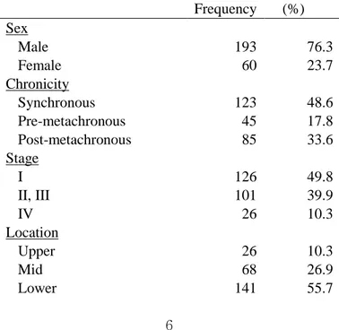
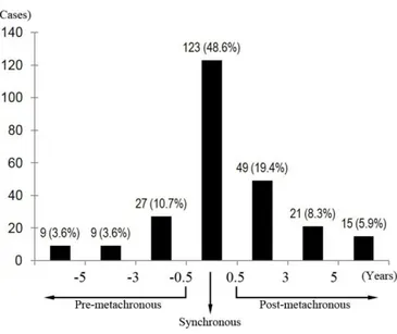
Of 8,839 patients with gastric cancer, 253 (2.9%) were found who had a SPC (24 vague cases and 13 triple primary cases were not included). Of these, 123 (48.6%) had a synchronous SPC, while 130 (51.4%) had a metachronous SPC (Table 1). In other words, 1.4% of all 8,839 gastric cancer patients were diagnosed with SPC synchronously, whereas 1.5% were diagnosed metachronously. Of the 130 metachronous cancer cases, 45 were discovered 6 months before diagnosis of gastric cancer, while 85 were discovered 6 months after diagnosis. The median age at diagnosis of gastric cancer in patients with SPC was 64 (range 31-83 years old) and the male-to-female ratio was 3.2 to 1. The median follow-up duration was 37 months.
Table 1. Clinicopathologic features of gastric cancer with SPC
Frequency (%) Sex Male 193 76.3 Female 60 23.7 Chronicity Synchronous 123 48.6 Pre-metachronous 45 17.8 Post-metachronous 85 33.6 Stage I 126 49.8 II, III 101 39.9 IV 26 10.3 Location Upper 26 10.3 Mid 68 26.9 Lower 141 55.7
7 Mixed 18 7.1 Multiplicity 1 240 94.9 ≥2 13 5.1 Cell type Adenocarcinoma 197 77.9 Signet ring cell 39 15.4 Mucinous carcinoma 2 2.0 Others 8 3.2 Unknown 7 2.8 Outcome Alive 84 33.2 Dead 169 66.8 Site distribution of SPC
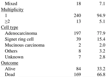
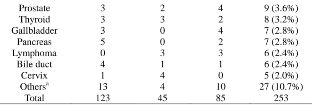
The most common site of SPC in gastric cancer was the colorectum (46 cases, 18.2%), followed by the liver (42 cases, 16.6%), lung (36 cases, 14.2%), esophagus (19 cases, 7.5%), and kidney (13 cases, 5.1%) (Table 2, Fig. 1). The occurrence of second primary colorectal cancers (CRC) was evenly distributed in the pre-metachronous, synchronous, and post-metachronous groups. Interestingly, most cases of SPCs in the liver, lung, esophagus, kidney, head, and neck occur synchronously or post-metachronously. In contrast, the majority of breast cancer and uterine cervical cancer were pre-metachronous cancers.
Table 2. Site distribution of SPC in gastric cancer patients
Synchronous Metachronous Total (%) Pre-M Post-M Colorectum 17 12 17 46 (18.2%) Liver (HCC) 26 4 12 42 (16.6%) Lung 14 3 19 36 (14.2%) Esophagus 15 0 4 19 (7.5%) Kidney 9 1 3 13 (5.1%)
Head & Neck 7 2 3 12 (4.7%)
8 Prostate 3 2 4 9 (3.6%) Thyroid 3 3 2 8 (3.2%) Gallbladder 3 0 4 7 (2.8%) Pancreas 5 0 2 7 (2.8%) Lymphoma 0 3 3 6 (2.4%) Bile duct 4 1 1 6 (2.4%) Cervix 1 4 0 5 (2.0%) Othersa 13 4 10 27 (10.7%) Total 123 45 85 253
HCC = Hpatocellular carcinoma, Pre-M = pre-metachronous, Post-M = post-metachronous
a : Urinary bladder (4), Ampulla of Vater (4), Leukemia (3), Brain (2), Endometrium (2), Ovary (2), Penis (2), Skin (2), Unknown primary (2), Gestational trophoblastic tumor (1), Thymus (1), Small ntestine (1), Multiple myeloma (1)
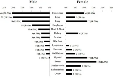
Figure 1. Site distribution of SPCs in gastric cancer patients
9
In males, the most common site for SPC was the colorectum (40 cases), followed by the liver (30 cases), lung (29 cases), and esophagus (19 cases) (Fig. 2). In females, the most common site was the breast (10 cases), followed by the thyroid (7 cases), lung (7 cases), colorectum (6 cases), and uterine cervix (5 cases). These patterns of site distribution in each sex were similar to the incidence of major cancers in the general population of Korea.
10 Time interval
The time interval between the diagnosis of gastric cancer and SPC was less than three years in 78.6% of the patients (Fig. 3). More than half (57.6%) of post-metachronous cancers were detected within three years of diagnosis of the gastric cancer. However, some were found after three years and even after more than 5 years, suggesting that SPC can occur at any time following the diagnosis of gastric cancer.
11
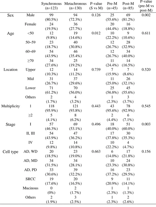
Comparison of synchronous and metachronous cancer
Table 3 showed the clinicopathologic features of synchronous and metachronous cancer in patients with gastric cancer. More patients were older at the diagnosis of gastric cancer in the metachronous group than in the synchronous group (p=0.012), although the sex, stage, location, multiplicity, and cell type of gastric cancer showed no differences between the synchronous and metachronous groups. When the metachronous group was divided into pre- and post-metachronous, more females were found in the pre-metachronous group (p=0.002). This was probably because of the age distribution pattern of breast, uterine cervix, and gastric cancers. In Korean female, peak incidence was observed at age 40-45 for breast and uterine cervix cancer, and 65-70 for gastric cancer, respectively.12 Also, the proportion of stage I gastric cancer was higher in the post-metachronous group than the pre-metachronous group (p=0.003). It has been suggested that better clinical outcomes due to earlier detection of gastric cancer may lengthen survival and consequently increase the risk of developing post-metachronous cancer.
12
Table 3. Clinicopathologic features of gastric cancer with synchronous and metachronous SPCs Synchronous (n=123) Metachronous (n=130) P-value (S vs M) Pre-M (n=45) Post-M (n=85) P-value (pre-M vs post-M) Sex Male 99 (80.5%) 94 (72.3%) 0.126 25 (55.6%) 69 (81.2%) 0.002 Female 24 (19.5%) 36 (27.7%) 20 (44.4%) 16 (18.8%) Age <50 12 (9.8%) 19 (14.6%) 0.012 10 (22.2%) 9 (10.6%) 0.611 50~59 23 (18.7%) 40 (30.8%) 12 (26.7%) 28 (32.9%) 60~69 54 (43.9%) 46 (35.4%) 12 (26.7%) 34 (40.0%) ≥70 34 (27.6%) 25 (19.2%) 11 (24.4%) 14 (16.5%) Location Upper 12 (10.3%) 14 (11.2%) 0.739 7 (15.9%) 7 (8.6%) 0.520 Mid 31 (26.7%) 37 (29.6%) 11 (25.0%) 26 (32.1%) Lower 71 (61.2%) 70 (56.0%) 25 (56.8%) 45 (55.6%) Others 2 (1.7%) 4 (3.2%) 1 (2.3%) 3 (3.7%) Multiplicity 1 118 (95.9%) 121 (93.8%) 0.443 43 (95.6%) 78 (92.9%) 0.545 ≥2 5 (4.1%) 8 (6.2%) 2 (4.4%) 6 (7.1%) Stage I 57 (46.3%) 69 (53.1%) 0.496 18 (40.0%) 51 (60.0%) 0.003 II, III 54 (43.9%) 47 (36.2%) 17 (37.8%) 30 (35.3%) IV 12 (9.8%) 14 (10.8%) 10 (22.2%) 4 (4.7%)
Cell type AD, WD 20 (18.5%) 23 (19.0%) 0.663 6 (14.0%) 17 (21.8%) 0.156 AD, MD 34 (31.5%) 34 (28.1%) 10 (23.3%) 24 (30.8%) AD, PD 33 (30.6%) 39 (32.2%) 16 (37.2%) 23 (29.5%) SRCC 19 (17.6%) 20 (16.5%) 9 (20.9%) 11 (14.1%) Mucinous 0 (0%) 2 (1.7%) 1 (2.3%) 1 (1.3%) Others 2 (1.9%) 3 (2.5%) 1 (2.3%) 2 (2.6%)
S = Synchronous, Pre-M = Pre-metachronous, Post-M = Post-metachronous. AD = Adenocarcinoma, WD = Well differentiated, MD = Moderately
13 Survival outcome and causes of death
The 5-year survival rate for all gastric cancer patients having a SPC was 39.5%. Interestingly, the Kaplan-Meier estimate of overall survival showed a better survival rate for patients with metachronous cancer than patients with synchronous cancer (Fig. 4). The median survival for patients with metachronous cancer was 63 months compared with 14 months for patients with synchronous cancer, and 5-year survival rates were 50.9% and 27.8%, respectively. When analyzing patients by the stage of gastric cancer, the survival of patients with metachronous cancer was higher than that of patients with synchronous cancer at all stages. Interestingly, the differences were significant in stage I (p=0.02), II and III (p<0.0001), while the difference at stage IV did not reach statistical significance (p=0.08). Within the metachronous patients, the post-metachronous group showed a better survival rate comparing to the pre-metachronous group. Median survival for patients with post-metachronous cancer was 80 months, compared with 41 months for patients with pre-metachronous cancer, and 5-year survival rates were 56.3% and 41.1%, respectively (Fig. 5). When metachronous patients were divided by the stage of gastric cancer, however, no differences were observed. Thus, the reason for the survival difference between pre-metachronous and post-metachronous groups is because there were more early stage gastric cancers in the post-metachronous group (Table 3).
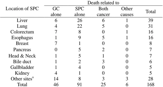
Of 253 SPC patients, a total of 168 (66.4%) patients died after the diagnosis of gastric cancer. Table 4 showed the common sites of the SPC and cause of death for those patients who died. A high incidence was seen in the liver, lung, colorectum, and esophagus as a SPC. Most causes of death (69.0%) were related mainly or
14
partially to SPC, except in the cases with breast cancer and kidney cancer of whom the major cause of death were gastric cancer.
Figure 4. The Kaplan-Meier survival curve for (A) all stage of gastric cancer, (B) stage I gastric cancer, (C) stage II and III gastric cancer, and (D) stage IV gastric cancer according to chronicity of SPC. Survival of gastric cancer
patients with metachronous SPC is better than that of synchronous SPC in all stage of gastric cancer. M = Metachronous, S = Synchronous.
15
Figure 5. The Kaplan-Meier survival curve for gastric cnacer with pre- and post- metachronous SPC. Survial of gastric cancer with post-metachronous SPC
is better than that with pre-metachronous SPC.
Pre-M = Pre-metachronous, Post-M = Post-metachronous, S = Synchronous
Table 4. Site distribution of the SPC and causes of death
Location of SPC Death related to GC alone SPC alone Both cancer Other causes Total Liver 6 26 6 1 39 Lung 4 22 5 0 31 Colorectum 7 8 0 1 16 Esophagus 1 9 5 1 16 Breast 7 1 0 0 8 Pancreas 0 5 2 0 7
Head & Neck 1 5 1 0 7
Bile duct 1 2 3 0 6
Gallbladder 1 4 0 0 5
Kidney 4 1 0 0 5
Other sitesa 14 8 3 3 28
Total 46 91 25 6 168
GC = gastric cancer, SPC = second primary cancer
a : Prostate (4), Ampulla of Vater (3), Leukemia (3), Lymphoma (3), Thyroid (3), Urinary bladder (3), Ovary (2), Skin (2), Brain (1), Cervix (1), Penis (1), Thymus (1), Unknown primary (1)
16
IV. DISCUSSION
Advances in diagnostic techniques and new drugs have improved the clinical outcome of cancer, and more patients are surviving longer after the diagnosis of cancer 13. In general, it is suggested that improved patient survival is now also associated with an increased risk of developing a SPC14. In the U.S., the 5-year survival rate for all cancer patients increased from 50% in 1975-1979 to 66% in 1996-2002 13. With improved clinical outcomes, the prevalence of SPCs has also increased. According to data from the National Cancer Institute’s Surveillance, Epidemiology, and End Results (SEER) program, about 7.9% (756,467 people) of US cancer survivors between 1975 and 2001 were diagnosed with a SPC 14,15.
Although the major reason for the increased prevalence of SPC is increased survival, and therefore an extended risk period, there are other possible explanations for this phenomenon. First, genetic vulnerability associated with specific genes may play a role in the development of SPCs. For example, hereditary nonpolyposis colorectal cancer (HNPCC), which is a syndrome associated with mutations in a class of genes such as MLH1, MSH2, MSH6, PMS1, and PMS2, is characterized by an increased susceptibility to other malignancies, especially of the uterus, ovary, urinary tract and stomach 16,17. Second, some carcinogenic environmental factors may induce multiple neoplasms of independent organs that were exposed to the same carcinogens. The field cancerization effect, which is associated with an increased risk of multiple cancers in aerodigestive organs after prolonged exposure to cigarette smoking and alcohol, is a well-known example of this phenomenon 18. Lastly, modalities used in the
17
treatment of the index cancer may also induce secondary cancers. For example, combined therapy with radiotherapy and alkylating agents has been reported to be related to an increased risk of gastric and colon cancer in long-term survivors of Hodgkin’s lymphoma 19,20
.
In gastric cancer, which is the fourth most common cancer in the world, the trend toward increasing SPC is similar. In the present study, of 8,839 patients with gastric cancer, 253 patients (2.9%) had been diagnosed with a SPC, which is consistent with the results of previous reports that ranged from 2.0 to 7.6% 7-10,21. A few previous studies reported the incidence and clinical pattern of SPC in gastric cancer patients who underwent curative gastrectomy. Eom et al. 7 reported that the most common SPC is CRC (20.8%), followed by lung cancer (11.9%), liver cancer (11.3%), and kidney cancer (7.5%) in Korea. Ikeda et al. 8 similarly reported that in Japan, the most common SPC is CRC (32.6%), followed by lung cancer (28.4%), liver cancer (8.4%), and esophageal cancer (7.4%). The majority of studies regarding SPC in gastric cancer have been performed in Eastern countries, with fewer studies performed in Western countries. Lundegardh et al. 21 reported that in Sweden, the most common site is the colorectum (19.9%), followed by lung (6.1%), kidney (5.3%), urinary bladder (4.5%), and pancreas (4.4%) in a cohort study that included SPC after the diagnosis of gastric cancer. Meanwhile, in the present study, the patients included were not only those who underwent gastrectomy but also those who were inoperable at the time of diagnosis. The most common site of SPC was the colorectum (18.2%), followed by liver (16.6%), lung (14.2%), esophagus (7.5%), and kidney (5.1%). This result was similar to previous studies of Eastern countries, but remarkably different from studies of Western
18
countries. The prominent differences from other Eastern reports were the relatively high incidence of hepatocellular carcinoma (HCC) and hematologic malignancies, such as leukemia and lymphoma, in the present study. An explanation might be that, unlike the previous studies, we included many inoperable cases in the present study. It is also probable that there are ethnic and epidemiological differences among the countries.
CRC, HCC, and lung cancer are the most common cancers in Korea. Therefore, the high occurrence of these cancers along with gastric cancer is not surprising. The proportion and order of the most common SPCs also roughly corresponded to their incidence in the general population in Korea.
A possible explanation for the highest incidence of CRC is that the colon is the same holoviscus organ, and shares the same carcinogens and genetic factors. Some reports suggested that microsatellite instability (MSI) plays an important role in the development of SPCs of the gastrointestinal tract 22,23. Actually, MSI has been reported more frequently in second primary gastrointestinal cancers than in sporadic single primary gastric or CRCs 22,23. In patients with CRC, 76% were diagnosed within three years of diagnosis of gastric cancer and 91.3% within five years. The proportion of CRC in patients with gastric cancer was also found to be rapidly increasing in this study. It accounted for 13.4% of SPCs in gastric cancer patients during the first five years from 1995 to 1999, whereas its occurrence increased to 20% during next five years from 2000 to 2004. Because second primary CRC showed the highest incidence with an increasing trend, is mainly diagnosed synchronously or post-metachronously, and is curable if diagnosed early, we suggest aggressive surveillance for second primary CRC in gastric cancer
19
patients. It is well known that screening for CRC in the general population by using the fecal occult blood test (FOBT) or sigmoidoscopy reduces mortality from CRC 24. Also, there is some evidence that screening by colonoscopy or CT colonography may be helpful 24,25. Though the FOBT is the least expensive and convenient screening tool, it appears less effective for screening second primary CRC in gastric cancer patients because of false positives generated by the gastric cancer itself. Thus, colonoscopy or CT colonography can be an another option. In particular, CT colonography can be performed relatively easily instead of conventional abdominal CT during the follow-up of gastric cancer survivors. However, there is no available data yet on the effectiveness of colonoscopy or CT colonography in screening second primary CRC. There is also the limitation that CT colonography is dependant on the CT scanner type and mode of imaging, and has a variable range of sensitivity, especially for detecting small polyps 25,26. Further data on the effectiveness of colonoscopy or CT colonography in screening second primary CRC are needed to determine an optimal surveillance program.
For cases of second primary HCC and lung cancer, they were not only the second and third most common SPCs but also the most common causes of death in gastric cancer patients with SPC. Moreover, 90.5% of second primary HCC and 91.7% of lung cancer developed synchronously or post- metachronously in this study. Thus, these cancers are also good candidates for active surveillance after the diagnosis of gastric cancer. Alpha-fetoprotein and abdominal ultrasonography for HCC, and low dose spiral CT for lung cancer are currently the most commonly used screening tools, although their efficacies have not yet been confirmed in randomized controlled trials.27,28 While screening programs could not evidently
20
show reduction in mortalities from HCC and lung cancer in the general population, screening and early detection can be beneficial for gastric cancer patients with high risk for developing HCC and lung cancer. It is because that earlier detection of second primary HCC and lung cancer is important to determine a gastric cancer treatment strategy. For example, in this study, among 14 gastric cancer patients who were diagnosed with synchronous lung cancer, 5 patients could have undergone curative surgery for both of gastric cancer and lung cancer. In addition, during the follow-up after curative resection of gastric cancer, 11 patients were diagnosed with post-metachronous HCC. Of these 11 patients, 3 patients could have undergone curative surgery for HCC. If all these patients were misdiagnosed as pulmonary or hepatic metastasis of gastric cancer, they might have been treated with palliative chemotherapy for gastric cancer, which eventually lead to adverse influence on overall survival. Thus, if single lesions were detected in the liver or lung at the time of gastric cancer diagnosis or follow-up after curative resection for gastric cancer, the possibility of the lesions being SPCs rather than a metastasis of gastric cancer should be considered for those who have high risk factors such as chronic B or C viral hepatitis, underlying liver cirrhosis, or a history of heavy smoking. Aggressive tissue biopsy can be helpful in differential diagnosis, and can play a decisive role in determining the treatment strategy. In addition, because abdominal CT scan is frequently performed during follow-up of gastric cancer survivors, and liver would be screened simultaneously in abdominal CT scan, we fully assume that there will be, in fact, more cases of overlooked post- metachronous HCC which were misdiagnosed as hepatic metastasis of gastric cancer.
21
Esophageal cancer and kidney cancer were found more frequently than expected in the general population, which might be because of increased detection rate from frequent use of diagnostic tools like endoscopy, abdominal CT, and positron emission tomography (PET) scan during initial staging and follow-up of gastric cancer. There are also possibilities that common carcinogens associated with gastric cancer might influence the carcinogenesis of esophageal cancer or kidney cancer. For example, tobacco smoking is a risk factor not only for gastric cancer, but also for esophageal squamous cell carcinoma, esophageal adenocarcinoma, and renal cell carcinoma 29-31.
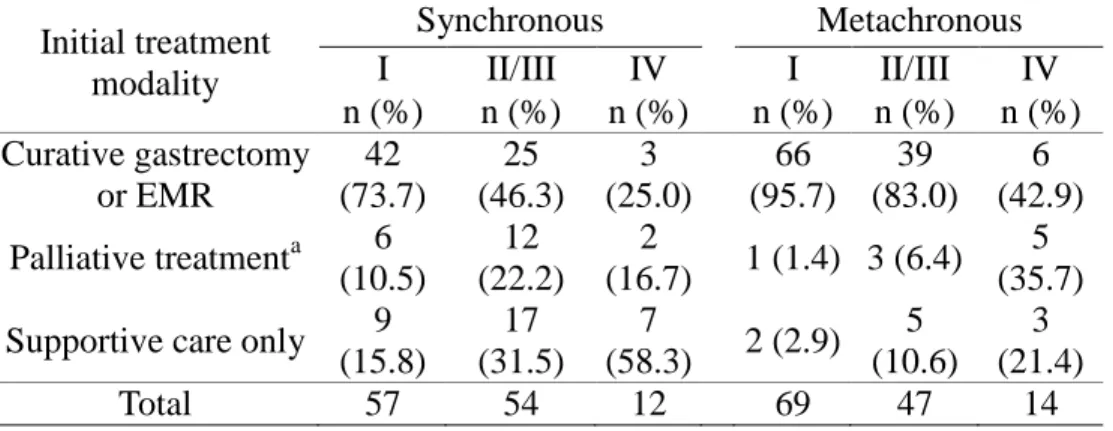
In the survival analysis of this study, the 5-year survival rate of gastric cancer patients with SPC was only 39.5%. It is a remarkably poor outcome, because the 5-year survival rate for all gastric cancer patients was 65.6% in our institute from 1987 to 2004. Moreover, for cases of stage I gastric cancer patients, whose 5-year survival rate is higher than 80~90% in Eastern and Western countries 2,32,33, those patients with SPC had a 5-year survival rate of only 53.2%. Therefore, SPC seems to adversely impact the overall outcome of gastric cancer treatment. The survival analysis also showed better median survival time and 5-year survival rate for metachronous cancer than for synchronous cancer. A possible explanation is that synchronous cancers like HCC and lung cancer may have negative influences on the general medical condition of the patient, thus hindering suitable therapeutic strategies. The findings shown in Table 5 are consistent with this explanation. In patients with stage I gastric cancer, 95.7% of patients in the metachronous group could underwent curative gastrectomy or endoscopic mucosal resection (EMR), whereas only 73.7% of patients in the synchronous group could underwent
22
curative treatment. The rest of 26.3% of stage I gastric cancer patients in the synchronous group were treated with palliative treatment or supportive care because of poor medical condition caused by synchronous SPC. This trend was also similar for the stage II and III, in which more patients were treated with curative surgery or EMR in the metachronous group than those in the synchronous group.
Table 5. Treatment modalities of gastric cancer with SPC according to the chronicity of SPC
Initial treatment
modality
Synchronous
Metachronous
I
II/III
IV
I
II/III
IV
n (%)
n (%)
n (%) n (%) n (%)
n (%)
Curative gastrectomy
or EMR
42
(73.7)
25
(46.3)
3
(25.0)
66
(95.7)
39
(83.0)
6
(42.9)
Palliative treatment
a6
(10.5)
12
(22.2)
2
(16.7)
1 (1.4) 3 (6.4)
5
(35.7)
Supportive care only
9
(15.8)
17
(31.5)
7
(58.3)
2 (2.9)
5
(10.6)
3
(21.4)
Total
57
54
12
69
47
14
I, II, III, IV = stage of gastric cancer, EMR = Endoscopic mucosal resection
a : Palliative treatment includes palliative chemotherapy (25 cases),
23
V. CONCLUSION
In conclusion, in this study, the incidence of SPC was 2.9% of the patients who were diagnosed with gastric cancer. We fully expect that the development of SPC will continue to increase due to improved survival of gastric cancer patients. Because SPCs can change the treatment strategy of gastric cancer and adversely impact the clinical outcome of gastric cancer, we need to try to detect common SPCs in gastric cancer patients earlier, especially CRC, HCC, and lung cancer. A thorough and regular surveillance program to discover SPC is warranted to improve the ultimate clinical outcome of gastric cancer.
24
REFERENCES
1. Kamangar F, Dores GM, Anderson WF. Patterns of cancer incidence, mortality, and prevalence across five continents: defining priorities to reduce cancer disparities in different geographic regions of the world. J Clin Oncol 2006; 24:2137-50.
2. Otsuji E, Yamaguchi T, Sawai K, Hagiwara A, Taniguchi H, Takahashi T, et al. Recent advances in surgical treatment have improved the survival of patients with gastric carcinoma. Cancer 1998; 82:1233-7.
3. Choi IJ. Screening and surveillance of gastric cancer. Korean J Gastroenterol 2007:15-22.
4. Korean Gastric Cancer Association. Nationwide gastric cancer report in Korea. J Korean Gastric Cancer Assoc 2002; 2:105-14.
5. Roukos DH. Current advances and changes in treatment strategy may improve survival and quality of life in patients with potentially curable gastric cancer. Ann Surg Oncol 1999; 6:46-56.
6. Lordick F, Siewert JR. Recent advances in multimodal treatment for gastric cancer: a review. Gastric Cancer 2005; 8:78-85.
7. Eom BW, Lee HJ, Yoo MW, Cho JJ, Kim WH, Yang HK, et al. Synchronous and metachronous cancers in patients with gastric cancer. J Surg Oncol 2008; 98:106-10.
8. Ikeda Y, Saku M, Kawanaka H, Nonaka M, Yoshida K. Features of second primary cancer in patients with gastric cancer. Oncology 2003; 65:113-7. 9. Ueno M, Muto T, Oya M, Ota H, Azekura K, Yamaguchi T. Multiple primary
25
cancer: an experience at the Cancer Institute Hospital with special reference to colorectal cancer. Int J Clin Oncol 2003; 8:162-7.
10. Wu CW, Lo SS, Chen JH, Hsieh MC, Li AF, Lui WY. Multiple primary cancers in patients with gastric cancer. Hepatogastroenterol 2006; 53:463-7. 11. Warren S, Gate O, Multiple primary malignant tumors: a survey of literature
and a statistical study. Am J Cancer 1932; 1358-1414.
12. Shin H-R, Jung K-W, Won Y-J, Kong H-J, Yim S-H, Sung J, et al. National cancer incidence for the year 2002 in Korea. Cancer Res Treat 2007; 39:139-49.
13. Jemal A, Siegel R, Ward E, Murray T, Xu J, Thun MJ. Cancer statistics, 2007. CA Cancer J Clin 2007; 57:43-66.
14. Hayat MJ, Howlader N, Reichman ME, Edwards BK. Cancer statistics, trends, and multiple primary cancer analyses from the Surveillance, Epidemiology, and End Results (SEER) Program. Oncologist 2007; 12:20-37. 15. Mariotto AB, Rowland JH, Ries LAG, Scoppa S, Feuer EJ. Multiple cancer
prevalence: A growing challenge in long-term survivorship. Cancer Epidemiol Biomarkers Prev 2007; 16:566-71.
16. Lynch HT, Chapelle A. Genetic susceptibility to non-polyposis colorectal cancer. J Med Genet 1999; 36:801-18.
17. Leon MP, Bertario L, Genuardi M, Lanza G, Oliani C, Ranzani GN, et al. Identification and classification of hereditary nonpolyposis colorectal cancer (Lynch syndrome): adapting old concepts to recent advancements. Report from the Italian Association for the study of hereditary colorectal tumors consensus group. Dis Colon Rectum 2007; 50:2126-34.
26
18. Braakhuis BJ, Tabor MP, Kummer JA, Leemans CR, Brakenhoff RH. A genetic explanation of Slaughter's concept of field cancerization: evidence and clinical implications. Cancer Res 2003; 63:1727-30.
19. Bassal M, Mertens AC, Taylor L, Neglia JP, Greffe BS, Hammond S, et al. Risk of selected subsequent carcinomas in survivors of childhood cancer: a report from the Childhood Cancer Survivor Study. J Clin Oncol 2006; 24:476-83.
20. Metayer C, Lynch CF, Clarke EA, Glimelius B, Storm H, Pukkala E, et al. Second cancers among long-term survivors of Hodgkin's disease diagnosed in childhood and adolescence. J Clin Oncol 2000; 18:2435-43.
21. Lundegardh G, Hansson LE, Nyrbn O, et al. The risk of gastrointestinal and other primary malignant diseases following gastric cancer. Acta Oncologica. 1991; 30:1-6.
22. Kim HS, Cho NB, Yoo JH, Shin K-H, Park J-G, Kim YI, et al. Microsatellite instability in double primary cancers of the colorectum and stomach. Mod Pathol 2001; 14:543-8.
23. Yamashita K, Arimura Y, Kurokawa S, Itoh F, Hirata K, Imamura A, et al. Microsatellite instability in patients with multiple primary cancers of the gastrointestinal tract. Gut 2000; 46:790-4.
24. Ransohoff DF. Colon cancer screening in 2005: Status and challenges. Gastroenterol 2005; 128:1685-95.
25. Aschoff A, Ernst A, Brambs H, Juchems MS. CT colonography: an update. Eur Radiol 2008; 18:429-37.
27
tomographic colonography. Ann Intern Med 2005; 142:635-50.
27. De Masi S, Tosti M, Mele A. Screening for hepatocellular carcinoma. Digest Liv Dis 2005; 37:260-8.
28. Field JK, Duffy SW. Lung cancer screening: the way forward. Br J Cancer 2008; 99:557-62.
29. Gonzalez CA, Pera G, Agudo A, Palli D, Krogh V, Vineis P, et al. Smoking and the risk of gastric cancer in the European Prospective Investigation Into Cancer and Nutrition (EPIC). Int J Cancer 2003; 107:629-34.
30. Enzinger PC, Mayer RJ. Esophageal cancer. N Engl J Med 2003; 349:2241-52.
31. Chow WH, Gridley G, Fraumeni JF, Jarvholm B. Obesity, hypertension, and the risk of kidney cancer in men. N Engl J Med 2000; 343:1305-11.
32. Kattan MW, Karpeh MS, Mazumdar M, Brennan MF. Postoperative nomogram for disease-specific survival after an R0 resection for gastric carcinoma. J Clin Oncol 2003; 21:3647-50.
33. Kitamura K, Yamaguchi T, Taniguchi H, Hagiwara A, Sawai K, Takahashi T, et al. Analysis of lymph node metastasis in early gastric cancer: Rationale of limited surgery. J Surg Oncol 1997; 64:42-7.
28
< ABSTRACT (IN KOREAN)>
위암 환자에서 발생한 이차암의 임상병리학적 특성
<지도교수 라선영>
연세대학교 대학원 의학과
김 찬
치료 성적과 생존률의 향상으로 인해 위암 환자에서 이차암이 발생할 위험도가 증가하게 되었다. 하지만 이러한 이차암의 빈도 및 발생 양상에 대해서는 지금까지 많은 연구가 이루어지지 않았다. 따라서 본 연구는 위암 환자에서 발생한 이차암의 임상병리적 특성과 치료 성적을 평가하고자 하였다. 1995년 1월부터 2004년 12월까지 연세 의료원 세브란스 병원에서 위암을 진단 받은 8,839명의 환자를 대상으로 의무 기록 및 통계청의 사망 데이터 베이스를 후향적으로 분석되었다. 총 8,839명의 환자 중 253명 (2.9%)이 이차암으로 진단받았다. 이 중 123명 (48.6%)은 동시성 이차암이었고, 나머지 130명 (51.4%)은 이시성 이차암이었다. 이차암이 가장 호발하는 부위는 대장직장 (18.2%), 간 (16.6%), 폐 (14.2%), 식도 (7.5%)의 순이었다. 남자에서 가장 흔한 이차암은 대장직장 (20.7%), 간 (20.2%), 폐 (15.0%). 식도 (9.8%)의 순이었다. 여성에서는 유방 (16.7%), 폐 (11.7%), 갑상선 (11.7%), 대장직장 (10.0%)의 순서를 보였다. 흔한 이차암의 비율 및 순서는 일반 인구 집단에서의 발생율과 비례하는 양상을 보였다. 동시성 이차암과 이시성 이차암 간에는 나이 외에 임상병리학적 변수들은 차이를 보이지 않았다. 동시성 이차암에서 위암 진단 당시29 환자의 나이가 더 많았다 (p=0.012). 위암 외에 이차암을 가지고 있는 환자는 역사적 대조에 비해 생존률이 더 불량한 양상을 보였다. 동시성 이차암 보다는 이시성 이차암을 진단 받은 환자의 경우 생존률이 더 양호하였다 (p<0.0001). 또한 후이시성 (Post-metachronous) 이차암은 전이시성 (Pre-metachronous) 이차암보다 더 좋은 예후를 보였다 (p<0.0001). 결론적으로 이차암의 발생은 위암의 치료 전략을 결정하는 데 영향을 미칠 수 있고, 임상 성적에도 좋지 않은 영향을 미치기 때문에, 위암 생존자들에서 흔한 이차암을 조기에 찾아내기 위한 관심이 필요하다. ---