ABSTRACT
Objective: A simultaneous detection of germline and somatic mutations in ovarian cancer (OC) using tumor materials is considered to be cost-effective for BRCA1/2 testing. However,
there are limited studies of the analytical performances according to various sample types. The aim of this study is to propose a strategy for routine BRCA1/2 next-generation sequencing
(NGS) screening based on analytical performance according to different sample types.
Methods: We compared BRCA1/2 NGS screening assay using buffy coat, fresh-frozen (FF) and
formalin-fixed paraffin-embedded (FFPE) from 130 samples.
Results: The rate of repeated tests in a total of buffy coat, FF and FFPE was 0%, 8%, and 34%, respectively. The accuracy of BRCA1/2 NGS testing was 100.0%, 99.9% and 99.9% in
buffy coat, FFPE and FF, respectively. However, due to the presence of variant allele frequency (VAF) shifted heterozygous variants, tumor materials (FFPE and FF) showed lower sensitivity (95.5%–99.0%) than buffy coat (100%). Furthermore, FFPE showed 51.4% of the positive predictive value (PPV) on account of sequence artifacts. When performed in the post-filtration process, PPV was increased by approximately 20% in FFPE. Buffy coat showed 100% of sensitivity, specificity and accuracy in BRCA1/2 NGS test.
Conclusions: On the comparison of the analytical performance according to different sample types, the buffy coat was not affected by sequencing artifacts and VAF shifted variants. Therefore, the blood test should be given priority in detecting germline BRCA1/2 mutation,
and tumor materials could be suitable to detect somatic mutations in OC patients without identifying germline BRCA1/2 mutation.
Keywords:BRCA1; BRCA2; Blood Buffy Coat; Tissue Preservation;
High-Throughput Nucleotide Sequencing
INTRODUCTION
High-grade serous ovarian cancer (HGSOC) patients with pathogenic germline or somatic mutations in either BRCA1 or BRCA2 are most responsive to poly (ADP-ribose) polymerase
(PARP) inhibitors, and PARP inhibitors combined with angiogenesis inhibition showed
Original Article
Received: Jan 23, 2019 Revised: Jul 17, 2019 Accepted: Jul 25, 2019 Correspondence to Kyung-A LeeDepartment of Laboratory Medicine, Yonsei University College of Medicine, 211, Eonju-ro, Gangnam-gu, Seoul, 06273, Korea. E-mail: KAL1119@yuhs.ac
*Yoonjung Kim and Chi-Heum Cho contributed equally to this work
Copyright © 2020. Asian Society of Gynecologic Oncology, Korean Society of Gynecologic Oncology
This is an Open Access article distributed under the terms of the Creative Commons Attribution Non-Commercial License (https:// creativecommons.org/licenses/by-nc/4.0/) which permits unrestricted non-commercial use, distribution, and reproduction in any medium, provided the original work is properly cited. ORCID iDs Yoonjung Kim https://orcid.org/0000-0002-4370-4265 Chi-Heum Cho https://orcid.org/0000-0002-0437-4099 Jung-Sook Ha https://orcid.org/0000-0002-6475-4886 Do-Hoon Kim https://orcid.org/0000-0002-9854-7850 Sun Young Kwon
https://orcid.org/0000-0002-8410-0185 Seoung Chul Oh
https://orcid.org/0000-0001-8211-5466
Yoonjung Kim ,1,* Chi-Heum Cho ,2,* Jung-Sook Ha ,3 Do-Hoon Kim ,3
Sun Young Kwon ,4 Seoung Chul Oh ,5 Kyung-A Lee 1
1Department of Laboratory Medicine, Yonsei University College of Medicine, Seoul, Korea 2Department of Obstetrics and Gynecology, Keimyung University School of Medicine, Daegu, Korea 3Department of Laboratory Medicine, Keimyung University School of Medicine, Daegu, Korea 4Department of Pathology, Keimyung University School of Medicine, Daegu, Korea
5Department of Laboratory Medicine, Gangnam Severance Hospital, Seoul, Korea
An optimized BRCA1/2 next-generation
sequencing for different clinical
improved anti-cancer response [1,2]. The recent United States Food and Drug Administration (USFDA) approval of olaparib applies a fourth line treatment for germline BRCA-mutant
ovarian cancer. Additionally, the European Medical Agency (EMA) obtained approval for olaparib maintenance in both germline and somatic BRCA-mutant platinum-sensitive ovarian
cancers in 2014 [2-4]. Treatment of ovarian cancers in individuals with somatic mutations of BRCA genes is similar to that of these cancers with germline mutations of BRCA genes.
However, currently available techniques are not sufficient to ensure identification of cancer-predisposing allelic variants in BRCA1 or BRCA2 from whole blood or tissue. Direct testing of
tumor samples is needed to identify additional patients with somatic mutations who might benefit from PARP inhibitor treatment. Recently, a few studies evaluated the practicality of next-generation sequencing (NGS) based vendor-developed BRCA1/BRCA2 testing or
open source testing with formalin-fixed paraffin-embedded (FFPE) tumor samples [5-8]. Many diagnostic laboratories are adopting NGS technology to increase screening capacity and reduce processing time and costs. However, NGS technology is neither simple nor homogeneous in NGS workflow (type of samples, NGS platforms, enrichment methods, and analytical procedures) [9].
Here, we compare the sequencing quality and variant call data obtained from buffy coat, fresh-frozen (FF) and FFPE samples of the same individual to determine the accuracy and concordance between the sample types using an Oncomine™ BRCA1/2 NGS panel (Thermo
Fisher Scientific, Carlsbad, CA, USA). We suggest acceptable quality measures of BRCA1/2
NGS test and cutoff values for variant calls to obtain reliable variant call information from different sample types in this NGS workflow.
MATERIALS AND METHODS
1. Study samples
We obtained a total of 50 ovarian cancer patients' sample sets (n=130) stored at Keimyung University Dongsan Medical Center Biobank, a member of the National Biobank of Korea. All samples derived from the National Biobank of Korea were obtained with informed consent under Institutional Review Board-approved protocols (DSMC IRB file No. 201609021). The sample size was determined to achieve a 95% confidence level in sensitivity and specificity analysis, with a less than 1% of error [10,11]. Ninety of the 130 samples were placed in subset A, which consisted of 30 matched buffy coat, FF, and FFPE samples, and 40 of the 130 samples were placed in subset B, which consisted of 20 matched FF and FFPE sample sets. All samples were obtained from pretreated OC patients who underwent surgical resection between 2008 and 2016. The ages of these samples range from 0.7 to 8.6 years (median 3.7 years). A board-certified pathologist reviewed hematoxylin and eosin (H&E)-stained sections from the FFPE blocks and the FFPE slides were macro-dissected to achieve sections that had at least 50% tumor content. All methods were performed in accordance with the approved protocols.
2. NGS sample preparation, library preparation, and ion S5 XL sequencing
Genomic DNA was extracted using the QIAamp DNA Blood Mini Kit (Qiagen, Venlo, The Netherlands), the QIAamp FFPE Tissue Kit (Qiagen), and the G-DEX™ Genomic DNA Extraction Kit (iNtRON Biotechnology, Seongnam, Korea) for buffy coats, FFPE, and FF tissues, respectively. Input DNA quantitation was performed using a Qubit 3.0 Fluorometer (Thermo Fisher Scientific) with 10 ng input per sample.
Kyung-A Lee
https://orcid.org/0000-0001-5320-6705
Funding
This research was conducted with support from AstraZeneca Korea Ltd. (ESR-16-12150).
Conflict of Interest
No potential conflict of interest relevant to this article was reported.
Author Contributions
Conceptualization: H.J.S., O.S.C., C.C.H., L.K.A.; Data curation: K.Y., O.S.C.; Formal analysis: K.Y., H.J.S., K.D.H.; Funding acquisition: C.C.H.; Investigation: K.Y., H.J.S., K.D.H., K.S.Y., L.K.A.; Methodology: K.Y., L.K.A.; Project administration: L.K.A.; Resources: H.J.S., K.D.H., K.S.Y., C.C.H.; Software: O.S.C.; Supervision: C.C.H., L.K.A.; Validation: K.Y., H.J.S., O.S.C.; Writing - original draft: K.Y., H.J.S.; Writing - review & editing: K.S.Y., C.C.H., L.K.A.
NGS libraries were constructed using an Oncomine™ BRCA1/2 panel (Thermo Fisher Scientific). The Oncomine™ BRCA1/2 panel was designed for 100% amplicon coverage of all targeted coding exons and exon-intron boundaries. And, the entire length of the region of interest (ROI) of this panel was 22,404 bp. The quality of final libraries was evaluated using the 4200 TapeStation (Agilent Technologies, Santa Clara, CA, USA). The average value (range) of final library concentration (pM) was 71.6 (17.3–207). The prepared libraries were then sequenced on an Ion S5 XL Sequencer using an Ion 520 Chip and an Ion 520 kit–Chef Kit (all Thermo Fisher Scientific).
3. Sequence alignment, variant calling, and annotation
Sequence data were aligned and mapping using the Torrent Mapping Alignment Program aligner implemented in v5.2 of the Torrent Suite software (Thermo Fisher Scientific). Variant calling was performed using Torrent Variant Caller v5.2 (Thermo Fisher Scientific) with the default setting of germline low-stringency parameters or somatic low-stringency parameters according to the type of samples. After variant calling, variants were annotated using ANNOVAR (http://www.openbioinformatics.org/annovar/) and Alamut version 2.10 (Interactive Biosoftware, Rouen, France). The reference sequences used were NM_007294.3 for BRCA1 and NM_000059.3 for BRCA2. The discovered variants are classified as s
pathogenic, likely pathogenic, uncertain significance, likely benign and benign according to American College of Medical Genetics and Genomics standards and guidelines [12].
4. Coverage analysis
Amplicon coverage data for targeted regions was generated from Torrent Coverage Analysis Plugin (Thermo Fisher Scientific). The details of the quality metrics of sequencing were described in Supplementary Table 1. The average read depth (>500×) and 100% of the targeted region covered with at least 20× and 100× were used to check the quality of
sequencing (Fig. 1). To obtain reliable sequencing data for the detecting germline and somatic variants, we have retested the specimens which did not satisfy both 100% of the targeted region >20× and an average read depth >500×. A minimum coverage of 20× is required to ensure covering all bases of the ROI for the detection of germline heterozygote variants [10,13]. Generally, candidate variants should be obtained only when a variant frequency at a given position of ≥20% and variant coverage of ≥20× [10]. In a case of somatic mutation, higher coverage is required (≥500×) since mutations are usually present at subclonal levels resulting in low percentages [14,15]. Minimum 100×1 is required for detecting clonal mutations in tumor samples at sufficient power (>0.8) [16].
5. Adaptive thresholds for variant calling in FFPE DNA
To balance the desirable sensitivity and PPV of FFPE samples, we set 2 different thresholds; 5% and 10%. To determine the values of metrics used for selecting allele frequency threshold (5% or 10%), we had adopted a threshold setting strategy for variant calls according to a previous study [17]. A threshold setting strategy consisted of 1) variants per kilobase, 2) percent annotation, and 3) transition (Ti)/transversion (Tv) ratio. The thresholds of the three metrics were calculated using receiver operating characteristic curve analysis or the non-parametric percentile method according to Clinical and Laboratory Standards Institute C28-A3. Transitional change (C:G>T:A) is the most frequent sequence artifact arising from FFPE DNA [18]. Ti/Tv ratio is used to check the risk of false positives [17].
6. Confirmatory study with Sanger sequencing and multiplex
ligation-dependent probe amplification (MLPA)
Sanger sequencing was performed to verify pathogenic variants of BRCA1/2. A MLPA assay
was applied for samples showing aberrant copies in copy number variation (CNV) analysis using NextGENe software (SoftGenetics, LLC, State College, PA, USA). MLPA was performed using P002-D1 BRCA1 and P045 BRCA2/CHEK2 probe mixes (MRC-Holland, Amsterdam, The
Netherlands) according to the manufacturer's instructions. MLPA results were analyzed using GeneMarker software (SoftGenetics, LLC).
7. Data analysis
Evaluation of analytic sensitivity, analytic specificity, and accuracy was performed with the candidate variants in the ROI which spanned all protein-coding regions and intron-exon boundaries (±20 bp). In the analytical performance analysis, “positive” indicates the case where the variants were detected in both a buffy and tumor samples (FFPE and/or FF) or confirmed by Sanger sequencing. “Negative” means that the variants were not detected in buffy or not confirmed by Sanger sequencing.
In a previous study, we deduced parameters corresponding to CNV analysis using NextGENe software (SoftGenetics, LLC). Similarly, CNV analysis was performed using NextGENe software (SoftGenetics, LLC).
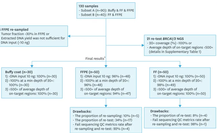
130 samples
- Subset A (n=90): Buffy & FF & FFPE - Subset B (n=40): FF & FFPE
Final results* 5 FFPE re-sampled
- Tumor fraction <30% in FFPE or
- Extracted DNA yield was not sufficient for DNA input (<10 ng)
Drawbacks:
- The proportion of re-sampling: 10% (n=5) - The proportion of re-test: 34% (n=17) - Fail sequencing QC metrics rate after re-sampling and re-test: 92% (n=4)
Drawbacks:
- The proportion of re-test: 8% (n=4) - Fail sequencing QC metrics rate after
re-sampling and re-test: 98% (n=1) 21 re-test BRCA1/2 NGS
- 20× coverage (%) <100% or
- Average depth of on-target regions <500× (details in Supplementary Table 1)
Buffy coat (n=30) 1) >DNA input 10 ng: 100% (n=30) 2) >100% at a min depth of 20×: 100% (n=30) 3) >500× of average depth of on-target regions: 100% (n=30) FF (n=50) 1) >DNA input 10 ng: 100% (n=50) 2) >100% at a min depth of 20×: 98% (n=49) 3) >500× of average depth of on-target regions: 100% (n=50) FFPE (n=50) 1) >DNA input 10 ng: 96% (n=48) 2) >100% at a min depth of 20×: 96% (n=48) 3) >500× of average depth of on-target regions: 94% (n=47)
Fig. 1. Summary of quality metrics in the different form of specimens. FF, fresh-frozen; FFPE, formalin-fixed paraffin-embedded; QC, quality control.
*Final results excluded the results of the 21 first replicates that were reexamined because they did not meet the criteria of quality metrics and included the re-tested results from these 21 replicates. Fail sequencing QC metrics rate included replicates that did not meet the requirements of quality metrics 1) a coverage of 100% at a minimum depth of 20× and 2) an average depth of on-target regions>500×.
All statistical analyses were performed using MedCalc Software (https://www.medcalc.org/), and p values less than 0.05 were regarded as significant. Mann-Whitney U was used to compare differences between two independent groups that were not normally distributed.
RESULTS
1. Comparison of quality metrics in different types of specimens
Quality metrics of buffy coat, FF, and FFPE are summarized in Supplementary Table 1 and
Supplementary Fig. 1. Five FFPE samples that did not have sufficient input DNA (10 ng, 0.67 ng/uL) for NGS were re-obtained from FFPE blocks. Two of the re-obtained FFPE samples were failed in DNA extraction (Fig. 1). Finally, the DNA yields from all 128 samples exceeded 0.67 ng/uL (Supplementary Fig. 1).
The mean of mapped reads for each type of specimen ranged from 318,663 to 503,020. The average depths of on-target region in buffy coat, FF, and FFPE were 1,442×, 2,243×, and 1,834×, respectively (Supplementary Table 1). A high degree of coverage uniformity (>98%) and on-target reads (>94%) were achieved on both buffy coat and FF samples. The overall values of quality metrics of FFPE including on-target (%), uniformity (%), and 20× coverage (%) were lower than those of buffy coat and FF (Supplementary Table 1). We repeated 21 tests (FF 4 samples; FFPE 17 samples) that did not satisfy 2 quality metrics simultaneously; 100% coverage of minimum read depth of 20× and more than 500× average read depth of on-target regions (Supplementary Table 2 and Supplementary Fig. 1). The rate of repeat testing was higher in FFPE (34%) than in buffy coat (0%) and FF (8%). In the initial results before 21 repeated tests, 91.5% showed a coverage 100% at a minimum depth of 20x, and only 86.9% had a sufficient average read depth of on-target regions (>500×). After 21 repeated tests, 97.7% of 130 samples showed a coverage 100% at a min depth of 20× and an average read depth of on-target regions (>500×), respectively (Supplementary Table 3 and Supplementary Fig. 1). Five samples were excluded in sequencing data analysis because they did not meet the criteria of acceptable quality metrics after repeated tests.
2. The storage period of the formalin-fixed paraffin-embedded tissues
The ages of these samples range from 0.7 years to 8.6 years (median, 3.7 years). The median storage time of FFPE (n=28) and re-tested FFPE (n=17) blocks were 5.6 and 4.0 years, respectively. The storage time of FFPE blocks has no significant effect on the quality of sequencing (p value: 0.0534, Mann-Whitney U test) (Supplementary Fig. 2 and
Supplementary Table 2).
3. Pathogenic variants
Pathogenic variants in BRCA1/2 genes were found in 12 of the 50 (24%) ovarian cancers.
Among them, 9 and 3 were in BRCA1 and BRCA2, respectively. We found only one somatic
pathogenic variant in this study (Fig. 2). Six pathogenic germline variants (20%) and one somatic variant (3.3%) were detected in subset A. And, six pathogenic variants were detected in subset B (Table 1), whether these variants arose from germline or somatic cells could not be assessed due to the absence of the matched buffy coat in subset B. The averages of variant allele frequency (VAF) (%) of pathogenic variants were approximately 50%, 75%, and 90% in buffy coat, FFPE, and FF, respectively (Supplementary Fig. 3). The VAF (%) of germline BRCA1
pathogenic variants (c.5080G>T, p.Glu1694*) in one case (ID: 130227014) in FFPE (11.1%)
BRCA1 pathogenic variant (c.5339T>C, p.Leu1780Pro) was called in FFPE with 373 coverage
depths and VAF 17%, while it was not observed in FF. Sanger sequencing was performed with DNA extracted from FFPE and FF. We could not confirm this sequence variation in FFPE due to PCR failure. However, through successfully performed sequencing with DNA from FF, the absence of variation (c.5339T>C) was confirmed in FF (data not shown). We conducted CNV analysis using NextGENe software (SoftGenetics, LLC) across all cases and carried out MLPA assay for cases that show indefinite calls. We did not observe pathogenic duplication/deletion of the BRCA gene in our cases.
(A) Sample type: Buffy
Wild type
Wild type
c.3309dup, p.Lys1104* c.3309dup, p.Lys1104*
c.3309dup, p.Lys1104*
(B) Sample type: FF (C) Sample type: FFPE
(D) Sample type: Buffy
(E) Sample type: FF Case 131127016, BRCA1 (NM_007294.3) c.3309dup, p.Lys1104*
Fig. 2. Somatic mutations detected by NGS and validated by Sanger sequencing.
On the left, the results of NGS where the reads are aligned to the reference genome as provided by NextGENe software (SoftGenetics, LLC). On the right, we performed sequencing to validate the somatic nature of the mutation (c.3309dup, p.Lys1104*) by its absence in the matching buffy coat DNA. This somatic nonsense mutation was shown in FF (B and E) and FFPE (C). Wild-type was demonstrated in buffy coat (A and D). FF, fresh-frozen; FFPE, formalin-fixed paraffin-embedded; NGS, next-generation sequencing.
Table 1. Identified pathogenic variants* and its VAF (%) in 30 matched buffy coat, FF, and FFPE samples (subset A) and 20 matched FF & FFPE samples (subset B)
Subset Case ID BRCA1 BRCA2
Germline-somatic Total Buffy coat FFPE FF
Cov. Allele Cov. VAF (%) Total Cov. Allele Cov. VAF (%) Total Cov. Allele Cov. VAF (%) A 110518001 (-) c.7480C>T, p.Arg2494* Germline 971 483 50 284 271 95 1,138 1,045 92 A 120725016 c.5496_5506delinsA, p.Val1833Serfs*7 (-) Germline 1,664 701 42 4,540 1,094 55.8 1,893 1,710 90 A 130227014 c.5080G>T, p.Glu1694* (-) Germline 1,726 855 50 463 51 11.1 1,995 1,824 91 A 131014030 c.5080G>T, p.Glu1694* (-) Germline 1,996 950 48 1,982 1,616 82 1,996 1,793 90 A 150522003 c.5080G>T, p.Glu1694* (-) Germline 1,989 998 50 494 410 83 1,989 1,881 95 A 150529001 (-) c.1399A>T, p.Lys467* Germline 1,607 765 48 1,999 1,756 88 1,000 897 90
A 131127016 c.3309dup, p.Lys1104* (-) Somatic ND ND ND 762 462 61 754 645 86
B 90217003 c.390C>A, p.Tyr130* (-) Unknown NT NT NT 1,157 1,048 91 1,427 1,254 88
B 90609005 c.981_982del, p.Cys328* (-) Unknown NT NT NT 1,512 970 64 315 265 84
B 90824007 c.5339T>C, p.Leu1780Pro (-) Unknown NT NT NT 756 611 81 270 231 86
B 100708008 (-) c.5574_5577del,
p.Ile1859Lysfs*3 Unknown NT NT NT 1,380 1,297 94 1,272 1,186 93 B 101206009 c.5509T>C, p.Trp1858Gly (-) Unknown NT NT NT 1,503 1,437 96 2,000 1,983 99 Cov., coverage; FF, fresh-frozen; FFPE, formalin-fixed paraffin-embedded; ND, not detected; NT, not tested; VAF, variant allele frequency.
*Pathogenic Variant was including pathogenic and likely pathogenic variants that were classified based on American College of Medical Genetics and Genomics standards and guidelines. GenBank accession numbers NM_007294.3 for BRCA1 and NM_000059.3 for BRCA2 were used as reference sequences.
4. False positive calls in FFPE
For reliable mutation detection across BRCA genes using FFPE samples, artifacts not of
biological origin should be filtered. Of the total, 290 false positive calls were observed in subset A, and the average allele frequency of false positive calls was 8.4% (range, 5%–36%). Among the 290 false positive calls, 47.9% consisted of non-synonymous variants
(41.7%, n=121) and protein-truncating variants (6.2%, n=18), while 85.8% presented as G>A or C>T Ti. Transitions (changes from A <-> G and C <-> T) occurred 35 times as frequently as Tv (Supplementary Fig. 4).
5. Filtering strategies for minimizing of sequence artifacts from FFPE DNA
The lowest limit of detection for low-frequency variants is approximately 5%–10% in tumor samples [19-22]. We used Torrent Variant Caller v5.2 after adjusting its somatic low-stringency parameters using AcroMetrix Oncology Hotspot Control at 5% allele frequency for Oncomine™
BRCA1/2 panel NGS testing. In FFPE samples, the average VAF of false positive calls was 8.6%
and sequence artifacts were apparent at the range of 5%–10% VAFs. And the positive predictive value (PPV) in subset A was 51.4% using a 5% allele frequency threshold. The average calls per kilobase (kb) were 0.6, 2.3, and 0.8 in buffy coat, FFPE, and FF, respectively. The percentage of annotated variants was 96.2%, 81.7%, and 97.5% in buffy coat, FFPE and FF, respectively. The average the Ti/Tv ratio was 0.6, 6.7, and 1.0 in buffy coat, FFPE, and FF, respectively (Table 2). The three metrics consisted of 1) variants per kilobase, 2) percent annotation, and 3) Ti/Tv ratio showed significant differences between samples with or without false positive calls (p<0.001, Mann-Whitney U test). To determine the values of metrics used for selecting allele frequency threshold (5% or 10%), the cut-offs of the three metrics as a set were modulated to achieve 100% sensitivity for identifying samples with false positives (Table 2).
Among the 125 samples, 16 (12.8%) including 2 FF and 14 FFPE, did not meet the criteria of 1) <1.2 calls/kb and >64.3% annotated, or 2) <3.8 Ti/Tv ratio. When the 14 FFPE samples were processed the post-filtration with a 10% allele frequency threshold, the PPV increased from 39.3% (95% confidence interval [CI]=36.6%–41.9%) to 71.0% (95% CI=66.3%–75.2%) and the sensitivity and negative predictive value (NPV) was 98.0% and 99.9%, respectively.
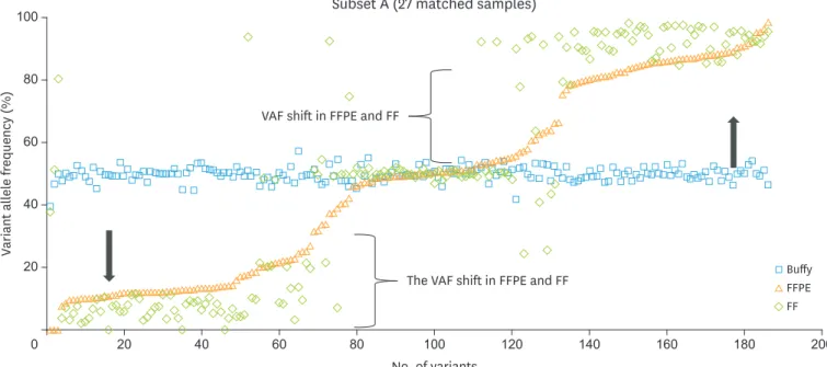
6. Sequence variants and VAF shifted heterozygous variants
In order to create a fair comparison, among the 310 single-nucleotide variant (SNV) calls from buffy coat in subset A, the VAF (%) of 186 heterozygous variants in buffy coat were compared with those of each SNV call from FFPE and FF in subset A. Of 186 heterozygous variants, the differences of allele frequency between buffy coat and matched tumor tissue were more than 10% at 152 heterozygous variants. This phenomenon was seen across BRCA1 or/and BRCA2
genes from 19 of the 27 matched samples in subset A. Interestingly, the disproportionate allele Table 2. The thresholds of 3 metrics for the post-filtration process in FFPE samples
Type of sample Buffy coat (n=30) FFPE (n=46) FF (n=49) Criterion Average±SD of Variants/kb 0.6±0.3 2.3±2.9 0.8±0.7 >1.2* Percent annotated 96.2%±5.9% 81.7%±34.8% 97.5%±4.7% <64.3% Ti/Tv ratio 0.6±0.5 6.7±13.4 1.0±1.1 >3.8 Sensitivity (95% CI) 100.0% (69.2–100.0) Specificity (95% CI) 94.8% (90.0–98.1) AUC (95% CI) 0.97 (0.928–0.994)
AUC, area under the receiver operating characteristic curve; SD, standard deviation; Ti, transition; Tv, transversion.
frequencies of variants occurred both in wild type cases and BRCA1/2 mutation harboring cases
(Fig. 3). Therefore, we referred to this phenomenon as ‘VAF shifted heterozygous variants.’
7. Analytical performance
We analyzed the analytical performance using subset A. The analytical performance of
BRCA1/2 NGS test was evaluated with or without VAF shifted heterozygous variants (Table 3). The sensitivity, specificity, and accuracy of BRCA1/2 NGS test were all 100% for buffy coat with
or without VAF shifted heterozygous variants. The sensitivity, specificity, and accuracy for FF including heterozygous variants with VAF shift was 95.5%,100%, and 100%, respectively. When excluding heterozygous variants with VAF shift in FF, the sensitivity was increased up to 100% (Table 3). In this study, VAF shifted heterozygous variants were frequently shown in tumor tissues. When using 5% allele frequency threshold in FF, 17 VAF shifted heterozygous variants (45% to 58% VAFs in buffy coat, 7% to 22% in FFPE) were filtered out. Therefore, these VAF shifted heterozygous variants could affect the analytical performance of BRCA1/2
NGS test with tumor materials, especially the sensitivity of FF.
No. of variants
Subset A (27 matched samples)
100 200 180 160 140 120 100 80 60 40 20 0 Variant all el e fr equency (%)
VAF shift in FFPE and FF
The VAF shift in FFPE and FF 80 60 40 20 Buffy FFPE FF
Fig. 3. Comparison of VAF (%) among 186 SNVs detected as “heterozygous variants” from buffy coat and other forms of samples including FFPE and FF in subset A. The variant allele frequency (%, VAF) of SNVs from buffy coat, FFPE, and FF are depicted as a square dots, triangle dots and diamond dots, respectively. The black arrow indicates the direction of change in variant allele frequency of the 186 VAF shifted heterozygous variants in FFPE and FF.
FF, fresh-frozen; FFPE, formalin-fixed paraffin-embedded; SNV, single-nucleotide variant; VAF, variant allele frequency. Table 3. Analytical performance of subset A
Threshold Sample
type shiftedVAF (n)FP (n)FN (n)TP (n)TN Sensitivity (95% CI) Specificity (95% CI) Accuracy (95% CI) (95% CI)PPV (95% CI)NPV Using a
5% allele frequency threshold*
Buffy coat† Include 0 0 310 478,373 100.0% (98.8–100.0) 100.0% 100.0% 100.0% 100.0%
Exclude 0 0 293 478,390 100.0% (98.8–100.0) 100.0% 100.0% 100.0% 100.0%
FFPE Include 290 3 307 478,083 99.0% (97.2–99.8) 99.9% (99.9–99.9) 99.9% (99.9–100.0) 51.4% (48.5–54.3) 100.0% Exclude 290 3 290 478,100 99.0% (97.0–99.8) 99.9% (99.9–100.0) 99.9% (99.9–100.0) 50.0% (47.1–52.9) 100.0%
FF Include 0 17‡ 290 478,376 95.5% (91.3–96.8) 100.0% 100.0% 100.0% 100.0%
Exclude 0 0 293 478,390 100.0% (98.8–100.0) 100.0% 100.0% 100.0% 100.0%
FF, fresh-frozen; FFPE, formalin-fixed paraffin-embedded; FP, false positive; FN, false negative; n, number of variants; NPV, negative predictive value; PPV, positive predictive value; TN, true negative TP, true positive; VAF, variant allele frequency; VAF shifted, variant allele frequency shifted heterozygous variants. *The somatic variant was called when the variant frequency at a given position was ≥5% and variant coverage was ≥100×; †The germline variant was called when
the variant frequency at a given position was ≥20% and variant coverage was ≥20×; ‡Seventeen VAF shifted heterozygous variants that detected in buffy coat with
DISCUSSION
Screening BRCA1 and BRCA2 germline variant using NGS is well established in clinical practice
and is used primarily to determine hereditary breast and ovarian cancer risk [10,13,23,24]. The screening methods using buffy coat are optimized in several literatures. In our study, we compared the sequencing quality and variant call data obtained in buffy, FF and FFPE samples from all 130 samples. 100% of buffy coat and 98% of FF achieved 100% coverage at a minimum depth of 20× and mean coverage depth of 500× without re-sampling and re-testing. However, when the BRCA1/2 test was performed using FFPE, the rate of resampling and re-testing were
10% and 34% of total FFPE cases, respectively, which might be due to the damage of FFPE DNA [17,25]. In a previous study, over 76% of FFPE samples were successfully analyzed, with >95% coverage of the BRCA1/2 coding regions and a mean average read depth >1,000-fold
using the GeneRead DNAseq Targeted Exon Enrichment Panel [8]. In this study, re-testing increased the quality of sequencing, and the failure rate decreased from 34% to 8%. Buffy coat, FFPE, and FF samples showed ≥99.9% of the accuracy in BRCA1/2 NGS testing.
However, FFPE showed 51.4% of the positive predictive value on account of sequence artifacts. Among the 290 false positive calls in FFPE, 47.9% consisted of non-synonymous variants (41.7%, n=121) and protein-truncating variants (6.2%, n=18). The average allele frequency of false positive calls was 8.4% (range: 5%–36%). To distinguish candidate somatic mutations from sequence artifacts with more than 20% allele frequency, Sanger sequencing was applied [26]. And, from that, we confirmed the variant with maximum allele frequency (36%) in NGS was negative. Most of the false positive calls of FFPE materials in NGS were failed to confirm with Sanger sequencing. Therefore, FFPE DNA could be limited to correctly assess the somatic mutations with low allele frequency [27,28]. And, tumor samples with loss of heterozygosity (LOH) [29], the allele frequency of germline BRCA pathogenic variation in tumor tissues is
higher than that in buffy coat. However, one of our FFPE showed 11.1% of the allele frequency of germline BRCA1 pathogenic variation (p.Glu1694*, ID: 130227014), which is close to the 10%
allele frequency threshold. In this study, the ranges of allele frequency (%) of true calls (5.3%– 100.0%) and false positive calls (5.0%–36.0%) were overlapped in FFPE samples. To be used in routine BRCA NGS assay, filtering strategies for minimizing of sequence artifacts from FFPE
DNA should be required to ascertain the reliability of the final report. Most artifactual changes in FFPE occur in the 1-10% allele frequency range [30]. When a 10% cut-off is applied to FFPE, clinically relevant somatic mutations with 5%–10% VAFs [31] will be missed. Therefore, we suggest applying not only a 5% cut-off strategy but also a 10% cut-off to minimize the false positive calls in FFPE. For example, when the 14 FFPE samples that did not meet the criteria (variants per kilobase, percent annotation, and Ti/Tv ratio) were proceeded using the 10% allele frequency thresholds, the PPV was increased from 39.3% to 71.0%.
In a previous study, the excellent quality of DNA extracted from FF was considered as an alternative to DNA from FFPE samples, and FF samples showed >99.7% sensitivity, specificity, and accuracy in BRCA1/2 testing [32]. However, in this study, the sensitivity of FF
was 95.46% using a 5% allele frequency threshold due to VAF shifted heterozygous variants with low VAF (%). When VAF shifted heterozygous variants were excluded in evaluating analytical performance, the sensitivity would increase to 100%. In tumor tissue (FFPE and FF), VAF shifted heterozygous variants were observed in both wild-type cases and BRCA1/2
mutation harboring cases. Majority of VAF shifted heterozygous variants phenomenon in this study could be explained by chromosomal abnormalities on 13q12-q14 or 17q where BRCA2
were showed copy number aberration on these regions [33,34]. And, the allele frequency of coexisted cis-variants with BRCA1/2 pathogenic variants also contributed to shift the allele
frequency based on LOH in tumor suppress gene [29]. And, this ‘VAF shifted heterozygous variants’ were observed 70% (n=19/27 pairs) cases of subset A in our study. As VAF shifted heterozygous variants showed a decreasing trend of allele frequency, the number of false negative calls increased in tumor tissue. These findings from FFPE and FF indicate that the false-positive calls and VAF shifted variants might be drawbacks of BRCA1/2 NGS assay using
only tumor tissue.
In December 2014, the EMA approved the PARP inhibitor olaparib as monotherapy treatment for relapsed platinum-sensitive HGSOC patients with pathogenic germline or somatic
mutations in BRCA1/2 [3]. Approximately 6%–25% of OC patients have a BRCA1/BRCA2 germline
mutation [35], and somatic BRCA mutations occur in approximately 3%–7% of ovarian cancer
cases [6,36,37]. Since a simultaneous detection of germline and somatic mutations in OC is cost-effective, there is an increasing clinical need for routinely screening BRCA using tumor materials [38]. In this study, buffy coat achieved 100% coverage at 20× and 100× without re-sampling or re-analysis, and they showed 100% of the sensitivity, specificity, and accuracy. The buffy coat was not affected by shifted allele frequency of variants or false calls which are intrinsic obstacles of tumor materials. However, due to VAF shifted heterozygous variants, tumor materials (FFPE and FF) showed lower sensitivity (95.5%–99.0%) than buffy coat (100%). Our results showed that the VAF shifted heterozygous variants with low VAF (%) can be neglected during the variant calling in tumor samples when the usual 5 allele frequency threshold were applied. Furthermore, sequence artifacts could not be distinguished from pathogenic variants because the ranges of VAF (%) of true calls and false positive calls are overlapped. According to recently updated guidelines for BRCA1/2 and above findings, it is
reasonable to test OC blood samples for BRCA mutations first, and then, if negative, carry
testing with a tumor sample to extend patients group who might potentially benefit from PARP inhibitors therapy [39,40].
We improved the performance of BRCA1/2 NGS assay through the post-filtration process using
the thresholds of metrics and exclusion of VAF shifted heterozygous variants from different tissue types. In conclusion, we suggest the strategy of germline screening BRCA1/2 from buffy
coat prior to testing from tumor materials to obtain clinically reliable results.
ACKNOWLEDGEMENTS
The biospecimens for this study were provided by Keimyung University Dongsan Hospital Korea Regional Biobank, a member of the National Biobank of Korea, which is supported by the Ministry of Health and Welfare.
SUPPLEMENTARY MATERIALS
Supplementary Table 1
Quality metrics in BRCA1/2 NGS (n=130) Click here to view
Supplementary Table 2
Twenty-one tests that did not meet the criteria of quality metrics of BRCA1/2 NGS assay Click here to view
Supplementary Table 3
Comparison of QC metrics between the different types of samples in BRCA1/2 NGS Click here to view
Supplementary Fig. 1
gDNA concentration plots from three different sample types. (A) Buffy coat, (B) FF, and (C) FFPE. Y-axis indicates the concentration of gDNA in nanograms per microliter, and the x-axis indicates sample types.
Click here to view
Supplementary Fig. 2
Comparison of the storage time of FFPE (n=28) and re-tested FFPE (n=17) blocks. FFPE samples (n=28) that passed the QC assay metrics 1) DNA input (>10 ng), 2) a coverage of 100% at a minute read depth of 20×, and 3) an average read depth of on-target regions >500×. Re-tested FFPE (n=17) that did not satisfy both a coverage of 100% at a minute read depth of 20× and an average read depth of on-target regions >500× were retested. Five FFPE samples that did not have sufficient input DNA (10 ng) were not included in this comparison. Data represented as median with interquartile range.
Click here to view
Supplementary Fig. 3
Identified 12 pathogenic variations and its VAFs (%) in 30 matched buffy coat, FF, and FFPE samples (subset A) and 20 matched FF & FFPE samples (subset B). The black colored dotted lines were indicated the average of VAF (%) in buffy coat, FF, and FFPE samples from top to bottom.
Click here to view
Supplementary Fig. 4
Transition and transversion sequence variation counts in 290 false positive calls. There was a trend of increasing G/A and C/T substitution rate along with total variation count.
Click here to view
REFERENCES
1. Ivy SP, Liu JF, Lee JM, Matulonis UA, Kohn EC. Cediranib, a pan-VEGFR inhibitor, and olaparib, a PARP inhibitor, in combination therapy for high grade serous ovarian cancer. Expert Opin Investig Drugs 2016;25:597-611.
2. Parkes EE, Kennedy RD. Clinical application of poly(ADP-ribose) polymerase inhibitors in high-grade serous ovarian cancer. Oncologist 2016;21:586-93. .
PUBMED | CROSSREF
3. Ledermann J, Harter P, Gourley C, Friedlander M, Vergote I, Rustin G, et al. Olaparib maintenance therapy in patients with platinum-sensitive relapsed serous ovarian cancer: a preplanned retrospective analysis of outcomes by BRCA status in a randomised phase 2 trial. Lancet Oncol 2014;15:852-61. PUBMED | CROSSREF
4. Liu JF, Matulonis UA. What is the place of PARP inhibitors in ovarian cancer treatment? Curr Oncol Rep 2016;18:29.
PUBMED | CROSSREF
5. Pennington KP, Walsh T, Harrell MI, Lee MK, Pennil CC, Rendi MH, et al. Germline and somatic mutations in homologous recombination genes predict platinum response and survival in ovarian, fallopian tube, and peritoneal carcinomas. Clin Cancer Res 2014;20:764-75.
PUBMED | CROSSREF
6. Mafficini A, Simbolo M, Parisi A, Rusev B, Luchini C, Cataldo I, et al. BRCA somatic and germline mutation detection in paraffin embedded ovarian cancers by next-generation sequencing. Oncotarget 2016;7:1076-83.
PUBMED | CROSSREF
7. Endris V, Stenzinger A, Pfarr N, Penzel R, Möbs M, Lenze D, et al. NGS-based BRCA1/2 mutation testing
of high-grade serous ovarian cancer tissue: results and conclusions of the first international round robin trial. Virchows Arch 2016;468:697-705.
PUBMED | CROSSREF
8. Ellison G, Huang S, Carr H, Wallace A, Ahdesmaki M, Bhaskar S, et al. A reliable method for the detection
of BRCA1 and BRCA2 mutations in fixed tumour tissue utilising multiplex PCR-based targeted next
generation sequencing. BMC Clin Pathol 2015;15:5. PUBMED | CROSSREF
9. Wallace AJ. New challenges for BRCA testing: a view from the diagnostic laboratory. Eur J Hum Genet 2016;24 Suppl 1:S10-8.
PUBMED | CROSSREF
10. Shin S, Kim Y, Chul Oh S, Yu N, Lee ST, Rak Choi J, et al. Validation and optimization of the Ion Torrent S5 XL sequencer and Oncomine workflow for BRCA1 and BRCA2 genetic testing. Oncotarget 2017;8:34858-66.
PUBMED | CROSSREF
11. Mattocks CJ, Morris MA, Matthijs G, Swinnen E, Corveleyn A, Dequeker E, et al. A standardized framework for the validation and verification of clinical molecular genetic tests. Eur J Hum Genet 2010;18:1276-88.
PUBMED | CROSSREF
12. Richards S, Aziz N, Bale S, Bick D, Das S, Gastier-Foster J, et al. Standards and guidelines for the interpretation of sequence variants: a joint consensus recommendation of the American College of Medical Genetics and Genomics and the Association for Molecular Pathology. Genet Med 2015;17:405-24. PUBMED | CROSSREF
13. Pilato B, Pinto R, De Summa S, Petriella D, Lacalamita R, Danza K, et al. BRCA1-2 diagnostic workflow
from next-generation sequencing technologies to variant identification and final report. Genes Chromosomes Cancer 2016;55:803-13.
PUBMED | CROSSREF
14. Pritchard CC, Salipante SJ, Koehler K, Smith C, Scroggins S, Wood B, et al. Validation and implementation of targeted capture and sequencing for the detection of actionable mutation, copy number variation, and gene rearrangement in clinical cancer specimens. J Mol Diagn 2014;16:56-67. PUBMED | CROSSREF
15. Kamps R, Brandão RD, Bosch BJ, Paulussen AD, Xanthoulea S, Blok MJ, et al. Next-generation sequencing in oncology: genetic diagnosis, risk prediction and cancer classification. Int J Mol Sci 2017;18:308. PUBMED | CROSSREF
16. Carter SL, Cibulskis K, Helman E, McKenna A, Shen H, Zack T, et al. Absolute quantification of somatic DNA alterations in human cancer. Nat Biotechnol 2012;30:413-21.
PUBMED | CROSSREF
17. Choudhary A, Mambo E, Sanford T, Boedigheimer M, Twomey B, Califano J, et al. Evaluation of an integrated clinical workflow for targeted next-generation sequencing of low-quality tumor DNA using a 51-gene enrichment panel. BMC Med Genomics 2014;7:62.
18. Do H, Dobrovic A. Limited copy number-high resolution melting (LCN-HRM) enables the detection and identification by sequencing of low level mutations in cancer biopsies. Mol Cancer 2009;8:82.
PUBMED | CROSSREF
19. Portier BP, Kanagal-Shamanna R, Luthra R, Singh R, Routbort MJ, Handal B, et al. Quantitative assessment of mutant allele burden in solid tumors by semiconductor-based next-generation sequencing. Am J Clin Pathol 2014;141:559-72.
PUBMED | CROSSREF
20. Fassunke J, Haller F, Hebele S, Moskalev EA, Penzel R, Pfarr N, et al. Utility of different massive parallel sequencing platforms for mutation profiling in clinical samples and identification of pitfalls using FFPE tissue. Int J Mol Med 2015;36:1233-43.
PUBMED | CROSSREF
21. Malapelle U, Vigliar E, Sgariglia R, Bellevicine C, Colarossi L, Vitale D, et al. Ion Torrent next-generation sequencing for routine identification of clinically relevant mutations in colorectal cancer patients. J Clin Pathol 2015;68:64-8.
PUBMED | CROSSREF
22. Singh RR, Patel KP, Routbort MJ, Reddy NG, Barkoh BA, Handal B, et al. Clinical validation of a next-generation sequencing screen for mutational hotspots in 46 cancer-related genes. J Mol Diagn 2013;15:607-22.
PUBMED | CROSSREF
23. Shin S, Hwang IS, Lee ST, Choi JR. Evaluation of an amplicon-based next-generation sequencing panel for detection of BRCA1 and BRCA2 genetic variants. Breast Cancer Res Treat 2016;158:433-40.
PUBMED | CROSSREF
24. Feliubadaló L, Lopez-Doriga A, Castellsagué E, del Valle J, Menéndez M, Tornero E, et al. Next-generation sequencing meets genetic diagnostics: development of a comprehensive workflow for the analysis of
BRCA1 and BRCA2 genes. Eur J Hum Genet 2013;21:864-70.
PUBMED | CROSSREF
25. Chen H, Luthra R, Goswami RS, Singh RR, Roy-Chowdhuri S. Analysis of pre-analytic factors affecting the success of clinical next-generation sequencing of solid organ malignancies. Cancers (Basel) 2015;7:1699-715.
PUBMED | CROSSREF
26. Thunnissen E, Kerr KM, Herth FJ, Lantuejoul S, Papotti M, Rintoul RC, et al. The challenge of NSCLC diagnosis and predictive analysis on small samples. Practical approach of a working group. Lung Cancer 2012;76:1-18.
PUBMED | CROSSREF
27. Arsenic R, Treue D, Lehmann A, Hummel M, Dietel M, Denkert C, et al. Comparison of targeted next-generation sequencing and Sanger sequencing for the detection of PIK3CA mutations in breast cancer. BMC Clin Pathol 2015;15:20.
PUBMED | CROSSREF
28. Ivanov M, Laktionov K, Breder V, Chernenko P, Novikova E, Telysheva E, et al. Towards standardization of next-generation sequencing of FFPE samples for clinical oncology: intrinsic obstacles and possible solutions. J Transl Med 2017;15:22.
PUBMED | CROSSREF
29. Welcsh PL, King MC. BRCA1 and BRCA2 and the genetics of breast and ovarian cancer. Hum Mol Genet
2001;10:705-13. PUBMED | CROSSREF
30. Wong SQ, Li J, Tan AY, Vedururu R, Pang JM, Do H, et al. Sequence artefacts in a prospective series of formalin-fixed tumours tested for mutations in hotspot regions by massively parallel sequencing. BMC Med Genomics 2014;7:23.
PUBMED | CROSSREF
31. Shin HT, Choi YL, Yun JW, Kim NK, Kim SY, Jeon HJ, et al. Prevalence and detection of low-allele-fraction variants in clinical cancer samples. Nat Commun 2017;8:1377.
PUBMED | CROSSREF
32. Badoer C, Garrec C, Goossens D, Ellison G, Mills J, Dzial M, et al. Performance of multiplicom's BRCA MASTR Dx kit on the detection of BRCA1 and BRCA2 mutations in fresh frozen ovarian and breast tumor
samples. Oncotarget 2016;7:81357-66. PUBMED | CROSSREF
33. Gras E, Cortes J, Diez O, Alonso C, Matias-Guiu X, Baiget M, et al. Loss of heterozygosity on chromosome 13q12-q14, BRCA-2 mutations and lack of BRCA-2 promoter hypermethylation in sporadic epithelial ovarian
tumors. Cancer 2001;92:787-95. PUBMED | CROSSREF
34. Garcia A, Bussaglia E, Machin P, Matias-Guiu X, Prat J. Loss of heterozygosity on chromosome 17q in epithelial ovarian tumors: association with carcinomas with serous differentiation. Int J Gynecol Pathol 2000;19:152-7.
PUBMED | CROSSREF
35. Eccles DM, Balmaña J, Clune J, Ehlken B, Gohlke A, Hirst C, et al. Selecting patients with ovarian cancer for germline BRCA mutation testing: findings from guidelines and a systematic literature review. Adv Ther
2016;33:129-50. PUBMED | CROSSREF
36. Moschetta M, George A, Kaye SB, Banerjee S. BRCA somatic mutations and epigenetic BRCA
modifications in serous ovarian cancer. Ann Oncol 2016;27:1449-55. PUBMED | CROSSREF
37. Cunningham JM, Cicek MS, Larson NB, Davila J, Wang C, Larson MC, et al. Clinical characteristics of ovarian cancer classified by BRCA1, BRCA2, and RAD51C status. Sci Rep 2014;4:4026.
PUBMED | CROSSREF
38. Pinto C, Bella MA, Capoluongo E, Carrera P, Clemente C, Colombo N, et al. Recommendations for the implementation of BRCA testing in the care and treatment pathways of ovarian cancer patients. Future Oncol 2016;12:2071-5.
PUBMED | CROSSREF
39. Vergote I, Banerjee S, Gerdes AM, van Asperen C, Marth C, Vaz F, et al. Current perspectives on recommendations for BRCA genetic testing in ovarian cancer patients. Eur J Cancer 2016;69:127-34. PUBMED | CROSSREF
40. Llort G, Chirivella I, Morales R, Serrano R, Sanchez AB, Teulé A, et al. SEOM clinical guidelines in Hereditary Breast and ovarian cancer. Clin Transl Oncol 2015;17:956-61.