http://ajs.sagepub.com/
Medicine
The American Journal of Sports
http://ajs.sagepub.com/content/40/12/2786
The online version of this article can be found at:
DOI: 10.1177/0363546512462678
2012 40: 2786 originally published online October 29, 2012
Am J Sports Med
Sung-Jae Kim, In-Sung Lee, Sung-Hwan Kim, Chan-Myoung Woo and Yong-Min Chun
Comparison With Biceps Tenotomy
Arthroscopic Repair of Concomitant Type II SLAP Lesions in Large to Massive Rotator Cuff Tears :
Published by:
http://www.sagepublications.com
On behalf of:
American Orthopaedic Society for Sports Medicine
can be found at:
The American Journal of Sports Medicine
Additional services and information for
http://ajs.sagepub.com/cgi/alerts Email Alerts: http://ajs.sagepub.com/subscriptions Subscriptions: http://www.sagepub.com/journalsReprints.nav Reprints: http://www.sagepub.com/journalsPermissions.nav Permissions:
What is This?
- Oct 29, 2012
OnlineFirst Version of Record
- Nov 28, 2012
Version of Record
>>
at YONSEI UNIV LIBRARY on July 14, 2013
ajs.sagepub.com
Type II SLAP Lesions in Large to Massive
Rotator Cuff Tears
Comparison With Biceps Tenotomy
Sung-Jae Kim,* MD, PhD, In-Sung Lee,* MD, Sung-Hwan Kim,* MD,
Chan-Myoung Woo,* MD, and Yong-Min Chun,*
yMD, PhD
Investigation performed at the Department of Orthopaedic Surgery, Arthroscopy and Joint
Research Institute, Severance Hospital, Yonsei University College of Medicine, Seoul, Korea
Background: There are no studies examining superior labrum anterior and posterior (SLAP) repair combined with repair of large to massive rotator cuff tears, and it is unclear whether a combined SLAP repair would lead to better outcomes than biceps tenotomy.Hypothesis: Tenotomy and rotator cuff repair would lead to better outcomes compared with those of combined SLAP and rotator cuff repair.
Study Design: Cohort study; Level of evidence, 2.
Methods: Our study population consisted of 36 patients who had undergone either combined SLAP and rotator cuff repair (when the biceps was too healthy to cut; group R = 16 patients) or tenotomy and rotator cuff repair (when any fraying or partial tear existed in the biceps tendon; group T = 20 patients) for concomitant type II SLAP lesions and large to massive rotator cuff tears. The cuff repair was performed in a single row for both groups. Outcomes were assessed by comparing range of motion as well as Simple Shoulder Test (SST), American Shoulder and Elbow Surgeons (ASES), and University of California, Los Angeles (UCLA) scores between the 2 groups.
Results: At the 2-year follow-up, both groups demonstrated significant improvements in functional shoulder scores and range of motion. However, group T had better SST scores (group T, 9.3 6 1.6; group R, 7.8 6 1.9; P = .012), ASES scores (group T, 88.6 6 8.9; group R, 80.4 68.9; P = .009), UCLA scores (group T, 29.6 6 3.0; group R, 26.0 6 4.2; P = .007), and forward flexion (group T, 145.9° 6 13.0°; group R, 132.5° 6 15.3°; P = .008). The mean tear size and the degree of preoperative muscle atrophy and fatty infiltration on magnetic resonance imaging were similar between the groups.
Conclusion: For patients with concomitant type II SLAP lesions and large to massive rotator cuff tears, the outcomes of simul-taneous arthroscopic SLAP and rotator cuff repair were inferior to those of arthroscopic biceps tenotomy and cuff repair in terms of functional shoulder scores and range of motion. Biceps tenotomy and rotator cuff repair may be a more reliable method to address concomitant type II SLAP lesions and large to massive rotator cuff tears in patients, although a randomized controlled trial is needed to confirm the results.
Keywords: shoulder; rotator cuff tear; SLAP; tenotomy; repair
Since Andrews et al4first described that superior glenoid labrum tears were related to the long head of the biceps, and Snyder et al32classified the superior labrum anterior and posterior (SLAP) lesion into 4 subtypes, the restora-tion of the biceps-labral complex by arthroscopic repair has been one of the optimal treatments for isolated type II SLAP lesions. Indeed, a number of clinical studies have demonstrated satisfactory outcomes after arthro-scopic repair of isolated type II SLAP lesions.3,9,13,20,28
However, it is unclear whether these favorable out-comes can also be expected for concomitant type II SLAP lesions and rotator cuff tears, and there is no consensus as to the optimal treatment for concomitant SLAP
yAddress correspondence to Yong-Min Chun, MD, PhD, Department
of Orthopaedic Surgery, Arthroscopy and Joint Research Institute, Sever-ance Hospital, Yonsei University College of Medicine, CPO Box 8044, 134, Shinchon-dong, Seodaemun-gu, Seoul 120-752, Korea (e-mail: [email protected]).
*Department of Orthopaedic Surgery, Arthroscopy and Joint Research Institute, Severance Hospital, Yonsei University College of Medicine, Seoul, Korea.
The authors declared that they have no conflicts of interest in the authorship and publication of this contribution.
The American Journal of Sports Medicine, Vol. 40, No. 12 DOI: 10.1177/0363546512462678
lesions.1,11,12Abbot et al1indicated that debridement alone for concomitant SLAP lesions produced better results than SLAP repair in the context of rotator cuff repair. Franceschi et al12reported that combined SLAP and rotator cuff repair
does not have any advantages nor does it lead to greater improvements compared with biceps tenotomy and rotator cuff repair. Recently, Forsythe et al11 noted that the out-comes of combined SLAP and rotator cuff repair were com-parable with those of repair for the isolated rotator cuff tears alone.
The above studies only examined isolated supraspina-tus tears with minimal retraction and minimal or no fatty infiltration, and the tear size or degree of muscle atrophy was not well defined. Also, there have been no studies examining SLAP repair combined with repair of large to massive rotator cuff tears, and it is unclear whether a com-bined SLAP repair would lead to better outcomes than other options, such as tenotomy, for patients with large to massive rotator cuff tears. Given the inferior postopera-tive outcomes for large to massive rotator cuff repairs com-pared with small or medium-sized rotator cuff repairs, the preserved long head of the biceps tendon might play a role as a pain generator after surgery.22,33
The purpose of this study was to compare the outcomes at the 2-year follow-up for patients with concomitant type II SLAP lesions and large to massive rotator cuff tears who underwent either combined arthroscopic SLAP and rotator cuff repair or tenotomy and rotator cuff repair. We hypoth-esized that tenotomy and rotator cuff repair would lead to better outcomes than those of the combined SLAP and rotator cuff repair, even though re-establishment of the torn superior labrum through anatomic repair might be theoretically better for type II SLAP lesions.
MATERIALS AND METHODS Patients
Between January 2005 and April 2010, 42 patients under-went arthroscopic rotator cuff repair with either SLAP repair or biceps tenotomy for large to massive rotator cuff tears and concomitant type II SLAP lesions at our institute. Among them, our study population consisted of 36 patients who had not only undergone either combined arthroscopic SLAP and rotator cuff repair (group R, n = 16) or arthro-scopic tenotomy and rotator cuff repair (group T, n = 20) for concomitant type II SLAP lesions and large to massive rotator cuff tears but also met the following inclusion and exclusion criteria. The inclusion criteria were (1) a large to massive rotator cuff tear with a diameter .3 cm, following the Cofield et al classification system10; (2) a rotator cuff tear amenable to complete repair; (3) a positive O’Brien active compression test result at the preoperative physical examination as well as a confirmed concomitant type II SLAP lesion on arthroscopic examination including the loss of attachment of the superior labrum with 5 mm or more of superior movement of the superior labrum when attempting to elevate away from the glenoid and significant fraying, hemorrhage, and granulation tissue27; and (4)
available at the 2-year follow-up after surgery (6 patients were excluded). Patients received SLAP repair if the long head of the biceps tendon was robust. Patients received tenotomy if any fraying or partial tear existed in the biceps tendon, regardless of the degree of fraying or tearing.
The exclusion criteria for both groups were (1) an irrep-arable rotator cuff tear followed by incomplete rotator cuff repair, (2) static superior migration of the humeral head on anteroposterior radiography, (3) subscapularis tear requir-ing repair, (4) previous biceps tenodesis for the SLAP lesion, (5) history of surgery on the affected shoulder, (6) adhesive capsulitis, (7) rotator cuff arthropathy, and (8) workers’ compensation claim. The data were collected ret-rospectively for all patients, and institutional review board approval was obtained with a waiver of informed consent.
Assessment
For the radiological assessment, all patients had preopera-tive standard anteroposterior plain radiographs taken in the neutral, axial, and outlet views, and they also under-went magnetic resonance imaging (MRI). On the antero-posterior plain radiograph, the acromiohumeral distance was measured before and after surgery by an independent examiner who was blinded to the surgical procedure. On MRI, the degree of fatty infiltration and muscle atrophy in the supraspinatus and infraspinatus was assessed in the most lateral oblique-sagittal T1-weighted view where the scapular spine was seen in contact with the scapular body (ie, the ‘‘Y-shaped view’’).24 For each patient, the degree of fatty infiltration and muscle atrophy in the cuff muscle was categorized as follows: An independent exam-iner reviewed the MRI scans and determined the stage of the supraspinatus and infraspinatus for each patient.
For the functional assessment, patients received Simple Shoulder Test (SST), American Shoulder and Elbow Sur-geons (ASES), and University of California, Los Angeles (UCLA) scores as assessed by an independent examiner to rate the preoperative and postoperative shoulder func-tion and pain. Preoperative and postoperative sports/ recreation activity was also evaluated; patients were asked to rate the sports/recreation activity level of their affected shoulder as a percentage of the premorbid level. The rating was divided into 4 grades: grade I represents no limitations in sports/recreation activity (100% of premorbid level), grade II represents mild limitation in sports/recreation activity (.90%), grade III represents moderate limitation in sports/recreation activity (.70% of premorbid level), and grade IV represents severe limitation (\70% of pre-morbid level) or inability to return to previous sports/ recreation activity.6An independent examiner also
evalu-ated the active range of motion including forward flexion in the scapular plane with a goniometer, external rotation with elbow at the side, and internal rotation. For internal rotation, we determined how far the patients could reach with the thumb, using the spinal segments as a reference point. To convert the data into continuous values, each spi-nal segment was assigned a number: T1-12 were given the numbers 1-12; L1-5, the numbers 13-17; and the sacrum, the number 18.30
Vol. 40, No. 12, 2012 SLAP Repair Versus Biceps Tenotomy 2787
at YONSEI UNIV LIBRARY on July 14, 2013
ajs.sagepub.com
Operative Technique
All arthroscopic procedures were performed with the patient in the lateral decubitus position with longitudinal 10-lb trac-tion under general anesthesia. First, a standard posterior portal was established to investigate the intra-articular lesions, and then, an anterior portal was created. To identify the type II SLAP lesion and check stability, the superior labrum was pulled away from the glenoid rim with a probe. We then determined whether the labrum was elevated by at least 5 mm and whether there was an absence of cartilage or any fraying, hemorrhage, or granulation tissue beneath the labrum.24 Then, the biceps tendon was examined in detail for tears and fraying by pulling into the joint with a probe.
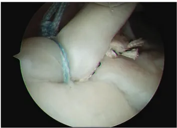
After preparation of the footprint with a shaver, an ante-rosuperior portal was created, and a smooth plastic cannula (Universal Cannula, Linvatec, Largo, Florida) was intro-duced. Through the cannula, a fish mouth drill guide (Lin-vatec) was placed on the midportion of the footprint of the biceps anchor, and a drill was introduced through the guide at an angle of 45° to the superior glenoid rim. After drilling, a 3.0-mm Bio-SutureTak anchor with double-loaded Fiber-Wire sutures (Arthrex, Naples, Florida) was introduced through the anterosuperior portal and inserted onto the gle-noid footprint. The suture limbs were shuttled just anterior and posterior to the biceps anchor, respectively, in a stan-dard technique with a No. 2 polydioxanone (PDS) suture (Ethicon, Somerville, New Jersey) and a Suture Hook (Lin-vatec) and tied in a simple suture configuration with the sliding SMC knot-tying technique19(Figure 1).
For the patients in whom the SLAP lesion extended to a more posterior portion of the labrum, an additional anchor was inserted through the posterosuperior lateral portal (port of Wilmington)26 after trial with a spinal needle to ensure an adequate angle of approach for anchor insertion onto the glenoid rim. The labrum was then repaired. For the patients undergoing tenotomy (Figure 2), the biceps
was cut at the junction between the superior labrum and the biceps.
After SLAP repair or tenotomy, the rotator cuff tear was addressed. Using the posterior and lateral portal as a viewing portal and the anterolateral portal as a working portal, we performed subacromial decompression and acromioplasty (limited to the impinged acromion undersurface). The cora-coacromial ligament was preserved as much as possible. The status and configuration of the rotator cuff tear were determined through the posterolateral and lateral portal, and the tear size was measured with a calibrated probe. The tear size was defined as the longest anteroposterior diameter of the cuff tear after debridement of the fibrous bur-sal tissue. In addition to appropriate release of the adherent fibrous tissue and capsular contracture to gain mobility of the retracted tendon, anterior interval slide (release of the cora-cohumeral ligament at the coracoid base) was performed if necessary. However, posterior or double interval slide was not performed.23 The footprint of the cuff on the greater Figure 1. Repair of a type II SLAP lesion. With use of a
double-loaded suture anchor, the biceps and superior labral complex were reattached on the footprint at both the anterior and posterior sides of the biceps anchor.
Figure 2. The long head of the biceps tendon. Note the fray-ing and partial tear of the biceps tendon. B, long head of the biceps tendon; H, humeral head.
tuberosity was prepared with a shaver. Bio-Corkscrew suture anchors (Arthrex) with double-loaded FiberWire sutures (Arthrex) were inserted through either the anterosuperior portal or through new portals created after trials with a spi-nal needle for an adequate angle of anchor insertion. In a sin-gle row with or without using the margin convergence technique, the cuff was repaired with a Scorpion suture passer (Arthrex) or a suture hook using the shuttle relay technique (Figure 3).
Postoperative Rehabilitation
All patients wore an abduction brace for 6 weeks after sur-gery, and pendulum exercise was begun on the first day after surgery. Self-assisted passive range of motion exer-cises were begun as tolerated 4 to 5 weeks postoperatively. Patients received instruction in these exercises from a cian before discharge, and they were supervised by a physi-cal therapist. Self-assisted active range of motion exercises were encouraged at 6 to 8 weeks postoperatively. Isotonic strengthening exercises using an elastic band were begun 12 weeks after surgery. After 6 months postoperatively, patients were permitted to return to full sports activity.
Statistical Analysis
Statistical analyses were conducted with SPSS (version 18.0, SPSS Inc, Chicago, Illinois). The Mann-Whitney U test was used to compare continuous data or ranked con-tinuous data such as the SST scores, ASES scores, UCLA scores, and range of motion between the 2 groups. Wil-coxon signed-rank tests were used to compare the preoper-ative and postoperpreoper-ative shoulder scores and the range of motion in each group. The x2test was used to compare cat-egorical data, including sex, dominant arm involvement, stage of fatty infiltration and muscle atrophy, and sports/ recreation activity between the 2 groups. The level of sta-tistical significance was set at P \ .05. Data are reported as mean 6 standard deviation.
RESULTS
Patient Demographics
Group R included 7 men and 9 women, and group T included 9 men and 11 women. The mean age at the time of surgery was 61.1 6 5.1 years in group R and 63.3 6 6.0 years in group T. The mean time period between symp-tom onset and surgery was 21.2 months (range, 6-72) in group R and 24.5 months (range, 6-60) in group T. The dominant arm was involved for 13 patients (81%) in group R and 16 patients (80%) in group T (Table 1). None of these characteristics were significantly different between the 2 groups.
Radiological Assessments
Preoperative Stage of Muscle Atrophy and Fatty Infiltra-tion on MRI. Based on the MRI findings for the most
lateral oblique-sagittal T1-weighted view (ie, ‘‘Y-shaped view’’), 4 patients in group R had stage 2, 9 had stage 3, and 3 had stage 4 in the supraspinatus; 8 had stage 2, 6 had stage 3, and 2 had stage 4 in the infraspinatus. Six patients in group T had stage 2, 10 had stage 3, and 4 had stage 4 in the supraspinatus; 11 had stage 2, 6 had stage 3, and 3 had stage 4 in the infraspinatus. There was no significant difference between the 2 groups (supra-spinatus, P = .926; infra(supra-spinatus, P = .890).
Acromiohumeral Distance. The mean preoperative acro-miohumeral distance was 8.9 6 0.9 mm in group R and 8.8 6 1.0 mm in group T. There was no significant differ-ence between the groups (P = .582). At the 2-year follow-up, the mean acromiohumeral distance was 8.6 6 1.0 mm in group R and 8.7 6 1.1 mm in group T. There was no sig-nificant difference between the groups (P = .440). Within each group, there were no significant differences between the preoperative and postoperative acromiohumeral dis-tance (group R, P = .083; group T, P = .102).
Arthroscopic Findings
The mean tear size (ie, the longest anteroposterior diame-ter) was 37.1 mm (range, 30-55) in group R and 38.4 mm (range, 30-55) in group T; there was no significant differ-ence between the groups (P = .460). In group R, a mean of 1.3 (range, 1-2) suture anchors were used for the SLAP repair. In 12 patients (75%), the type II SLAP lesion was repaired with a single suture anchor with double-loaded sutures. In 4 patients (25%), an additional anchor was inserted at the 10- to 11-o’clock position (in the right shoul-der) or the 1- to 2-o’clock position (in the left shoulshoul-der) to repair the posterior extension of the SLAP lesion. In group T, in addition to the SLAP lesion, the fraying or partial tear in the biceps tendon was identified, and biceps tenot-omy was performed. Anterior interval slide was performed in 12 patients (75%) in group R and 14 patients (70%) in group T. The cuff could be repaired onto the tuberosity in 6 patients (37%) in group R and 7 (35%) in group T. For the remaining patients, the cuff was repaired at the artic-ular margin.
Clinical Outcomes and Range of Motion
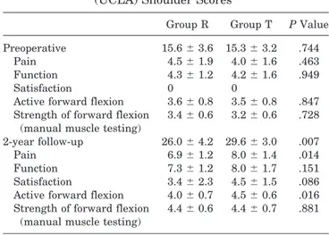
The mean preoperative SST score was 5.0 6 1.3 in group R and 4.6 6 1.1 in group T (P = .757). At the 2-year follow-up, the mean SST score improved to 7.8 6 1.9 in group R (P \ .001) and 9.3 6 1.6 in group T (P \ .001); the postoperative difference between the groups was significant (P = .012). The mean preoperative ASES score was 40.7 6 8.5 in group R and 38.7 6 8.2 in group T (P = .467). At the 2-year follow-up, the ASES score improved to 80.4 6 8.9 in group R (P \ .001) and 88.6 6 8.9 in group T (P \ .001); the postoperative difference was significant between the groups (P = .009) (Table 2). The UCLA score also improved significantly after surgery, from 15.6 6 3.6 to 26.0 6 4.2 in group R (P \ .001) and from 15.3 6 3.2 to 29.6 6 3.0 in group T (P \ .001). While the preoperative difference was not significant (P = .744), the postoperative difference was significant between the groups (P = .007) (Table 3).
Vol. 40, No. 12, 2012 SLAP Repair Versus Biceps Tenotomy 2789
at YONSEI UNIV LIBRARY on July 14, 2013
ajs.sagepub.com
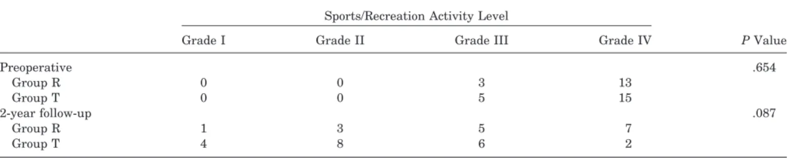
Regarding preoperative sports/recreation activity level, in group R, there were 3 patients with activity grade III and 13 with grade IV; in group T, there were 5 with grade III and 15 with grade IV activity levels. Neither group had patients with grade I or II preoperatively, and there was no signifi-cant difference between groups (P = .654). At the 2-year follow-up, group R had 1 patient with grade I, 3 with grade II, 5 with grade III, and 7 with grade IV activity; group T had 4 with grade I, 8 with grade II, 6 with grade III, and 2 patients with grade IV activity. There was no significant dif-ference between groups (P = .087) (Table 4).
With regard to the active range of motion, the mean for-ward flexion improved significantly, from 119.4° 6 10.6° to 132.5° 6 13.5° in group R (P = .022) and from 117.0° 6 10.1° to 145.9° 6 13.0° in group T (P \ .001) at the 2-year follow-up. While the preoperative difference was not significant (P = .496), the postoperative difference between the groups was significant (P = .004). The mean external rotation with the arm at the side also improved significantly, from 41.6° 6 7.0° to 52.2° 6 9.5° in group R (P = .001) and from 42.0° 6 9.7° to 60.0° 6 6.8° in group T (P \ .001). While the pre-operative difference was not significant (P = .988), the post-operative difference between the groups was significant (P = .009). The mean internal rotation also improved signifi-cantly, from 12.2° 6 2.9° to 10.0° 6 2.6° in group R (P = .003) and from 12.2° 6 2.7° to 9.2° 6 2.4° in group T (P = .001). Both preoperative and postoperative differences between the groups were not significant (P = .966, preoper-ative; P = .233, postoperative) (Table 5). In group T, 3 of the 20 patients (15%) developed the Popeye deformity, but no patients had cramping pain in the affected arm.
DISCUSSION
The purpose of this retrospective study was to compare functional outcomes between patients undergoing repair or tenotomy for concomitant type II SLAP lesions and large to massive rotator cuff tears. In combined type II SLAP lesions with rotator cuff tears that had only minimal mus-cle atrophy, fatty infiltration, or isolated supraspinatus tears with minimal retraction, most investigators have reported satisfactory outcomes after simultaneous repair of both lesions comparing preoperative pain and function, although Abbot et al1 and Franceschi et al12 reported biceps tenotomy or debridement for the concomitant SLAP lesion was better than repair.11However, the out-comes of treatment for concomitant SLAP lesions and large to massive rotator cuff tears in patients have not been investigated, so it is unclear whether the favorable out-comes associated with simultaneous repair of both lesions can be expected in this population.
Although many authors have reported satisfactory improvements after repair of large to massive rotator cuff tears, these outcomes cannot be comparable with those of repair for small to medium-sized tears with relatively TABLE 2
Simple Shoulder Test (SST) and American Shoulder and Elbow Surgeons (ASES) Scoresa
Group R Group T P Value SST score Preoperative 5.0 6 1.3 4.6 6 1.1 .757 2-year follow-up 7.8 6 1.9 9.3 6 1.6 .012 ASES score Preoperative 40.7 6 8.5 38.7 6 8.2 .467 2-year follow-up 80.4 6 8.9 88.6 6 8.9 .009
aValues are expressed as mean 6 standard deviation. Group R,
simultaneous repair for type II SLAP lesion and rotator cuff tear; group T, tenotomy for concomitant type II SLAP lesion and repair for rotator cuff tear.
TABLE 3
University of California, Los Angeles (UCLA) Shoulder Scoresa
Group R Group T P Value Preoperative 15.6 6 3.6 15.3 6 3.2 .744
Pain 4.5 6 1.9 4.0 6 1.6 .463 Function 4.3 6 1.2 4.2 6 1.6 .949 Satisfaction 0 0
Active forward flexion 3.6 6 0.8 3.5 6 0.8 .847 Strength of forward flexion
(manual muscle testing)
3.4 6 0.6 3.2 6 0.6 .728 2-year follow-up 26.0 6 4.2 29.6 6 3.0 .007 Pain 6.9 6 1.2 8.0 6 1.4 .014 Function 7.3 6 1.2 8.0 6 1.7 .151 Satisfaction 3.4 6 2.3 4.5 6 1.5 .086 Active forward flexion 4.0 6 0.7 4.5 6 0.6 .016 Strength of forward flexion
(manual muscle testing)
4.4 6 0.6 4.4 6 0.7 .881
aValues are expressed as mean 6 standard deviation. Group R,
simultaneous repair for type II SLAP lesion and rotator cuff tear; group T, tenotomy for concomitant type II SLAP lesion and repair for rotator cuff tear.
TABLE 1 Patient Demographicsa
Group R (n = 16) Group T (n = 20) P Value Sex, male/female, n 7/9 9/11 .942 Age, mean 6 SD, y 61.1 6 5.1 63.3 6 6.0 .238 Symptom period before surgery, mean (range), mo 21.2 (6-72) 24.5 (6-60) .210 Dominant arm involvement, n (%) 13 (81) 16 (80) .932
aGroup R, simultaneous repair for type II SLAP lesion and rotator cuff tear; group T, tenotomy for concomitant type II SLAP lesion and
healthy and robust cuff muscle.7,14,16,17,22,36Large to mas-sive rotator cuff tears also have relatively high retear rates, and although a re-established transverse force cou-ple should help improve functional outcomes in spite of retears, these situations may put the biceps under an unusual load. Given that biceps lesions are often associ-ated with large to massive rotator cuff tears and are responsible for patients’ pain and disability,8,34we hypoth-esized that the biceps tendon of the repaired SLAP lesion might have a detrimental effect on overall outcomes in large to massive rotator cuff repair, even when the biceps appears healthy at the time of surgery.
In the current study, we attempted to preserve the biceps by repairing the concomitant SLAP lesion and rota-tor cuff tear if the biceps tendon appeared to be robust (ie, without any tears and fraying) during the arthroscopic assessment. However, if the SLAP lesion had a coexisting biceps lesion, we performed tenotomy regardless of the degree of fraying and tearing of the biceps. Some investiga-tors have suggested a partial biceps tendon tear of \25% is best treated with simple debridement for the unstable fiber
and preservation of the biceps.2,21However, when consid-ering the patients’ age and the severity of the rotator cuff tears in our study, we suspected that the diseased biceps tendon would likely cause problems in the future, and we chose to cut the biceps tendon rather than preserve it.
The biceps–superior labral complex functions as a humeral head depressor, superior stabilizer, and resister to torsion and strain from the inferior glenohumeral liga-ment, and these functions may be reduced by cutting the biceps.5,31,35 However, it is unclear whether preserving the functional role of the biceps should be the highest pri-ority when treating patients with large to massive rotator cuff tears. Rather, it may be more beneficial to cut the dis-eased biceps tendon to relieve the patients’ pain rather than preserve the biceps and its functional role.
While rotator cuff repair generally leads to successful outcomes, the repair of large to massive rotator cuff tears seems to have less favorable outcomes with relatively high retear rates.14,16,25Furthermore, many investigators report that well-healed rotator cuffs have better clinical outcomes than retears,15,18,25,29 even when clinical improvement for the patients with retears is achieved post-operatively. Given that large to massive rotator cuff tears are more often accompanied by advanced muscle atrophy and fatty infiltration and their outcomes are less favorable than those of small to medium-sized tears, we hypothe-sized that simultaneous repair of the SLAP lesion and cuff tear might not be beneficial.
It seems likely that the inferior outcomes of patients undergoing SLAP repair (ie, group R) can be attributed to the repair of the SLAP lesion because the group did not otherwise differ in demographics, shoulder functional scores, range of motion, degree of muscle atrophy and fatty infiltration, or acromiohumeral distances before surgery. Rather, the difference between groups was the method used to address the SLAP lesion, which suggests that the technique used to repair the SLAP lesion might not be opti-mal. We prefer the technique in which an anchor with double-loaded sutures is inserted at the midportion of the footprint of the biceps anchor and reattached to the supe-rior labrum both antesupe-riorly and postesupe-riorly to the biceps anchor in a simple suture configuration. When the SLAP TABLE 4
Preoperative and Postoperative Sports/Recreation Activity Scoresa
Sports/Recreation Activity Level
Grade I Grade II Grade III Grade IV P Value
Preoperative .654 Group R 0 0 3 13 Group T 0 0 5 15 2-year follow-up .087 Group R 1 3 5 7 Group T 4 8 6 2 a
Grade I, no limitation in sports/recreation activity (100% of premorbid level); grade II, mild limitation in sports/recreation activity (.90%); grade III, moderate limitation in sports/recreation activity (.70% of premorbid level); grade IV, severe limitation (\70% of premor-bid level) or inability to return to previous sports/recreation activity. Group R, simultaneous repair for type II SLAP lesion and rotator cuff tear; group T, tenotomy for concomitant type II SLAP lesion and repair for rotator cuff tear.
TABLE 5 Active Range of Motiona
Group R Group T P Value Forward flexion, deg
Preoperative 119.4 6 10.6 117.0 6 10.1 .496 2-year follow-up 132.5 6 13.5 145.9 613.0 .004 External rotation, deg
Preoperative 41.6 6 7.0 42.0 6 9.7 .988 2-year follow-up 52.2 6 9.5 60.0 6 6.8 .009 Internal rotation, deg
Preoperative 12.2 6 2.9 12.2 6 2.7 .966 2-year follow-up 10.0 6 2.6 9.2 6 2.4 .233
aValues are expressed as mean 6 standard deviation. For
inter-nal rotation, the spiinter-nal segments as reference were converted into continuous numbers: T1-12 = 1-12, L1-5 = 13-17, and sacrum = 18. Group R, simultaneous repair for type II SLAP lesion and rotator cuff tear; group T, tenotomy for concomitant type II SLAP lesion and repair for rotator cuff tear.
Vol. 40, No. 12, 2012 SLAP Repair Versus Biceps Tenotomy 2791
at YONSEI UNIV LIBRARY on July 14, 2013
ajs.sagepub.com
lesion extends in a more posterior direction, we prefer to insert an additional anchor and repair the lesion using the same simple suture configuration. Although there are several different techniques for SLAP lesion repair, and any of them could affect outcomes, this possibility was beyond the scope of the current study.
Our study has several limitations. First, the assignment of patients into groups was not randomized. Instead, group assignments were based on whether the arthroscopic find-ings indicated that the biceps was robust and healthy. Therefore, 2 groups had different injuries; group R had a robust biceps tendon, and group T had a biceps tendon lesion. This was an inherent potential bias and may have contributed to the different postoperative outcomes, even though the preoperative values between groups were not significantly different. Second, we measured the muscle strength manually, not with a dynamometer. Although an independent examiner measured and compared the strength of the affected arm with that of the contralateral side, our results may have been more accurate if we evalu-ated strength with a dynamometer. Third, our sample size was relatively small, so future studies will be needed with a larger sample size. Finally, we could not analyze the data for the follow-up MRI findings because the number of follow-up MRI scans was too small to compare outcomes. If we could have compared preoperative and postoperative MRI findings, our results would have been stronger.
In conclusion, at the 2-year follow-up after surgery for concomitant type II SLAP lesions and large to massive rotator cuff tears in patients, the outcomes of simultaneous arthroscopic SLAP and cuff repair were less satisfactory than those of arthroscopic biceps tenotomy and rotator cuff repair in terms of functional shoulder scores and range of motion, although both groups experienced significant improvements after surgery. Biceps tenotomy combined with cuff repair may be a more reliable method to address concomitant type II SLAP lesions and large to massive rotator cuff tears in patients, although a randomized con-trolled trial is needed to confirm this possibility.
ACKNOWLEDGMENT
The authors are grateful to Young-Jun Cho for his help with the figures.
REFERENCES
1. Abbot AE, Li X, Busconi BD. Arthroscopic treatment of concomitant superior labral anterior posterior (SLAP) lesions and rotator cuff tears in patients over the age of 45 years. Am J Sports Med. 2009;37:1358-1362.
2. Ahmad CS, ElAttrache NS. Arthroscopic biceps tenodesis. Orthop
Clin North Am. 2003;34:499-506.
3. Alpert JM, Wuerz TH, O’Donnell TF, Carroll KM, Brucker NN, Gill TJ. The effect of age on the outcomes of arthroscopic repair of type II superior labral anterior and posterior lesions. Am J Sports Med. 2010;38:2299-2303.
4. Andrews JR, Carson WG Jr, McLeod WD. Glenoid labrum tears related to the long head of the biceps. Am J Sports Med. 1985;13:337-341.
5. Barber FA, Byrd JW, Wolf EM, Burkhart SS. How would you treat the partially torn biceps tendon? Arthroscopy. 2001;17:636-639. 6. Bartl C, Scheibel M, Magosch P, Lichtenberg S, Habermeyer P. Open
repair of isolated traumatic subscapularis tendon tears. Am J Sports
Med. 2011;39:490-496.
7. Bjorkenheim JM, Paavolainen P, Ahovuo J, Slatis P. Surgical repair of the rotator cuff and surrounding tissues: factors influencing the results. Clin Orthop Relat Res. 1988;236:148-153.
8. Boileau P, Baque F, Valerio L, Ahrens P, Chuinard C, Trojani C. Iso-lated arthroscopic biceps tenotomy or tenodesis improves symp-toms in patients with massive irreparable rotator cuff tears. J Bone
Joint Surg Am. 2007;89:747-757.
9. Brockmeier SF, Voos JE, Williams RJ 3rd, Altchek DW, Cordasco FA, Allen AA. Outcomes after arthroscopic repair of type-II SLAP lesions.
J Bone Joint Surg Am. 2009;91:1595-1603.
10. Cofield RH, Parvizi J, Hoffmeyer PJ, Lanzer WL, Ilstrup DM, Rowland CM. Surgical repair of chronic rotator cuff tears: a prospective long-term study. J Bone Joint Surg Am. 2001;83:71-77.
11. Forsythe B, Guss D, Anthony SG, Martin SD. Concomitant arthro-scopic SLAP and rotator cuff repair. J Bone Joint Surg Am. 2010;92:1362-1369.
12. Franceschi F, Longo UG, Ruzzini L, Rizzello G, Maffulli N, Denaro V. No advantages in repairing a type II superior labrum anterior and posterior (SLAP) lesion when associated with rotator cuff repair in patients over age 50: a randomized controlled trial. Am J Sports
Med. 2008;36:247-253.
13. Friel NA, Karas V, Slabaugh MA, Cole BJ. Outcomes of type II supe-rior labrum, antesupe-rior to postesupe-rior (SLAP) repair: prospective evalua-tion at a minimum two-year follow-up. J Shoulder Elbow Surg. 2010;19:859-867.
14. Galatz LM, Ball CM, Teefey SA, Middleton WD, Yamaguchi K. The outcome and repair integrity of completely arthroscopically repaired large and massive rotator cuff tears. J Bone Joint Surg Am. 2004;86:219-224.
15. Gazielly DF, Gleyze P, Montagnon C. Functional and anatomical results after rotator cuff repair. Clin Orthop Relat Res. 1994;304:43-53.
16. Gerber C, Fuchs B, Hodler J. The results of repair of massive tears of the rotator cuff. J Bone Joint Surg Am. 2000;82:505-515.
17. Hanusch BC, Goodchild L, Finn P, Rangan A. Large and massive tears of the rotator cuff: functional outcome and integrity of the repair after a mini-open procedure. J Bone Joint Surg Br. 2009;91:201-205. 18. Harryman DT 2nd, Mack LA, Wang KY, Jackins SE, Richardson ML, Matsen FA 3rd. Repairs of the rotator cuff: correlation of functional results with integrity of the cuff. J Bone Joint Surg Am. 1991;73:982-989.
19. Kim SH, Ha KI. The SMC knot: a new slip knot with locking mecha-nism. Arthroscopy. 2000;16:563-565.
20. Kim SH, Ha KI, Choi HJ. Results of arthroscopic treatment of superior labral lesions. J Bone Joint Surg Am. 2002;84:981-985.
21. Koh KH, Ahn JH, Kim SM, Yoo JC. Treatment of biceps tendon lesions in the setting of rotator cuff tears: prospective cohort study of tenotomy versus tenodesis. Am J Sports Med. 2010;38:1584-1590.
22. Lafosse L, Brozska R, Toussaint B, Gobezie R. The outcome and structural integrity of arthroscopic rotator cuff repair with use of the double-row suture anchor technique. J Bone Joint Surg Am. 2007;89:1533-1541.
23. Lo IK, Burkhart SS. Arthroscopic repair of massive, contracted, immobile rotator cuff tears using single and double interval slides: technique and preliminary results. Arthroscopy. 2004;20:22-33. 24. Mellado JM, Calmet J, Olona M, et al. Surgically repaired massive
rotator cuff tears: MRI of tendon integrity, muscle fatty degeneration, and muscle atrophy correlated with intraoperative and clinical find-ings. AJR Am J Roentgenol. 2005;184:1456-1463.
25. Miller BS, Downie BK, Kohen RB, et al. When do rotator cuff repairs fail? Serial ultrasound examination after arthroscopic repair of large and massive rotator cuff tears. Am J Sports Med. 2011;39:2064-2070.
26. Morgan CD, Burkhart SS, Palmeri M, Gillespie M. Type II SLAP lesions: three subtypes and their relationships to superior instability and rotator cuff tears. Arthroscopy. 1998;14:553-565.
27. Nam EK, Snyder SJ. The diagnosis and treatment of superior labrum, anterior and posterior (SLAP) lesions. Am J Sports Med.
2003;31:798-810.
28. Neuman BJ, Boisvert CB, Reiter B, Lawson K, Ciccotti MG, Cohen SB. Results of arthroscopic repair of type II superior labral anterior poste-rior lesions in overhead athletes: assessment of return to preinjury playing level and satisfaction. Am J Sports Med. 2011;39:1883-1888. 29. Nho SJ, Adler RS, Tomlinson DP, et al. Arthroscopic rotator cuff repair: prospective evaluation with sequential ultrasonography. Am
J Sports Med. 2009;37:1938-1945.
30. Oh JH, Kim SH, Shin SH, Chung SW, Kim JY, Kim SJ. Outcome of rotator cuff repair in large-to-massive tear with pseudoparalysis: a comparative study with propensity score matching. Am J Sports
Med. 2011;39:1413-1420.
31. Rodosky MW, Harner CD, Fu FH. The role of the long head of the biceps muscle and superior glenoid labrum in anterior stability of the shoulder. Am J Sports Med. 1994;22:121-130.
32. Snyder SJ, Karzel RP, Del Pizzo W, Ferkel RD, Friedman MJ. SLAP lesions of the shoulder. Arthroscopy. 1990;6:274-279.
33. Szabo I, Boileau P, Walch G. The proximal biceps as a pain generator and results of tenotomy. Sports Med Arthrosc. 2008;16:180-186. 34. Walch G, Edwards TB, Boulahia A, Nove-Josserand L, Neyton L,
Szabo I. Arthroscopic tenotomy of the long head of the biceps in the treatment of rotator cuff tears: clinical and radiographic results of 307 cases. J Shoulder Elbow Surg. 2005;14:238-246.
35. Warner JJ, McMahon PJ. The role of the long head of the biceps bra-chii in superior stability of the glenohumeral joint. J Bone Joint Surg
Am. 1995;77:366-372.
36. Zumstein MA, Jost B, Hempel J, Hodler J, Gerber C. The clinical and structural long-term results of open repair of massive tears of the rotator cuff. J Bone Joint Surg Am. 2008;90:2423-2431.
For reprints and permission queries, please visit SAGE’s Web site at http://www.sagepub.com/journalsPermissions.nav
Vol. 40, No. 12, 2012 SLAP Repair Versus Biceps Tenotomy 2793
at YONSEI UNIV LIBRARY on July 14, 2013
ajs.sagepub.com