- 107 -
Serially Passaged Normal Human Nasal Epithelial Cells:
:
:
:
Morphology and Mucous Secretory Differentiation
Hwan-Jung Roh, M.D.
1, Eui-Kyung Goh, M.D.
1, Soo-Geun Wang, M.D.
1,
Kyong-Myong Chon, M.D.
1, Joo-Heon Yoon, M.D.
2and Yoo-Sun Kim, M.D.
3ABSTRACT
Background and Objectives:The purpose of this study was to develop a subculturing technique that allows the formation
of large amounts of normal human nasal epithelial (NHNE) cells without compromising the cells’ ability to differentiate into secretory and ciliated cells. Materials and Methods:Freshly isolated nasal epithelial cells, collected from normal inferior tur-binates, were subcultured repeatedly in a serum-free medium on plastic culture dishes. The subcultured cells were tested for growth curve and electromicroscopic characteristics in air-liquid interface (ALI) cultures and for mucous secretory differentiation.
Results:The cultures grew rapidly during the first three passages, demonstrating a 20- to 40-fold expansion with each subculture.
Ciliogenesis usually started on day 12 and the cultured cells formed cellular sheets exhibiting microvilli in the apical membrane, intracytoplasmic secretory granules, electron lucent, scant endoplasmic reticulum and complex tight junctions on the basolateral side. Passage-1 (P-1) and passage-2 (P-2) cells maintained their potential to differentiate into mucin-secretory and ciliated epi-thelial cells, and this potential was confirmed by immunocytochemistry with H6C5 and transmission electron microscopy. Alt-hough the number of NHNE cells on day 16 of culture decreased as the passage progressed from P-1 to P-3, the relative number of secretory cells did not significantly change. Conclusion:In conclusion, P-2 NHNE cell cultures retain the features of normal epithelium and are suitable for conducting many studies on upper airway cell biology.
KEY WORDS:Human nasal epithelial cells·Subculture·Mucin·Morphology.
INTRODUCTION
Culturing airway epithelial cells is a very useful me-ans of studying the effects of environmental pollution, viral and bacterial infections, and chemical carcinogens on the functions of differentiated airway epithelial cells. At present, there are many kinds of epithelial cell cult-ure methods:submerged,1) suspension,2) floating3) and air-liquid interface (ALI)4-7) techniques. Until now, most studies have used animal airway epithelial cells collected from guinea pigs,8) rats9) and hamsters.10) Some studies
have used primary culture cells taken from human air-way epithelium, but using a primary culture from human airway epithelial cells involves problems related to such factors as the limited amount of material available from one donor, contamination with pathogens and significant donor-to-donor variability. Therefore, instead of normal human specimens, tumor cells, which are relatively easy to grow, or cell lines such as BEAS-2B or 9HTE have often been used for studies on cellular response or phy-siologic characteristics. However, there remains a cont-inuing demand for normal human airway epithelial cell cultures due to the fact that tumor cells cannot appropr-iately represent the physiologic characteristics and cell-ular responses of normal airway epithelial cells. In 1996, Gray et al,11) succeeded in subculturing passage-1 nor-mal human tracheobronchial epithelial cells and compl-eted their characterization.
Although the incidence of airway disease is much hi-gher in the upper airway than in the lower airway, only minor progress has been made in the field of research on upper airway epithelial cells. We wanted to obtain enough normal human nasal epithelial cells to conduct *Supported by a grant from the Pusan National University
Hospital Research Fund (PNUH 97-05)
1Department of Otorhinolaryngology, College of medicine,
Pusan National University, Pusan, 2Otorhinolaryngology,
Col-lege of Medicine, Yonsei University ColCol-lege of Medicine, Seoul,
3Medical Research Institue of Pusan National University
Ho-spital, Pusan, Korea
Address correspondences and reprint requests to Hwan-Jung Roh, MD, Department of Otohinolaryngology, College of Me-dicine, Pusan National University, Pusan 602-739, Korea Tel:82-51-248-1248, Fax:82-51-240-7333
E-mail:[email protected] Accepted for publication on September 19, 1999
studies on upper airway cell biology. The purposes of this study were 1) to develop a subculturing technique that allows the formation of large amounts of normal human nasal epithelial (NHNE) cells without compro-mising their ability to differentiate into secretory and ciliated cells and 2) to study mucous secretory differen-tiation with immunocytochemistry and morphological ch-aracteristics with transmission electron microscopy.
MATERIALS AND METHODS
Expansion, subculturing and cryopreservation
of NHNE cells
Human nasal epithelial cells were obtained during rgery from the inferior turbinate mucosa of patients su-ffering from septal deviation or chronic sinusitis. The tissues were treated with 1.0% Pronase (type XIV pro-tease, Sigma, St. Louis, MO, USA) in a 1:1 mixture of Dulbecco’s modified Eagle’s medium and Ham’s nutri-ent F12 (DMEM/F12) supplemnutri-ented with penicillin (50 IU/mL) and streptomycin (50 μg/mL) for 16 to 20 hours at 4℃. Dissociated epithelial cells were washed three times in DMEM/F12 containing antibiotics and suspended in DMEM/F12 supplemented with antibiotics and 10% fetal bovine serum. Cells were preplated on a plastic dish at 37℃ for 1 hour in order to eliminate fi-broblasts, endothelial cells and myoblasts by differential attachment to plastic. Suspended epithelial cells were seeded at 3×10⁴ cells/dish (500 cells/cm2) in 10cm, pl-astic tissue culture dishes (passage-1). The culture me-dium was a bronchial epithelial growth meme-dium (BEGM; Clonetics Corp., San Diego, CA) containing hydrocor-tisone (0.5 μg/ml), insulin (5 μg/ml), transferrin (10 μg/ml), epinephrine (0.5 μg/ml), triiodothyronine (6.5 ng/ml), gentamycin (50 μg/ml), and amphotericin B (50 μg/ml), all supplied by Clonetics, and further su-pplemented with epidermal growth factor (25 ng/ml; Collaborative Research, Bedford, MA), all-trans retinoic acid (10-7M;Sigma Chemical Co., St. Louis, MO), bo-vine serum albumin (1.5 μg/ml;Sigma), and bobo-vine pituitary extract (1% vol/vol; Pel-Freez Biologicals, Rogers, AR). Cultures were maintained at 37℃ in an atmosphere of 5% CO2 in air. The culture medium was changed on day 1 after seeding and every other day th-ereafter until the cultures reached 50-60% confluency, at which time they were dissociated with trypsin/EDTA treatment using the methods and reagents supplied by
Clonetics Corp.
Suspended epithlial cells were seeded at 3×10⁴ cells/ dish (500 cells/cm2) in 10cm, plastic tissue culture dis-hes (passage-2). At passage-2, aliquots of cells not used for subculturing and reestablishing cultures were either suspended in supplemented BEGM containing 10% di-methyl sulfoxide at 1 to 2×10⁶ cells/ml and stored fro-zen in liquid nitrogen for future use or tested for their differentiation competence. When the cultures reached 50-60% confluency, they were subcultured (passage-3). The subculture was seeded as described above for subsequent passage (passage-4). The cells were obser-ved under contrast microscopy and photographed serially, and the cell number was determined with a hemocyto-meter.
ALI culture
Serially passaged NHNE cells (105 cells/culture, 2× 10⁴ cells/cm2) were seeded in 0.5ml of culture medium onto the surface of rat tail, collagen type 1 gel-coated (3.0 mg/ml;Collaborative Research), 24.5 mm, 0.45 μm pore size, Transwell-clear (Costar Co., Cambridge, MA) culture inserts. The cells were cultured in a 1:1 mixture of BEGM:DMEM containing the same conc-entrations of all supplements described above except 0.5 ng/ml of EGF was used. The cultures were grown sub-merged for the first 7 days, during which time the cult-ure medium was changed on day 1 and every other day thereafter. The ALI was created on day 7 by removing the apical medium and feeding the cultures only from the basal compartment. The culture medium was changed daily after the creation of an ALI. To dissociate cells grown on collagen gels in ALI cultures, the culture me-dium was removed and both the apical and basal comp-artment were washed with 1.5 ml of phosphate buffered saline (PBS). One millimeter of a 3×trypsin-EDTA (10 ×stock;GIBCO-BRL, Grand Island, NY) was added to the apical compartment. Cultures were incubated at 37℃ until cell detachment occurred. The cells were then resuspended in PBS, visually counted with a hemocyt-ometer and placed in cytospin slides for immunocytoc-hemical staining.
Immunocytochemistry
Cytospin slides were made using 2×10⁴ cells per sl-ide and fixed in a 1:1 mixture of acetone and methanol
and stored at 4℃ until stained. Immunocytochemical staining for detecting secretory cells were performed. Briefly, a primary antibody, a mouse monoclonal anti-mucin antibody (H6C5),7) was applied on the slides overnight at 4℃.
The slides were then rinsed with Ca/Mg-free PBS be-fore a 1-hr incubation with a secondary antibody (Bio-tinylated horse anti-mouse IgG:Vector Laboratories, Burlingame, CA). After an incubation in methanol/H2 O2, conducted to inhibit endogenous peroxidase, and Ca/Mg-free PBS rinses, they were incubated for 1 hr at room temperature in avidin-biotinperoxidase complex (Vector Laboratories, Burlingame, CA). The peroxidase reaction was developed by preincubation with 0.05% 3,3-diaminobenzidine (DAB) with 0.1 M sodium azide in Ca/Mg-free PBS for 10 min, followed by incubation with a Ca/Mg-free PBS solution containing 0.05% DAB, 0.1% H2O2, 0.1 M imidazole, and 0.1 M sodium azide for 5 min, and a final incubation with 0.5% CuSO4 for 5 min. The mean number of positive cells with H6C5 antibody was determined by scoring 3,000 cells on each slide, and statistical comparisons were made with a St-udent’s t-test.
Transmission electron microscopy
For electron microcopy, the cells on the transwell in-serts were gently washed with PBS and fixed with 2.5% glutaldehyde at 4℃ for 4-6 hr and treated with 1% OsO4 (osmium teroxide) at 4℃ for 1 hr. They were su-bsequently dehydrated in graded ethanols and embedded
in Epon 812. Semithin secretion (80 nm in thickness) were stained with toluidine blue and observed by light microscopy. Appropriate areas were selected and ultra-thin secretions were made. They were treated with ura-nyl acetate at room temperature for 6 min and lead citr-ate for 3 min. These were studied under a JEM 1200 EX II electron microscope.
RESULTS
Expansion of NHNE cells
Within 24 hr of primary culture, epithelial cells adh-ered to the culture dish. They then formed cellular islands at 3 days and thereafter expanded rapidly to cover the culture dish. By the 7th day they formed a confluent mo-nolayer (passage-1, Fig. 1A). The cultures grew rapidly for the first three passages, demonstrating a 20- to 40-fold expansion with each subculture. However, passage-4 cells grew very poorly and there was a large vacuole in each cell. In total, the original cell inoculum under-went more than a 500-fold expansion, including pass-age-3 cells.
Growth curve of passage-2 NHNE cells in ALI
cultures as a function of time
Passage-2 cells were seeded on permeable membranes at a seeding density of 105 cells/membrane, i.e., 2×10⁴ cells/cm2 in a 1:1 mixture of BEGM and DMEM. The cells grew rapidly and reached 90% confluency on day
Fig. 1. Phase contrast micrographs of the cultured nasal epithelial cells at 7 th day of passage-1 (A) and passage-2 cells in ALI cultures (B).
A. The primary cutured cells of 7 th day showed the typical shapeof epithelial cells (×200). B. Passage-2 cells were seeded on perm-eable membranes at a seeding density of 105 cells/membrane. The cells proliferated rapidly and reached 90% confluency on day 9
and formed multilayered pattern (×200).
B BB B A A A A
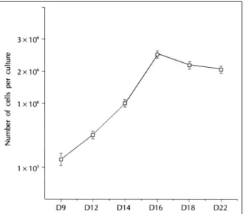
9 (Fig. 1B). ALI was created on day 9 for all subsequent experiments. The number of cells increased in a time-dependent manner between days 9 and 16 and thereafter decreased (Fig. 2). Peak cell densities of >2×10⁶ cells per culture between days 16 and 22 were noted.
Mucin-secretory cell differentiation of serially
passaged NHNE cells on day 16 culture
The cytospin slides for immunostaining were made on day 16. The mean number of positive cells with an anti-mucin antibody (H6C5) was determined by scoring 3,000 cells on each slide using passaged cells. The nu-mber of cells on day 16 using passage-1, -2, and -3 cells were 4.1±0.4 (×106), 2.6±0.5 (×106) and 1.9±0.3 (×106), respectively. The number of cells decreased slightly with succeeding passages and there was a stati-stically significant difference in cell number between passage -1 and passage -3. However, there was no sign-ificant change in secretary cell percentage among each Table 1. Growth and secretory cell differentiation of serially
pa-ssaged normal human nasal epithelial cells on day 16 culture Passage Number of cells per culture
(×106) Secretory cells (%)
p-1 4.1±0.4* 34.3±3.1 p-2 2.6±0.5 36.6±1.5 p-3 1.9±0.3* 32.9±2.4 p-4 cells did not grow
Secretory cells:anti-mucin antibody (H6C5)-positive cells *p<0.05 between passage-1 and passage-3
Fig. 2. Growth curve of passage-2 normal human nasal
epith-elial cells in air-liquid culture. 105 cells were plated in BEGM:
DMEM(1:1);duplicate cultures per time points.
Fig. 3. Transmission electron micrographs of human nasal epithelial cells on 12 days after air-liquid interface culture.
A. The cultured cells formed cellular sheets exhibiting cilia in the apical membrane, intracytoplasmic secretory granules and electron lucent and tight junctions in the basolateral side (×5,000). B. Apical membrane contained microvilli and basolateral membranes were complex featuring numerous interdigitations between adjacent cells. Some myelin figures and intracytoplasmic vacuoles were obs-erved (×8,000). B B B B A A A A
passage (Table 1). Passage-4 cells attached to plastic dishes, but failed to grow.
Transmission electron microscopy
Ciliogenesis, confirmed by transmission electron mi-croscopy, usually started on day 12 in some areas in which focal high cell density existed (Fig. 3A). The cu-ltured cells formed cellular sheets exhibiting microvilli in the apical membrane, intracytoplamic secretory gra-nules, electron lucent, scant endoplasmic reticulum and complex tight junctions in the basolateral side featuring numerous interdigitations between adjacent cells (Fig. 3B). Some myelin figures and intracytoplasmic vacuo-les were also observed.
DISCUSSION
Techniques for culturing the airway epithelium have improved over the last ten years. Owing to developments such as a serum-free hormone-supplemented culture dium, collagen that serves as growth supports, filter me-mbranes and culture dishes suitable for epithelial cell culture and an air interface method, it is presently poss-ible to induce the differentiation of airway epithelial cells with similar morphologic, physiologic as well as bioch-emical characteristics into in vivo airway epithelial cells. However, most studies to date have been conducted with animal airway epithelial cells or with primary cultures that were limited by such factors as the small amount of material available, contamination and donor to donor variability. Currently the main drawback of research on human airway epithelial cells comes from the difficulty of obtaining a sufficient number of epithelial cells with the potential to differentiate according to the patterns of normal epithelial cells in vivo. This limitation can be overcome by subculturing the airway epithelial cells wh-ile maintaining their ability to differentiate into secretory and ciliated epithelial cells.
The authors succeeded in subculturing normal human nasal epithelial cells down to passage-4 on plastic dishes containing serum-free culture media using cells from di-fferent donors. On day 16 of culture, the growth of pas-sage-2 cells from each donor was sufficient and the di-fferentiation had reached its plateau phase. Although the number of nasal epithelial cells on day 16 of culture decreased as the passage progressed from P-1 to P-4, the
percentage of secretory cell did not change significantly, as confirmed by immunocytochemistry using anti-mucin antibody H6C5. Thus, day 16 of culture can be considered to be the the appropriate time to collect the cells for analysis.
Cells grown under ALI conditions morphologically showed a marked increase in depth and presented a mu-ltilayered appearance as well as evidence of secretory cellular differentiation as demonstrated by the presence of cilia and intracytoplasmic secretory granules. There are two types of airway secretion by two different types of epithelial cells, serous and mucous. Mucin is the ma-jor airway secretion produced by mucous cells, and non-mucin secretions such as lysozyme, lactoferrin, secretory IgA, secretory leukocyte protease inhibitor are secreted by serous cells.12) The authors selected mucin as the ma-jor mucous secretion to study secretary differentation and were unable to evaluate serous secretion in this study. Given that a sufficient number of cells secreting mucin was detected using immunocytochemistry with H6C5 in serially subcultured nasal epithelial cells, the cultured nasal epithelial cells can be thought to be capable of se-creting mucin similar to airway secretions in vivo.
With regard to the characteristics of the cells in each passage, we noticed that P-1 and P-2 cells maintained their potential to differentiate into mucinsecretory and ciliated epithelial cells and this potential was confirmed by immunocytochemistry of NHNE cells with H6C5 and transmission electron microscopy. In addition, after se-veral passage, the number of cells on day 16 of culture demonstrated a decrease, which may be due to the aging of the cells. Differentiated P-1 and P-2 cells showed no significant morphological or functional difference com-pared to in vivo cells. In contrast, P-4 cells could not differentiate even though the cells were initially able to attach to the plastic culture dishes, and there was a large vacuole in each P-4 cell (data not shown). Therefore, P-2 cells are believed to be the the most appropriate subcultured cells for multipurpose research, including studies on nasal secretion and differentiation of upper airway epithelial cells.
In conclusion, the authors succeeded in subculturing human nasal epithelial cells without affecting their abi-lity to differentiate. As well, a method of differentiating P-2 human nasal epithelial cells using the ALI culture technique was shown to be highly effective in providing an experimental model for studies on the differentiation
of human upper airway epithelial cells and their secret-ions.
■ Acknolwedgement
We thank Joo-Heon Yoon for his advice and his don-ations of purified anti-mucin antibody (H6C5)
REFERENCES
1) Ostrowski LE, Nettesheim P. Inhibition of ciliated cell differentia-tion by fluid submersion. Exp Lung Res 1995;21:957-70. 2) Bridges MA, Walker DC, Harris RA, Wilson BR, Davison AGF.
Cultured human nasal epithelial multicellular spheroids: polar cyst-like model tissues. Biochem Cell Biol 1991;69:102-8.
3) Emerman JT, Pietelka DR. Maintenance and induction of morph-ological differentiation in dissociated mammary epithelium on fl-oating collagen membrane. In Vitro 1977;13:316-28.
4) Adler KB, Schwarz JE, Whitcutt MJ. A new chamber system for maintaining differentiated guinea pig respiratory epithelial cells between air and liquid phases. Biotechniques 1987;5:145-54. 5) Yoon JH, Gray T, Guzman K, Koo JS, Nettesheim P. Regulation
of the secretory phenotype of human airway epithelium by retinoic acid, triiodothyronine, and extracellular matrix. Am J Respir Cell
Mol Biol 1997;16:724-31.
6) Yoon JH, Kim SS, Park IY, Nettesheim P. Regulation of mucin and non-mucin secretions and gene expression by retinoic acid in human airway epithelium. Korean J Otolaryngol 1998;41:474-80. 7) Yoon JH, Kim KS, Kim SS, Kim JW, Lee JG, Park IY. Comparison of mucin and lysozyme expression between human in vivo nasal epithelial cells and cultures nasal epithelial cells. Korean J Otolar-yngol 1999;42:317-21.
8) Adler KB, Cheng PW, Kim KC. Characterization of guinea pig tracheal epithelial cells maintained in biphasic organotypic culture: cellular composition and biochemical analysis of released glycoc-onjugates. Am J Respir Cell Mol Biol 1990;2:145-54.
9) Kaartinen L, Nettesheim P, Adler KB, Randel SH. Rat tracheal epithelial cell differentiation in vitro. In Vitor Cell Dev Biol 1993; 29A:481-92.
10) Niles R, Kim KC, Hyman B, Christensen T, Wasano K, Brody J. Characterization of extended primary and secondary cultures of hamster tracheal epithelial cells. In Vitro Cell Dev Biol 1988;24: 457-63.
11) Gray T, Guzman K, Davis CW, Abdullah L, Nettesheim P. Muco-ciliary differentiation of serially passaged normal human trache-obronchial epithelial cells. Am J Respir Cell Mol Biol 1996;14: 104-12.
12) Basbaum CB, Jany B, Finkbeiner WE. The serous cell. Annu Rev Physiol 1990;52:97-113.