의학
의학
의학
의학 박사학위
박사학위
박사학위
박사학위 논문
논문
논문
논문
Roles of Interferon Lambda on
Hepatitis B Virus Replication
아
아
아
아 주
주
주
주 대
대
대
대 학
학
학 교
학
교 대
교
교
대
대
대 학
학
학
학 원
원
원
원
의
의
의
의 학
학
학
학 과
과
과
과
홍
홍
홍
홍 승
승
승
승 호
호
호
호
Roles of Interferon Lambda on
Hepatitis B Virus replication
by
Seung-Ho Hong
A Dissertation Submitted to The Graduate School of Ajou University
in Partial Fulfillment of the Requirements for the Degree of
DOCTOR OF PHILOSOPHY
Supervised by
Sun Park, M.D., Ph.D.
Department of Medical Sciences
The Graduate School, Ajou University
-ABSTRACT-
Roles of Interferon Lambda on Hepatitis B Virus Replication
Background/Aims: Chronic hepatitis B virus (HBV) infection is a major cause
of liver disease. Only interferon-α (IFN-α) and the nucleosidic inhibitors of the viral
polymerase, lamivudine and adefovir, are approved for therapy. However, these
therapies are limited by the side effects of interferon and by the substantial resistance
of the virus to nucleosidic inhibitors. New antiviral molecules suitable for
monotherapy or combination therapy are highly desired. Recently described IFN-λ
family utilizing its distinct cellular receptor has been reported to exert inhibitory
effect on replication of several viruses. In this study, I wanted to know whether
IFN-λ1 inhibits HBV replication in human hepatoma cell lines and whether HBV
modulates expression of CRF2-12, a subunit of IFN-λ1 receptor. In addition, I
wanted to establish ELISA for IFN-λ1 to examine HBV mediated modulation of
IFN-λ1 production.
Methods: I produced IFN-λ1 using E.coli expression system. For antiviral
activity of IFN-λ1, I used a functional form of MBP-IFN-λ1 and for generation of
antibody against IFN-λ1, I used His-IFN-λ1. I analyzed HBV replication in WT10
and PEB8 human hepatoma cell lines supporting HBV replication, after treatment of
these cells with IFN-λ1 by real-time PCR and Southern blotting. After treatment of
and the amount of secretory viral Ag by ELISA. I performed the RT-PCR to
investigate the induction of antiviral proteins, MxA and 2’5’-OAS, by IFN-λ1. I
investigated the expression of CRF2-12 by RT-PCR and Western blotting. I generated
monoclonal Abs against IFN-λ1 using splenocytes from immunized mice and
determined the detection limit of ELISA using these Abs.
Results: HBV replication in PEB8 but not in WT10 was suppressed by IFN-λ1
treatment. In both cell lines, similar amount of CRF2-12 to that in their parental cell
line was expressed, and similar amount of transcripts of MxA as well as 2’5’-OAS
were induced by IFN-λ1. In PEB8, neither HBV transcripts nor secretory Ag was
affected by IFN-λ1 treatment. IFN-λ1 was detected by ELISA using monoclonal Abs
and polyclonal Ab generated in this study with the detection limit of 40 ng/ml.
Conclusions: Antiviral activity of IFN-λ1 on HBV was demonstrated in one of
two human hepatoma cell lines suggesting that the effect of IFN-λ1 may be
dependent on the cellular factors, and/or viral factors. My results showed that
CRF2-12 expression was not regulated by HBV replication. Finally, an assay for IFN-λ1
production was established.
TABLE OF CONTENTS
ABSTRACT ··· i
TABLE OF CONTENTS ··· iii
LIST OF FIGURES ··· vi
LIST OF TABLES ··· vii
ABBREVIATION ··· viii I. INTRODUCTION ··· 1 A. Background ··· 1 1. Hepatitis B virus ··· 1 2. Interferon alpha ··· 5 3. Interferon lambda ··· 8 B. Aims ··· 12
II. MATERIALS AND METHODS ··· 13
A. Expression vectors for IFN-λ1 and its receptor subunit CRF2-12 ··· 13
B. Purification of the recombinant proteins ··· 14
1. Expression and purification of MBP-IFN-λ1 in E. coli ··· 14
2. Expression and purification of His-IFN-λ1 and His-CRF2-12 in E. coli ··· 16
3. Expression and purification of CRF2-12-mIg in COSM6 ··· 17
C. SDS-PAGE ··· 17
E. RNA preparation ··· 19
F. Reverse transcription-polymerase chain reaction ··· 19
G. Isolation of HBV core DNA ··· 22
H. Real-time PCR ··· 23
I. Southern blotting ··· 24
J. Northern blotting ··· 24
K. Enzyme-linked immunosorbent assay ··· 25
1. Secretion of HBsAg and HBeAg ··· 25
2. Screening of monoclonal anti-IFN-λ1 Ab ··· 25
3. Sensitivity of anti-IFN-λ1 Abs against IFN-λ1 ··· 25
L. Cell fusion··· 26
M. Limiting dilution ··· 27
N. Isotyping of monoclonal Abs ··· 28
O. Large production of monoclonal anti-IFN-λ1 Ab ··· 28
P. The production of polyclonal anti-IFN-λ1 Ab and anti-CRF2-12 Ab ··· 29
III. RESULTS ··· 30
A. Expression and purification of IFN-λ1 ··· 30
B. Functional activity of IFN-λ1 ··· 32
C. Expression of CRF2-12 in HBV replicating human hepatoma cell lines ··· 34
D. Induction of antiviral transcripts in HBV replicating human hepatoma cell lines by IFN-λ1··· 38
E. The effect of IFN-λ1 on HBV replication ··· 40
F. The effect of IFN-λ1 on HBV transcription ··· 43
G. The effect of IFN-λ1 on the secretion of HBsAg and HBeAg ··· 43
H. Generation of the polyclonal and the monoclonal Abs against IFN-λ1 ··· 46
I. Establishment of ELISA for IFN-λ1 ··· 48
IV. DISCUSSION ··· 50
V. CONCLUSION ··· 54
REFERENCES ··· 56
LIST OF FIGURES
Fig. 1. HBV life cycle ··· 3
Fig. 2. Transcriptional induction of MxA and 2’5’-OAS by IFN-λ1 ··· 11
Fig. 3. Expression vectors for IFN-λ1 and CRF2-12 ··· 15
Fig. 4. The purification of IFN-λ1 in E. coli ··· 31
Fig. 5. The functional activity of purified IFN-λ1 ··· 33
Fig. 6. The transcription level of CRF2-12 in HBV replicating cell ··· 35
Fig. 7. The expression of CRF2-12 in WT10 and PEB8 ··· 37
Fig. 8. The transcription of antiviral proteins by IFN-λ1 in WT10 and PEB8··· 39
Fig. 9. Real-time PCR analysis of antiviral activity against HBV ··· 41
Fig. 10. Southern blot analysis of antiviral activity against HBV ··· 42
Fig. 11. The effect of IFN-λ1 on HBV transcription in PEB8 ··· 44
Fig. 12. The effect of IFN-λ1 on the secretion of HBV Ags by PEB8 ··· 45
Fig. 13. The reactivity of monoclonal Abs against IFN-λ1 ··· 47
Fig. 14. The sensitivity of ELISA for IFN-λ1 using polyclonal and monoclonal anti-IFN-λ1 Abs ··· 49
LIST OF TABLES
Table 1. Primers used for the amplification of MxA, 2’5’-OAS and
ABBREVIATION
s 2’5’-OAS: 2’5’-Oligoadenylate Synthetase
s Ab: Antibody
s BSA: Bovine Serum Albumin
s CRF2: Type II Cytokine Receptor Family
s ECL: Enhanced Chemical Luminescence
s FPLC: Fast Protein Liquid Chromatography
s HBV: Hepatitis B Virus
s HCMV: Human Cytomegaly Virus
s HCV: Hepatitis C Virus
s HRP: Horseradish Peroxidase
s IFN-α: Interferon alpha
s IFN-λ: Interferon lambda
s Ig: Immunoglobulin
s MBP: Maltose Binding Protein
s MxA: Myxovirus resistance A
s p-NPP: p-Nitrophenyl Phosphate
s PBS: Phosphate Buffered Saline
s RT-PCR: Reverse Transcription-Polymerase Chain Reaction
I. INTRODUCTION
A. Background
1. Hepatitis B Virus (HBV)
HBV belongs to the family of hepadnaviruses, a group of small-enveloped
viruses with major liver tropism (Seeger and Mason, 2000). HBV infection may lead
to acute liver disease, chronic hepatitis, liver cirrhosis and hepatocellular carcinoma
(HCC).
HBV consists of an envelope, a nucleocapsid core and a viral genome. The
outer envelope encloses the nucleocapsid core of the virus, within which lies the viral
genome. The viral genome is a relaxed circular, partially double-stranded DNA
molecule of 3.2 kb in length, and contains four partially overlapping open reading
frames (ORFs) (Karayiannis, 2003). The Pre-S/S ORF encodes the three envelope
glycoproteins that are known as the large (L), middle (M) and small (S) HBsAgs.
The precore/core ORF yields two translation products, the precore polypeptide being
the precursor of the soluble hepatitis B e antigen (HBeAg), and the nucleocapsid or
core protein. One of the other two ORFs encodes for the X protein and the other one
the polymerase, which acts as a reverse transcriptase and DNA polymerase. The core
and polymerase genes are essential for viral DNA replication, and the envelope
proteins are essential for envelopment of nucleocapsids. Hepatitis X protein (HBx)
and Mason, 2000).
The life cycle of HBV is characterized by the synthesis of a 3.2 kb partially
double-strand, relaxed-circular DNA (rcDNA) genome by reverse transcription of a
3.5 kb pregenomic RNA. HBV initiates infection with attachment on the receptor of
hepatocytes which is the primary site of viral DNA replication. During initiation of
infection the nucleocapsids are imported into nucleus and the viral genome in the
nucleocapsid is released (Rabe et al., 2003). The viral relaxed circular DNA genome,
with the reverse transcriptase attached to the 5’ end of the minus DNA strand and a
short RNA attached to the 5’ end of the plus DNA strand, is converted into a
covalently closed circular DNA (cccDNA). The cccDNA serves as the template for
transcription of viral mRNAs (Fig. 1). The pregenome serves as the mRNA for the
synthesis of core protein and the viral reverse transcriptase. The reverse transcriptase
binds to the 5’ end of its own mRNA template, and the complex is then packaged into
nucleocapsids, where viral DNA synthesis occurs. Once partially double-stranded
DNA has been produced, nucleocapsids can undergo a maturation event that
facilitates their acquisition of an outer envelope via budding into the ER. These
nucleocapsids can also migrate to the nucleus to increase the copy number of
cccDNA (Seeger and Hu, 1997). Enveloped particles containing all three envelope
proteins are thought to be transported through the ER into the Golgi complex
(Huovila, 1992). Glycosylation at an asparagine residue located in the S domain of
the envelope proteins occurs during the phase of the assembly process, which is
Fig. 1. HBV life cycle. The virion is internalized and uncoated in the cytosol,
whence the genome translocates to the nucleus, where it is converted into a double-stranded covalently closed circular DNA (cccDNA) molecule, by completion of the synthesis of the shorter plus DNA strand and repair of the nick in the minus DNA strand. cccDNA serves as a template for synthesis of viral transcripts. The reverse transcriptase binds to the 5’ end of its own mRNA, pregenomic RNA, and the resulting complex is then packaged into nucleocapsids, where viral DNA synthesis occurs. Once partially double-stranded DNA has been produced, nucleocapsids can undergo a maturation event that facilitates their acquisition of an outer envelope via budding into the ER. These nucleocapsids can also migrate to the nucleus to increase the copy number of cccDNA.
HBV infects more than 350 million people worldwide. As these patients are at
increased risk of developing cirrhosis, hepatic decompensation and hepatocellular
carcinoma, therapeutic intervention offers the only means of interrupting this
progression. The ultimate goals of treatment are to achieve sustained suppression of
HBV replication and remission of liver disease. The agents currently available for the
treatment of chronic HBV infection are divided into two main groups: Interferon-α
(IFN-α), and the nucleoside/nucleotide analogues, among which lamivudine,
adefovir dipivoxil, and famciclovir are the most well-known. At present however,
only IFN-α, lamivudine and adefovir dipivoxil are approved for chronic HBV
treatment. Nucleoside analogues are chemically synthesized drugs that are able to
mimic natural nucleosides (Karayiannis, 2003). As such, they are incorporated into
newly synthesized HBV-DNA causing chain termination, and thus inhibiting viral
replication. The drug contains a sulphur atom instead of carbon at the 3′ position of
the sugar ring, which does not allow chain elongation by phosphodiester bond
formation, in the absence of the normal 3′ hydroxyl group. Since lamivudine acts by
terminating viral DNA synthesis and competitively inhibiting the viral
polymerase/reverse transcriptase, it is equally effective in patients of any race.
Adefovir dipivoxil is a nucleotide analogue of adenosine monophosphate that
inhibits both HBV reverse transcriptase and DNA polymerase activity. Adefovir has
been shown to be effective in suppressing not only wild-type HBV but also
2. Interferon alpha (IFN-α)
The IFNs are a large family of multifunctional secreted proteins involved in
antiviral defense, cell growth regulation and immune activation. By gene homology
and binding receptor as well as cellular source, the IFNs are classified into two major
groups: type I IFNs and type II IFNs. Type I IFNs are produced in direct response to
virus infection and consist of the products of the IFN-α multigene family and the
IFN-β. While IFN-α is predominantly synthesized by leukocyte, IFN-β is synthesized
by most cell types but particularly by fibroblasts. On the other hand, type II IFN
consists of the product of the IFN-γ gene and, rather than being induced directly by
virus infection, is synthesized in response to the recognition of infected cells by
activated T lymphocytes and natural killer cells(Goodbourn et al., 2000).
The biological activities of IFN-α are initiated by the binding of IFN-α/β to
their cognate receptors on the surface of cells. The IFN-α/β receptor is composed of
two major subunits, IFNAR1 and IFNAR2. Each subunit consists of the extracellular
domain for binding of IFN, the transmembrane domain and the intracellular domain
for binding for Janus kinase (JAK) family, which transduces the intracellular signal.
Prior to stimulation, the cytoplasmic domains of IFNAR1 and IFNAR2 are
respectively associated with the Janus tyrosine kinase Tyk2 and Jak1, which also
associate with signal transducer and activator of transcription 2 (STAT2). On IFN-α/β
binding, IFNAR1 and IFNAR2 associate, facilitating the tyrosine-phosphorylation
and activation of Tyk2 and Jak1 (Novick et al., 1994). Activated Tyk2
docking site for STAT2 (Colamonici et al., 1994). STAT2 is then phosphorylated by
Tyk2 at Tyr690 and serves as a platform for the recruitment of STAT1 (Qureshi et al.,
1996), which is subsequently phosphorylated on Tyr701 (Shuai et al., 1993). The
phosphorylated STAT1/STAT2 heterodimers formed dissociate from the receptor and
are translocated to the nucleus, where they associate with the DNA-binding protein
p48 to form a heterotrimeric complex called IFN-α/β-stimulated gene factor 3
(ISGF3), which binds interferon-stimulated response element (ISRE) and induces
transcription of genes under ISRE (Pestka S et al., 2004).
Of genes induced by IFN-α, only a few genes are reported to be involved in
viral replication. The well-characterized proteins involved in anti-viral response by
IFN-α are 2’5’ oligoadenylate synthetase (2’5’-OAS) and Myxovirus resistance A
(MxA).
2’5’-OAS is activated by binding to dsRNA and synthesizes oligoadenylates
with 2’5’-phosphodiester linkages and with varying chain length from two to more
than twenty of adenosine, which are collectively called 2-5A. It activates RNase L by
binding to the enzyme. The activated RNase L catalyzes the cleavage of
single-stranded RNA including mRNA, leading to inhibition of protein synthesis. RNase L
also cleaves 28S ribosomal RNA in a site-specific manner, leading to ribosomal
inactivation and thus translational inhibition of viral genes (Iordanov et al., 2000).
Human MxA is a 76 kDa GTPase protein belonging to the dynamin
superfamily of large GTPases. MxA is selectively up-regulated by type I IFNs but
accepted as a sensitive marker of IFN activity (Roers et al., 1994; von Wussow et al.,
1990). It accumulates in the cytoplasm of the cells at 4 h after type I IFN induction.
Its half-life (about 2 days) is considerably longer than that of the IFNs. Experimental
evidences have established that MxA is able to inhibit a broad spectrum of negative
and positive strand RNA viruses (Haller and Kochs, 2002), including influenza,
parainfluenza, vesicular stomatitis virus (Schwemmle et al., 1995), thogoto, and
measles virus. The antiviral activity of MxA is not restricted to RNA viruses, but
also includes a DNA virus (HBV) (Gordien et al., 2001). In vitro, MxA is able to
inhibit HBV replication a post-transcriptional level and acts, at least in part, by
inhibiting the nucleocytoplasmic export of viral mRNAs. Recent reports have shown
that antiviral mechanism of MxA may involve its direct interaction with HBV
nucleocapsid (Fernandez et al., 2003; Schwemmle et al., 1995). In the liver of female
HBV/MxA double transgenic mice, MxA expression moderately downregulates the
expression of viral proteins and HBV replication by reducing the synthesis of HBV
DNA without affecting the steady-state levels of HBV RNAs (Peltekian et al., 2005).
It therefore suggests a post-transcriptional inhibitory effect of MxA on HBV.
However, it was also published that IFN-α suppressed HBV replication in
MxA-deficient HEp2 cells (Rang A et al., 2002). It indicates that MxA is not essential for
these activities. The role of MxA in the host defense against HBV is still
3. Interferon lambda (IFN-λ)
In 2003, Kotenko group and Sheppard group have reported new cytokine
family and its receptor (Kotenko et al., 2003; Sheppard et al., 2003). While Kotenko
group designated these cytokines as IFN-λ1, IFN-λ2 and IFN-λ3, Sheppard group
designated them as IL-28A, IL-28B and IL-29. Their distinct receptor complex is
composed of two subunits, CRF2-12 (IL-28R) and IL-10R2. At the same time,
CRF2-12 was identified and termed likely interleukin or cytokine receptor-2 (LICR2)
by Dumoutier group through screening genomic DNA databases for similarity with
IL-22R (Dumoutier et al., 2003).
The subtypes of IFN-λ family is IFN-λ1 (IL-29), IFN-λ2 (IL-28A) and IFN-λ3
(IL-28B). The open reading frame (ORF) size of IFN-λ1, IFN-λ2 and IFN-λ3 is 856
bp, 734 bp, 595 bp, respectively. IFN-λ1, IFN-λ2 and IFN-λ3 are composed of all
200 amino acids. The predicted molecular weight of IFN-λ1, IFN-λ2 and IFN-λ3 is
21.90 kDa, 22.29 kDa, 22.19 kDa, respectively. While IFN-λ2 and IFN-λ3 are
identical with 96% amino acid identity, IFN-λ1 and IFN-λ2/IFN-λ3 are identical with
81% amino acid identity. Both genes are located on chromosome 19. Also IFN-λ1
and IFN-λ2/IFN-λ3 are composed of 5 exons and 6 exons, respectively, but type I
IFN is composed of one exon (Kotenko et al., 2003; Sheppard et al., 2003).
Although the biological activities of IFN-λ have not fully investigated, its
antiviral activity has been demonstrated. For antiviral activity, some researchers have
demonstrated that IFN-λ reduced cytopathogenic effect (CPE) by vesicular stomatitis
replication of several viruses, including Hepatitis C virus (HCV) (Robek et al., 2005)
and human cytomegaly virus (HCMV) (Vlotides et al., 2004). Like IFN-α, IFN-λ
was capable of inducing transcription 2’5’-OAS and MxA protein. It was suggested
that the antiviral potency of IFN-λ is comparable to that of IFN-α.
IFN-λ receptor is a heterodimer of IL-10R2 and CRF2-12. At RNA level,
CRF2-12 expression was observed in various normal tissues such as pancreas,
thyroid, skeletal muscle, heart, prostate and testis and in several cell lines such as
Raji, HL-60, SW480, HepG2 and HuH7 (Sheppard et al., 2003)Brand et al., 2005). It
showed that CRF2-12 appears to be constitutively expressed across a broad range of
cell lines and tissues. CRF2-12 forms three different transcripts by alternative
splicing: 28Rαv1, 28Rαv2 and 28Rαv3. The ORF of 28Rαv1,
IL-28Rαv2 and IL-28Rαv3 is 1,563 bp, 1476 bp and 674 bp, respectively. IL-28Rαv1,
IL-28Rαv2 and IL-28Rαv3 are composed of 520 amino acids (aa), 491 aa, and 211
aa, respectively. The predicted molecular weight of 28Rαv1, 28Rαv2 and
IL-28Rαv3 are 57.65 kDa, 54.41 kDa and 23.69 kDa, respectively. While IL-28Rαv1 is
consisted of cytokine-binding domain, transmembrane and intracellular region,
28Rαv2 differs only in the intracellular domain of 29 amino acids deletion from
IL-28Rav1. IL-28Rαv3 is a soluble form of IL-28Ra that transmembrane and
intracellular region is deleted (Sheppard et al., 2003). The gene is located together
with IL-22 on chromosome 1. The gene encoding CRF2-12 is composed of 7 exons.
The first exon contains the 5’-UTR and the signal peptide. Exons 2, 3, 4 and 5 and a
transmembrane domain and the beginning of the intracellular domain. Exon 7 covers
the rest of the intracellular domain the 3’-UTR. Both CRF2-12 and IL-10R2 required
for IFN-λ binding. Although intracellular signal transduction of IFN-λ receptor
remains to be elusive, some signaling pathways are known. IFN-λ receptor mediates
the tyrosine phosphorylation of STAT1, STAT2, STAT3, STAT4 and STAT5. STAT2
tyrosine phosphorylation is dependant on tyrosines 343 and 517 of CRF2-12
(Dumoutier et al., 2004). STAT activation induced by CRF2-12 was mediated by
JAK1. Therefore, CRF2-12 associates with JAK1, which activates STAT, and
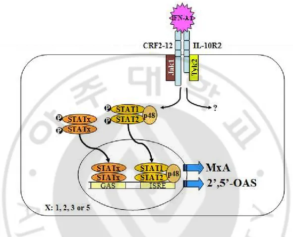
Fig. 2. Transcriptional induction of MxA and 2’5’-OAS by IFN-λ1. The IFN-λ
receptor complex consists of CRF2-12 and IL-10R2 chains. Upon binding of IFN-λ receptor with IFN-λ, STAT1 and STAT2 are phosphorylated, then associated with p48, translocated to the nucleus and activated genes, including MxA and 2’5’-OAS that contain the IFN-stimulated response element (ISRE).
B. Aims
In spite of the several reports regarding the antiviral activity of IFN-λ against
several RNA viruses, there was no report about the antiviral activity of IFN-λ
against HBV when started this study. Although this year Robeck et al. showed that
HBV replication in a murine hepatocyte cell line was inhibited by IFN-λ treatment
(Robek et al., 2005), it has not been investigated that HBV replication in a human
cell line is affected by IFN-λ1.
It is conceivable that HBV or its protein may modulate antiviral activity of
IFN-λ. HBx inhibits proteasome function, which is required for the inhibitory action
of IFN-α on HBV replication (Zhang et al., 2004). By interfering with the NF-kB
pathway, recombinant HBs inhibited LPS-induced IL-18 production, which has
been shown to inhibit HBV infection in vivo (Cheng et al., 2005). However, except
for HCV, the antagonistic effect of virus to IFN-λ1 activity has not been studied.
I wanted to know whether IFN-λ1 suppresses HBV replication in two HBV
replicating human hepatoma cell lines, and which stage of viral replication is
affected by IFN-λ1.
2. I wanted to know whether HBV regulates expression of the receptor for
IFN-λ1, and planned to establish ELISA system for analyzing the production of
II. MATERIALS AND METHODS
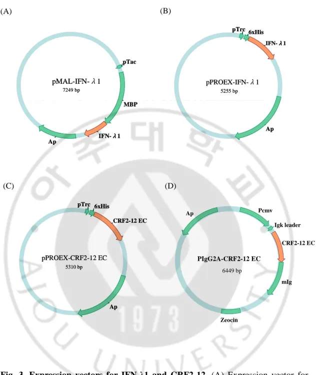
A. Expression vectors for IFN-λ1 and its receptor subunit CRF2-12
For the production of functional form of IFN-λ1, prokaryotic expression vector
pMAL-IFN-λ1 (kindly provided by Dr, Kotenko, New Jersey Medical School,
USA) containing DNA sequences encoding human IFN-λ1 gene adjacent to the 3’
end of a gene for maltose binding protein (MBP) was used because it was reported
that MBP-IFN-λ1 produced by E.coli transformed with this vector retains functional
activity (Fig. 1A) (Kotenko et al., 2003).
In order to produce a large amount of IFN-λ1 with a tag of low antigenicity,
pPROEX-IFN-λ1 was constructed. A DNA segment of IFN-λ1 produced by digestion
of pMAL-IFN-λ1 with XmnI and EcoRI was subcloned into pPROEX1 (Life
Technologies Inc., Gaithersburg, MD, USA) using EheI and EcoRI recogintion sites
(Fig. 1B). Because the expressed IFN-λ1 is fused with histidine tag, it can be purified
using Ni-NTA column.
In order to produce a large amount of CRF2-12 with a tag of low antigenicity,
pPROEX-CRF2-12 EC was constructed A DNA segment of extracellular domain of
CRF2-12 produced by digestion of pEF2-CRF2-12 (kindly provided by Dr, Kotenko)
with XhoI and EcoRI was subcloned into pPROEX1 (Life Technologies Inc.,
In order to produce CRF2-12 protein expressed from eukaryotic cell,
pIgG2A-CRF2-12 EC was constructed. The extracellular domain gene of pIgG2A-CRF2-12 was
amplified by PCR with sense primer CRF2-12 EX5(BamHI)
(5’-CGGGATCCCAGGGAGGCCCCGTCTG-3’) and antisense primer
CRF2-12EX3(EcoRI) (5’-CGGAATTCGTTGGCTTCTGGGACCTCCA-3’). A DNA
segment of extracellular domain of CRF2-12 produced by digestion of PCR product
with BamHI and EcoRI was subcloned into pIgG2A (kindly provided by Dr. Chwae,
Pochon Cha University College of Medicine, South Korea), which has secretory
signal sequence and was derived from pSecTag2C (Invitrogen, Carsbade, CA, USA)
(Fig. 1D). Because the expressed CRF2-12 is fused with mouse immunoglobulin
(mIg), it can be purified using Protein A column.
B. Purification of the recombinant proteins
1. Expression and purification of MBP-IFN-λ1 in E. coli. pMAL-IFN-λ1
was transformed into E. coli TB1. A single colony was incubated in 1 L LB
containing 100 ug/ml ampicillin at 37℃ with 250 rpm until A600 reaches 0.8~1.0.
Isopropyl-β-D-thiogalactopyranoside (IPTG) at a concentration of 0.3 mM was added to the culture to induce the expression of IFN-λ1. The culture was incubated at
37℃ for 4 h and was centrifuged at 5,500 rpm for 20 min. The cell pellet was
resuspended in 100 ml of 30 mM Tris-HCl, 20 % sucrose (pH 8.0) and 200 ul of 0.5
Fig. 3. Expression vectors for IFN-λ1 and CRF2-12. (A) Expression vector for
MBP-IFN-λ1. (B) Expression vector for IFN-λ1. (C) Expression vector for His-CRF2-12. (D) Expression vector for CRF2-12-mIg.
(A) (B) (C) pPROEX-CRF2-12 EC 5310 bp Ap pTrc 6xHis CRF2-12 EC pPROEX-CRF2-12 EC 5310 bp Ap pTrc 6xHis CRF2-12 EC pMAL-IFN-λ1 7249 bp MBP Ap pTac IFN-λλλλ1 pMAL-IFN-λ1 7249 bp MBP Ap pTac IFN-λλλλ1 pPROEX-IFN-λ1 5255 bp 6xHis pTrc Ap IFN-λλλλ1 pPROEX-IFN-λ1 5255 bp 6xHis pTrc Ap IFN-λλλλ1 (D) PIgG2A-CRF2-12 EC 6449 bp mIg Igk leader Pcmv Zeocin Ap CRF2-12 EC
was resuspended in ice-cold 100 ml of 5 mM MgSO4. After centrifugation, 8 ml of 1
M Tris-HCl (pH 7.4) was added into the supernatant, which was loaded into amylose
column. The column was washed with column buffer (20 mM Tris-HCl (pH 7.4), 200
mM NaCl, 1 mM EDTA). The protein was eluted with 10 mM maltose. The eluted
MBP-IFN-λ1 was applied to Fast Protein Liquid Chromatography (FPLC)
(Amersham Biosciences Co., Piscataway, NJ, USA) in order to remove MBP protein.
2. Expression and purification of His-IFN-λ1 and His-CRF2-12 in E. coli.
pPROEX-IFN-λ1 or pPROEX-CRF2-12 EC was transformed into E. coli PRT. A
single colony was incubated in 1 L LB containing 100 ug/ml ampicillin at 37℃ with
250 rpm until A600 reaches 0.8~1.0. Isopropyl-β-D-thiogalactopyranoside (IPTG) at
a concentration of 0.3 mM was added to the culture to induce the expression of
IFN-λ1. The culture was incubated at 37℃ for 4h and was centrifuged at 5,500 rpm for 20
min. The cell pellet was stored at -20℃ overnight and thawed for 15 min on ice. It
was resuspended in 20 ml lysis buffer (100 mM NaH2PO4, 10 mM Tris·Cl, 8 M urea,
pH 8.0). The lysate was centrifuged at 9,000 rpm for 30 min at room temperature.
The supernatant was loaded into Ni-NTA column (Qiagen, Valencia, CA, USA). The
column was washed with 30 ml of washing buffer (100 mM NaH2PO4, 10 mM
Tris·Cl, 8 M urea, pH 6.3). The protein was eluted with 20 ml of elution buffer (100
3. Expression and purification of CRF2-12-mIg in COSM6. Sixteen
micrograms of pIgG2A-CRF2-12 EC were transfected into COSM6 (2x 10⁶) using lipfectamin 2000 (Invitrogen, Carsbade, CA, USA). Next day the media were
exchanged with IgG-depleted FBS-DMEM. At fourth day posttransfection, the
culture supernatant was collected and centrifuged at 1,000 rpm for 15 min. The
supernatant was mixed with nProtein A Sepharose (Amersham Biosciences Co.,
Piscataway, NJ, USA) at 4℃ for 1 hr. The mixture was centrifuged at 5,000 rpm for
2 min. The pellet was washed 3 times with phosphate buffered saline (PBS) by
centrifugation of 5,000 rpm for 5 min. CRF2-12-mIg protein was eluted with 200 ul
of 0.1 M citrate (pH 3.0), followed by neutralization of 40 ul of 0.1M Tris-HCl (pH
9.0).
C. SDS-PAGE
The concentration of the purified recombinant proteins was measured and the
proteins were separated by 12% SDS-PAGE. The acrylamide gels were incubated
with Coomassie Blue staining solution (Amersham Biosciences Co., Piscataway, NJ,
USA) for 4 h. It was incubated with destaining solution (30% Methanol, 10% glacial
acetic acid, 60% distilled water) for 12 h.
To examine the purity of recombinant proteins, the purified protein was
separated by 12% SDS-PAGE, and transferred to a polyvinylidene fluoride (PVDF)
membrane (Millipore co. Bedford, MA, USA). The membrane was kept for 1 h in
3% Bovine Serum Albumin (BSA) in phosphate buffered saline (PBS) at room
temperature, and reacted for 1 h with primary Ab such as rabbit anti-MBP antiserum
(New England Biolabs Inc., Beverly, MA, USA) (1:10,000) or mouse anti-His Ab
(Qiagen Inc., Valencia, CA, USA) (1:1,000). Then the membrane was washed with
PBS containing 0.1% Tween 20 for 1 h. The membrane was incubated for 1 h in 3 %
BSA containing horseradish peroxidase (HRP)-goat anti-rabbit IgG (H+L) conjugate
or HRP-goat anti-mouse IgG (H+L) conjugate (Zymed, SanFrancisco, CA, USA)
(1:20,000). Then the membrane was washed with PBS containing 0.1% Tween 20 for
1 h. After washing, the membrane was incubated in enhanced chemical luminescence
(ECL) solution (Amersham Biosciences Co., Piscataway, NJ, USA) and developed
with X-ray.
To examine the reactivity of monoclonal Abs against IFN-λ1, Western blottig
was performed as above except followings. Purified His-IFN-λ1 was separated by
SDS-PAGE. The membrane was kept for 1 h in 10% skim milk in phosphate buffered
saline (PBS), and reacted for 1 h with culture supernatants of anti-IFN-λ1 Ab
producing hybridoma. The membrane was incubated for 1 h in 10 % skim milk
containing horseradish peroxidase (HRP)-goat anti-mouse IgG (H+L) conjugate
E. RNA preparation
Hepatoma cell lines (HuH7 and HepG2) and HBV replicating cell lines (WT10
and PEB8) were lysed by the addition of 200 ul of RNAzol Bee (Tel-Test, Inc,
Friendswood, TX, USA) followed by the addition of 20 ul of chloroform. After
vigorous shaking, the cell lysate was incubated on ice for 5 min and centrifuged at
12,000 rpm for 15 min at 4℃. The aqueous phase was transferred to the fresh tube.
The RNA was precipitated by adding the equal volume of isoporpanol and incubated
for 1 h at 4℃. After centrifugation, the pellet was washed with 75% ethanol. The
pellet was dried and then dissolved in diethyl pyrocarbonate (DEPC)-treated distilled
water.
F. Reverse transcription-polymerase chain reaction (RT-PCR)
The cDNA was synthesized from 500 ng of total RNA in a reaction mixture
containing 0.5 ul of RNase H- reverse transcriptase (Invitrogen, Carsbade, CA, USA), 0.5 ul of oligo (dT)12-18 (500 ug/ml), 2 ul of 5x first-strand buffer and 1 ul of 0.1 M
DTT, 0.25 ul 10mM dNTP mixture. The cDNA was amplified using specific primers
(MxA(5) and MxA(3) for MxA, OAS(5) and OAS(3) for 2’5’-OAS, IL-28RA(R) and
IL-28RA(F) for CRF2-12, GAPDH(5) and GAPDH(3) for GAPDH) (Table 1). PCR
reaction mixture contained 2 ul of cDNA, 2.5ul of 10x buffer, 2 ul of dNTP (2.5mM),
units/ul) and 17.25 ul of distilled water. PCR reaction condition was 94℃ for 5 min,
30 cycles at 94℃ for 30s, 55℃ for 30s, 72℃ for 1 min; final extension was 72℃ for
7min. Amplified cDNA products were separated by electrophoresis in 1% agarose
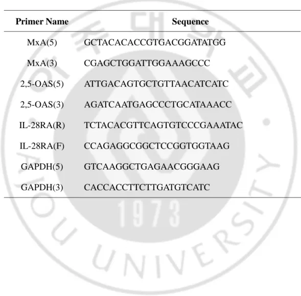
Table 1. Primers used for amplificating of MxA, 2’5’-OAS and CRF2-12 cDNAs
Primer Name Sequence
MxA(5) GCTACACACCGTGACGGATATGG MxA(3) CGAGCTGGATTGGAAAGCCC 2,5-OAS(5) ATTGACAGTGCTGTTAACATCATC 2,5-OAS(3) AGATCAATGAGCCCTGCATAAACC IL-28RA(R) TCTACACGTTCAGTGTCCCGAAATAC IL-28RA(F) CCAGAGGCGGCTCCGGTGGTAAG GAPDH(5) GTCAAGGCTGAGAACGGGAAG GAPDH(3) CACCACCTTCTTGATGTCATC
G. Isolation of HBV core DNA
WT10 or PEB8 cells (5x 10⁶) were seeded in 10 cm dishes. After 1 day or 2 days, the cells were treated with 0.5 ug/ml of poly(I:C) (Amersham Biosciences Co.,
Piscataway, NJ, USA), which was transfected by lipofectamin 2000 (Invitrogen,
Gaithersburh, MD, USA), or IFN-α (10,000 U/ml) (PBL Biomedical Laboratories,
Piscataway, NJ, USA), MBP-IFN-λ1 (3,000 ng/ml; equivalent of 1,000 ng/ml of
IFN-λ1) and MBP (1,000 ng/ml). At 1 day or 2 days after treatment, WT10 or PEB8
cells were harvested and lysed in 1% NP-40 containing 10mM Tris-HCl (pH 8.0), 50
mM NaCl and 1 mM EDTA at 4°C for 5 min followed by centrifuging for 5 min at
12,500 rpm. The cleared cell lysate was treated by 20 U DNase I (SIGMA, St. Louis,
MO, USA), 60 U of micrococcal nuclease (Calbiochem, San diego, CA, USA) with
10 mM MgCl2 and 8 mM CaCl2 at 37°C for 4 h. The virus in the cleared culture
medium was precipitated by polyethylene glycol (PEG) 8000 (40%) in 50 mM NaCl
and 50 mM EDTA at 4°C overnight. After centrifugation for 15 min at 12,000 rpm,
the virus was resuspended in 20 mM Tris-HCl (pH 8.0), 10 mM EDTA, 100 mM
NaCl and 2 % sodium dodecyl sulfate (SDS), and digested with 4 ug of proteinase K
for 4 h at 37°C. After phenol extraction, the intracellular virion DNAs were
H. Real-time PCR
Primers and probes were designed against a highly conserved region among 25
published HBV genome sequences in GenBank and EMBL, representing genotypes
A-F (Li et al., 2005). As the primers, HBV(sens) (5’-AGTGTGGATTCGCAC
TCCT-3’) and HBV(anti) (5’-GAGTTCTTCTTCTAGGGGACCTG-3’) was used.
The Taqman HBV probe (5’-CCAAATGCCCCTATCTTATCAACACTTCC-3’) was
labeled with a reporter dye FAM (6-Carboxy-uorescein) at its 5’ end and with a
quencher dye TAMRA (6-Carboxy-tetramethyl-rhodamine) at its 3’ end. The
amplification was performed in a 25 ul reaction mixture containing 12.5 ul TaqMan
Universal Master Mix (Applied Biosystems, Foster City, CA, USA), 0.7 mM of
HBV(sens) and HBV(anti) primers, and 0.15 mM of TaqMan HBV probe (Li et al.,
2005). Five microliters of DNA template were added to each reaction. The product
was first digested with uracil-N-glycosylase at 50℃ for 2 min to destroy the potential
carry-over contamination. An incubation of 10 min at 95℃ allowed the activation of
the AmpliTaq Gold DNA polymerase and the denaturation of the nucleic acids.
Forty-five cycles of denaturation at 95℃ for 15 sec and annealing-extension at 53℃
for 1 min were carried out allowing the amplification detection of HBV genomes.
The detection of amplified product (120 bp fragment) was carried out in an ABI
I. Southern blotting
Isolated HBV core DNA was separated by 1% agarose gel electrophoresis. The
gel was washed with denaturation solution (1.5 M NaCl, 0.5 M NaOH) for 1 h, and
then with neutralization solution (1.5 M NaCl, 0.5 M Tris-Cl (pH 7.0)). HBV core
DNA in the gel was transferred through 20x SSC (3 M NaCl, 0.3 M Na3Citrate2H2O,
pH 7.0) to nylon membrane (Schleichr & Schuell, Keene, NH, USA). Alpha-[32 P]-labeled probe specific for the HBV sequences was random-primed for 12 h in 50 ul
of reaction buffer, including 10 ul of 5x labeling buffer (Promega co., Madison, WI,
USA), 5 mM dNTP without dATP, 1x BSA, 150 ng of HBV DNA and 2 ul of DNA
polymerase I large (Klenow) fragment (Promega co., Madison, WI, USA). The
membrane-associated HBV DNA was hybridized to α-[32P]-labeled probe specific for the HBV sequences at 68℃ for 12 h. The membrane was washed with washing
solution (0.1% SDS, 2x SSC) for 1 h. The autoradiogram was scanned using the
imaging analyzer FLA-3000 (Fuji Photo Film co., Tokyo, Japan).
J. Northern blotting
Total RNA (10 ug) was denatured and electrophoressed on a 1% agarose gel
containing 40% formaldehyde, 1x MOP and 0.1% diethyl pyrocarbonate
(DEPC)-distilled water. RNA in the gel was transferred to a nylon membrane (Schleichr &
performed as above.
K. Enzyme-linked immunosorbent assay (ELISA)
1. Secretion of HBsAg and HBeAg. ELISA was performed using ELISA kit
specific for HBsAg (GENDIA, GreenCross, Kyunggi, Korea) and for HBeAg
(DiaSorin S.p.A., Saluggia, Italy) according to the manufacturer’s protocol,
respectively.
2. Screening of monoclonal anti-IFN-λ1 Ab. Microtiter plate was coated with
His-IFN-λ1, MBP-IFN-λ1 and MBP at concentration of 10 ug/ml, followed by being
blocked with 3% BSA. Then the hybridoma culture supernatant was added to each
well and incubated at room temperature. The microtiter plate was incubated with
alkaline phosphatase-goat anti-mouse IgG (H+L) conjugate (PIERCE, Rockford, IL,
USA) in 3% BSA (1:5,000). After washing, p-nitrophenyl phosphate (p-NPP)
(SIGMA, St. Louis, MO, USA) was used as a substrate and absorbance at 415nm
was determined using Microplate reader model 680 (Bio-Rad, Melville, NY, USA).
3. Sensitivity of anti-IFN-λ1 Abs against IFN-λ1. Microtiter plate was coated
with IFN-λ1 (R&D systems Inc., Minneapolis, MN, USA) at the concentration of 1
ng/well to 10 ng/well, followed by being blocked with 3% BSA. After washing, the
ug/ml to each well and incubated overnight at 4℃. After washing microtiter plate
was incubated with alkaline phosphatase-goat anti-mouse IgG (H+L) conjugate
(PIERCE, Rockford, IL, USA) in 3% BSA (1:1,000). After washing, p-nitrophenyl
phosphate (p-NPP) (SIGMA, St. Louis, MO, USA) was used as a substrate and
absorbance at 415nm was determined using Microplate reader model 680 (Bio-Rad,
Melville, NY, USA).
L. Cell fusion
For a primary immunization, 50 ug of the purified His-IFN-λ1 protein was
mixed with an equal volume of complete Freund’s adjuvant (SIGMA, St. Louis, MO,
USA). The emulsified solution was injected into three Balb/c mice (Daehan Biolink
co., LTD, Chungbuk, Korea) intravenous. For secondary and third immunization, 25
ug of the purified His-IFN-λ1 protein was mixed with an equal volume of incomplete
Freund’s adjuvant (SIGMA, St. Louis, MO, USA) after 2 and 4 weeks. The
emulsified solution was injected into the Balb/c mice intravenous. After 2 weeks, 25
ug of the purified MBP-IFN-λ1 protein was injected into the Balb/c mice
intraperitoneal. After 3 days, final boosted Balb/c mouse was sacrificed and its
spleen was removed. The spleen was teased apart using a couple of slide glasses. The
cells were resuspended in incomplete RPMI1650 and centrifuged at 1,500 rpm for 10
min. The cell pellet was resuspended in 5 ml of red blood cell (RBC) lysis buffer for
by centrifugation at 1,500 rpm for 10 min. The splenocytes and the F0 myeloma cells
were mixed in 5 ml of incomplete RPMI1650 in the ratio of 3 to 1. Cell mixture was
centrifuged at 1,500 rpm for 15 min. One milliliter of 50% polyethylene glycol
(PEG) (SIGMA, St. Louis, MO, USA) was slowly added to the cell pellet for 1 min
while resuspending the cells with complete RPMI1650 by stirring the tube. The cell
mixture was centrifuged at 1,500 rpm for 10 min and then cell pellet was
resuspended in 30 ml complete RPMI1650. One hundred microliter of cells was
dispensed into the wells of 96-well plate, which was placed at 37℃ in a CO2
incubator. Next day, the culture media were exchanged with HAT media (SIGMA, St.
Louis, MO, USA).
M. Limiting dilution
The anti-IFN-λ1 Ab-producing hybridoma cell was transferred from 96-well
plate to 24-well plate. The cell was incubated for 3 days in 24-well plate. The
number of the cell was counted, and the cell was diluted to be one cell per 3 wells.
One hundred microliter of the diluted cell was added into 96-well plate. It was
incubated at 37℃ for 10 days. When colony was appeared, the supernatant was
N. Isotyping of monoclonal Abs
The supernatant of anti-IFN-λ1 Ab was collected from the selected hybridomas.
The isotyping of the monoclonal Abs was performed according to the protocol of
immunotype mouse monclonal antibody isotyping kit (SIGMA, St. Louis, MO,
USA).
O. Large production of monoclonal anti-IFN-λ1 Ab
Three Balb/c mice (Daehan Biolink co., LTD, Chungbuk, Korea) were primed
by injecting 500 ul of pristane (2,6,19,14-tetraamethyldecanoic acid) into the
peritoneum. After 7 days, the mice were injected with HL2 hybridoma cell (1x 10⁶) intraperitorial. Within 2 weeks following the injection of the cells, the mice were
noticeably large. Ascite was withdrawn with 18 gauge needle attached to a 5 ml
syringe. The fluid was incubated at 37℃ for 1 h and then transferred to 4℃
overnight. It was centrifuged at 3,000g for 10 min. The supernatant was removed
from the cell pellet. Anti-IFN-λ1 Ab was eluted through nProtein A Sepharose
P. The production of polyclonal anti-IFN-λ1 Ab and anti-CRF2-12 Ab
For a primary immunization, 250 ug of the purified His-IFN-λ1 or
His-CRF2-12 protein was mixed with an equal volume of complete Freund’s adjuvant (SIGMA,
St. Louis, MO, USA). The emulsified solution was injected into each rabbit (Daehan
Biolink co., LTD, Chungbuk, Korea) subcutaneous. For secondary and third
immunization, 125 ug of the purified His-IFN-λ1 or His-CRF2-12 protein solution
was mixed with an equal volume of incomplete Freund’s adjuvant (SIGMA, St.
Louis, MO, USA) after 2 and 4 weeks. The emulsified solution was injected into
each rabbit subcutaneous. After 2 weeks, 125 ug of the purified MBP-IFN-λ1 or
His-CRF2-12 protein was mixed with an equal volume of incomplete Freund’s adjuvant.
The emulsified solution was final boosted into each rabbit subcutaneous. After 3 days
of the injection, each rabbit was sacrificed. The serums were collected and then
III. RESULTS
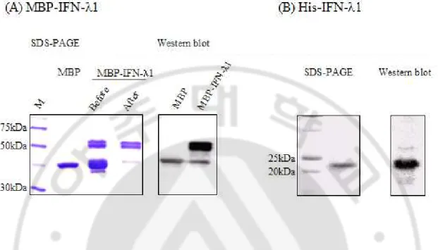
A. Expression and purification of IFN-λ1
For the experiment of antiviral activity of IFN-λ1, MBP-IFN-λ1 was purified based on the report that this form of IFN-λ1 retains functional activity (Kotenko et al., 2003). The purified MBP-IFN-λ1 using amylose column affinity chromatography and Fast Protein Liquid Chromatography (FPLC) was subjected to
SDS-PAGE and Western blotting (Fig. 4A). In SDS-PAGE, the major band of
around 65 kDa, which corresponds to MBP-IFN-λ1, and the minor band of 43 kDa,
which corresponds to MBP, were observed. In Western blotting, all of two bands
were detected with anti-MBP Ab. Thus the major band was confirmed as
MBP-IFN-λ1 and the minor one as MBP.
As antigen for the generation of monoclonal Ab specific to IFN-λ1, IFN-λ1
with histidine tag (His-IFN-λ1) was expressed in E. coli and purified due to the
lower antigenicity of histidine tag than MBP. Another advantage of His-IFN-λ1 was
the improved expression level. The yield of purification of His-IFN-λ1 was two
times greater than that of MBP-IFN-λ1. In SDS-PAGE of His-IFN-λ1 purified using
Ni-NTA column, the expected size (22 kDa) of single band was detected, and in
Western blotting this protein reacted with antibody specific to histidine tag (Fig.
Fig. 4. The purification of IFN-λ1 in E. coli. (A) IFN-λ1 fused with MBP was
purified using FPLC after amylose column chromatography and analyzed by SDS-PAGE (left) and Western blotting (right). Western blotting was performed with rabbit anti-MBP antibody and HRP conjugated anti-rabbit Ig antibody. Before: the protein before being purified using FPLC, After: the protein after being purified using FPLC(B) IFN-λ1 with histidine was purified using Ni-NTA column and analyzed by SDS-PAGE (left) and Western blotting (right). Western blotting was performed with mouse anti-His antibody and HRP conjugated anti-mouse Ig antibody.
B. Functional activity of IFN-λ1
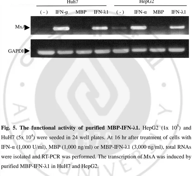
Next the functional activity of purified MBP-IFN-λ1 was examined. Human
hepatoma cell lines, HepG2 and HuH7, were treated with MBP-IFN-λ1, and the
transcriptional activation of MxA was analyzed by RT-PCR (Fig. 5). As a positive
control IFN-α was used, and as a negative control MBP was used. In MBP-treated
HuH7 cells as well as untreated cells, the transcription of MxA was not induced. In
HepG2 cells without any treatment or with MBP treatment, low level of
transcription of MxA was induced. As expected, IFN-λ1, like IFN-α, induced
transcription of MxA both in HuH7 and HepG2 cells. This indicates that the
purified MBP-IFN-λ1 has functional activity and that induction of MxA mRNA by
Fig. 5. The functional activity of purified MBP-IFN-λ1. HepG2 (1x 10⁵) and HuH7 (5x 10⁴) were seeded in 24 well plates. At 16 hr after treatment of cells with IFN-α (1,000 U/ml), MBP (1,000 ng/ml) or MBP-IFN-λ1 (3,000 ng/ml), total RNAs were isolated and RT-PCR was performed. The transcription of MxA was induced by purified MBP-IFN-λ1 in HuH7 and HepG2.
MxA
GAPDH
( - ) IFN-α MBP IFN-λ1 ( - ) IFN-α MBP IFN-λ1
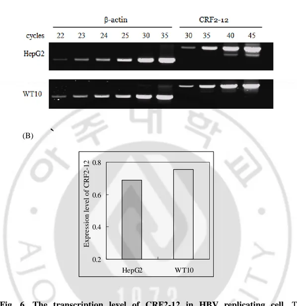
C. Expression of CRF2-12 in HBV replicating human hepatoma cell lines
IFN-λ receptor is a heterodimer of IL-10R2, ubiquitously expressed, and
CRF-2-12. Expression of CRF2-12 mRNA was demonstrated in normal liver and
human hepatic cancer derived cell lines (HepG2, HuH7 and HEP3B) (Sheppard et
al., 2003; Vlotides et al., 2004), but not in cells supporting HBV replication.
Semiquantitative RT-PCR was performed using HBV replicating human
hepatoma cell lines, WT10 (Imanaka et al., 2005) (Fig. 6). Both in HepG2 and
WT10 cells, the predicted length (799 bp) of PCR product was amplified. With
increased cycle numbers, smaller PCR product was appeared, which seems to be
derived from alternative splicing form of CRF2-12 since the predicted length of
alternative form was small by 87 bp than major band. Normalized band intensity of
CRF2-12 in WT10 cells was similar to that in HepG2 cells. This result demonstrates
`
Fig. 6. The transcription level of CRF2-12 in HBV replicating cell. The
transcripts of CRF2-12 were amplified by semiquantitative RT-PCR using RNAs isolated from HepG2 and WT10. (A) Amplified PCR products were analyzed by agarose gel electorphoresis. (B) The expression levels of CRF2-12 normalized to the expression levels of β-actin were comprared. It was calculated as intensity of CRF2-12 (35 cycles)/intensity of β-actin (23 cycles). Two independent experiments were done. (A) (B) 0.2 0.4 0.6 0.8 HepG2 WT10 E x p re ss io n l ev el o f C R F 2 -1 2
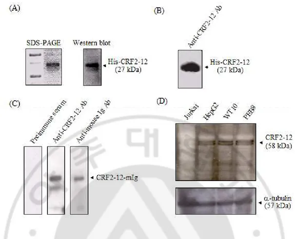
To examine the expression of CRF2-12 at protein level, rabbit polyclonal
anti-CRF2-12 Ab was generated by immunization of extracellular domain of anti-CRF2-12,
which was expressed in E. coli and purified using Ni-NTA column. The purity of
His-CRF2-12 used as immunogen was shown in SDS-PAGE (Fig. 7A). The
reactivity of rabbit polyclonal anti-CRF2-12 Ab was determined by Western blotting
using the immunogen. Like anti-histidine tag Ab, anti-CRF2-12 Ab reacted with
extracellular domain of CRF2-12 with histidine tag (Fig. 7B). To know whether
anti-CRF2-12 Ab may recognize anti-CRF2-12 expressed in eukaryotic cells, fusion protein of
CRF2-12 and mouse Ig was expressed in COSM6 cells and purified using nProtein A
Sepharose column. The binding activity of rabbit polyclonal anti-CRF2-12 Ab to this
protein was examined by Western blotting (Fig. 7C). The expected band was detected
at size of 56 kDa. Anti-CRF2-12 Ab but not normal rabbit Ig reacted with
CRF2-12-mIg. With this anti-CRF2-12 Ab, the CRF2-12 expression in hepatoma cell lines was
explored. In all these cells CRF2-12 expression was detected at similar levels as 58
kDa size (Fig. 7D). Consistent with finding of RT-PCR, this finding indicates that
Fig. 7. The expression of CRF2-12 in WT10 and PEB8 at protein level. (A) The
purified His-CRF2-12 in E. coli was visualized by SDS-PAGE (left) and Western blotting (right). Western blotting was performed with mouse anti-His Ab and HRP conjugated anti-mouse Ig Ab. (B) The reactivity of rabbit polyclonal anti-CRF2-12 Ab with His-CRF2-12 was examined by Western blotting using HRP conjugated anti-rabbit Ig Ab. (C) The reactivity of rabbit polyclonal anti-CRF2-12 Ab with CRF2-12 expressed in eukaryotic cells was examined by Western blotting using recombinant CRF2-12 containing mouse Ig as a fusion partner secreted from COSM6 cells. As a positive control, CRF2-12-mIg was detected by HRP-conjugated goat anti-mouse Ig Ab, and as the negative control it was reacted with preimmune rabbit serum and HRP-conjugated goat anti-rabbit Ig Ab. (D) The expression of CRF2-12 in HepG2, WT10 and PEB8 was analyzed by Western blotting using the rabbit polyclonal anti-CRF2-12 Ab. As a negative control, Jurkat T cell line was used.
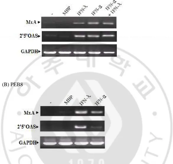
D. Induction of antiviral proteins in HBV replicating human hepatoma cell lines by IFN-λ1
To know whether IFN-λ1 induces transcription of MxA and 2’5’-OAS in HBV
replicating human hepatoma cell lines, RT-PCR was performed. Compared to
controls (untreated and MBP-treated cells), transcriptional levels of MxA and
2’5’-OAS were increased both in WT10 and PEB8 treated with IFN-λ1 (Fig. 8). These
findings indicate that HBV replication may not affect the transcriptional activation of
Fig. 8. The transcription of antiviral proteins by IFN-λ1 in WT10 and PEB8.
WT10 (A) and PEB8 (B), respectively, were treated with MBP (1,000 ng/ml), MBP-IFN-λ1 (3,000 ng/ml), IFN-α (1,000 U/ml) or both IFN-α (1,000 U/ml) and MBP-IFN-λ1 (3,000 ng/ml) for 18 h. The induction of transcription of MxA and 2’5’-OAS was analyzed by RT-PCR. MxA: 289 bp, 2’5’-OAS: 400 bp, GAPDH: 600 bp.
(A) WT10
E. The effect of IFN-λ1 on HBV replication
To determine the effect of IFN-λ1 on HBV replication in human hepatoma cells,
real-time PCR was performed (Fig. 9). In HBV replication, HBV DNA synthesis
occurred in nucleocapsid, where the HBV pregenomic RNA and reverse transcriptase
complex is packaged. HBV DNA in the nucleocapsid was isolated from cells treated
with IFN-λ1 for 24 h or 48 h. For the comparison, poly(I:C) or IFN-α was used.
Poly(I:C) for induction of natural IFN-α was transfected into PEB8, because HepG2
does not respond to extracellular poly(I:C) (Li et al., 2005). Amplification plot
showed that the level of HBV DNA in intracellular nucleocapsids isolated from
WT10 treated with IFN-λ1 was almost same as that seen in WT10 treated with MBP.
Even 3 times higher concentration of IFN-λ1 did not influence the level of HBV
DNA in WT10 (data not shown). However, the level of HBV DNA in intracellular
nucleocapsids isolated from PEB8 treated with IFN-λ1 was reduced to 73% of that
seen in PEB8 treated with MBP. The reduction in HBV DNA level by IFN-λ1 was
comparable to that by poly(I:C). These findings indicate that IFN-λ1 is able to inhibit
HBV replication in certain human cell lines albeit its inhibitory effect is not so great.
To support these findings, Southern blotting was done (Fig. 10). IFN-λ1 reduced the
level of HBV DNA in intracellular nucleocapsids isolated from PEB8 at 48 hr after
treatment of IFN-λ1 to about 70% of the level of MBP-control. But the level of HBV
DNA isolated at 24 hr after treatment of IFN-λ1 was similar to that of MBP-control.
Fig. 9. Real-time PCR analysis of antiviral activity against HBV. WT10 (A) and
PEB8 (B) were treated with MBP (1,000 ng/ml), MBP-IFN-λ1 (3,000 ng/ml), IFN-α (1,000 U/ml) or poly(I:C) (0.5 ug/ml) for 48 h. HBV core DNAs were prepared from cell lysates and subjected to real-time PCR with HBV Taqman probe.
(A) WT10 (B) PEB8 St andard Curve 18 20 22 24 26 28 30 32 8 9 10 11 LogCo C t St andard Curve 12 14 16 18 20 22 24 26 28 30 32 3 4 5 6 7 8 LogCo C t Delta Rn vs Cycle 0.01 0.1 1 10 1 3 5 7 9 11 13 15 17 19 21 23 25 27 29 31 33 35 37 39 Cycle Number D e lt a R n -poly(I:C) IFN-λ MBP Delta Rn vs Cycle 0.01 0.1 1 10 123456789 10 11 12 13 14 15 16 17 18 19 20 21 22 23 24 25 26 27 28 29 30 31 32 33 34 35 36 37 38 39 40 Cycle Number D e lt a R n (-) MBP IFN-λ1 IFN-α 0.E+00 1.E+00 2.E+00 3.E+00 4.E+00 5.E+00 MBP IFN-λ1 H B V D N A (g e q ) 3.E+00 4.E+00 5.E+00 6.E+00 7.E+00 8.E+00 MBP IFN-λ1 H B V D N A ( g e q )
Fig. 10. Southern blot analysis of antiviral activity against HBV. PEB8 was
treated with MBP (1,000 ng/ml), MBP-IFN-λ1 (3,000 ng/ml) and poly(I:C) (0.5 ug/ml). After final 24 h or 48 h, HBV core DNAs were prepared from cell lysates and subjected to Southern blotting. RC: relaxed circular DNA, SS: single stranded DNA. The figure is representative of three independent experiments.
F. The effect of IFN-λ1 on HBV transcription
To determine whether the inhibitory effect of IFN-λ1 on HBV replication
occurs in RNA level, Northern blotting was performed (Fig. 11). The amount of 3.5
kb HBV transcript isolated from cells treated with IFN-λ1 was similar to controls
(untreated cells or MBP-treated cells). It indicates that the inhibition of HBV
replication by IFN-λ1 may not occur at HBV transcriptional level.
G. The effect of IFN-λ1 on the secretion of HBsAg and HBeAg
The effect of IFN-λ1 on the production of HBsAg and HBeAg by PEB8 was
examined by ELISA (Fig. 12). Compared to control, IFN-λ1 did not have significant
effect on the secretion of HBsAg and HBeAg both at 24 h and 48 h, but poly(I:C)
significantly reduced the secretion of HBeAg at both times and HBsAg at 24 h,
suggesting that antiviral mechanism by IFN-λ1 may be different from that by
Fig. 11. The effect of IFN-λ1 on HBV transcription in PEB8. PEB8 was treated
with MBP (1,000 ng/ml), MBP-IFN-λ1 (3,000 ng/ml) and poly(I:C) (0.5 ug/ml). After 24 h or 48 h, total HBV RNAs were isolated from the cells and subjected to Northern blotting. Upper panel: three transcripts of HBV; lower panel: ribosomal RNA (rRNA). The figure is representative of two independent experiments.
(A) HBsAg
(B) HBeAg
Fig. 12. The effect of IFN-λ1 on the secretion of HBsAg and HBeAg by PEB8.
PEB8 was treated with MBP (1,000 ng/ml), MBP-IFN-λ1 (3,000 ng/ml) or poly(I:C)
(0.5 ug/ml). After 24 h or 48 h, the production of HBsAg (A) and HBeAg (B) was
analyzed by ELISA. Relative Ag production was calculated as (OD of each
H. Generation of the polyclonal and the monoclonal Ab against IFN-λ1
Although it was reported that IFN-λ1 mRNA expression was induced or
enhanced by IFN-α and other stimulators, the amount of IFN-λ1 was not quantitated
at protein level (Siren et al., 2005; Osterlund et al., 2005). The reason may be
because polyclonal or monoclonal anti-IFN-λ1 Ab was not commercially available at
that time. In order to establish a quantitation system to measure the production of
IFN-λ1, the polyclonal and the monoclonal anti-IFN-λ1 Abs were generated.
First, from three immunized mice 25 hybridoma cells producing Ab against
IFN-λ1 were selected and three clones of these, designated as HL1, HL2 and HL3,
were established through limiting dilution. The binding activity of these monoclonal
Abs to IFN-λ1 was revealed by ELISA using MBP-IFN-λ1 as well as His-IFN-λ1
(Fig. 13A) and by Western blotting using His-IFN-λ1 (Fig. 13B). The isotype of each
monoclonal Ab was determined; for HL1, G1 and for HL2 and HL3, G2b.
Next, the reactivity of polyclonal rabbit anti-IFN-λ1 Abs to native and
denatured form of IFN-λ1 was shown in Westren blotting and ELISA (Fig. 13B and
(A)
(B)
Fig. 13. The reactivity of monoclonal Abs against IFN-λ1. (A) The reactivity of
supernatants of monoclonal Abs to IFN-λ1 was examined by ELISA using
recombinant MBP-IFN-λ1 (1 ug/well) and His-IFN-λ1 (1 ug/well). (B) The reactivity
of supernatants of monoclonal Abs to denatured IFN-λ1 was examined by Western
blotting using His-IFN-λ1 (500 ng/lane) and HRP conjugated anti-mouse Ig Ab. (-):
myeloma cell culture supernatant. (+): rabbit polyclonal anti-IFN-λ1 Ab.
0 1 2 3 4 HL1 HL2 HL3 Hybridoma clones His-IFN-λ1 MBP-IFN-λ1 O D (4 1 5 n m ) HL1 HL2 HL3 (-) (+) His-IFN-λ1
I. Establishment of ELISA for IFN-λ1
Indirect ELISA for IFN-λ1 was established using polyclonal anti-IFN-λ1 Ab
(Fig. 14A) and HL2 monoclonal anti-IFN-λ1 Ab (Fig. 14B). The detection limits
were 40 ng/ml for both polyclonal and monoclonal anti-IFN-λ1 Abs. Sandwich
ELISA using both polyclonal and monoclonal Abs was performed but the detection
(A) Polyclonal anti-IFN-λ1 Ab
(B) HL2 monoclonal anti-IFN-λ1 Ab
Fig. 14. The sensitivity of ELISA for IFN-λ1 using polyclonal and monoclonal anti-IFN-λ1 Abs. Serially diluted IFN-λ1 was detected by ELISA (A) using
polyclonal rabbit anti-IFN-λ1 Ab (1:1,000) or (B) using HL1 monoclonal anti-IFN-λ1 Ab (10 ug/ml). IFN-λ1 expressed in eukaryotic cells was purchased from R & D systems Inc. Values represent the mean ± S.D. of
quadriplicates. 0.0 0.5 1.0 1.5 2.0 1 2 4 6 8 10
The amount of coated IFN-λ1 (ng/well)
O D (4 0 5 n m ) 0.0 0.2 0.4 0.6 0.8 1 2 4 6 8 10
The amount of coated IFN-λ1 (ng/well)
O D (4 0 5 n m )
IV. DISCUSSION
In this study, it was demonstrated that IFN-λ1 can inhibit HBV replication in
PEB8 but not in WT10. Additionally, it was shown that HBV does not modulate the
expression of CRF2-12, an IFN-λ1 receptor subunit. Also, ELISA for analyzing of
production of IFN-λ1 was established.
Although antiviral activity of IFN-λ1 against several RNA viruses and
cytomegalovirus has been reported (Robek et al., 2005), the effect of IFN-λ1 on
HBV replication in human cells has not. A recent report showed that HBV replication
in a murine hepatocyte cell line was inhibited by IFN-λ treatment (Robek et al.,
2005). They treated murine hepatocyte cells with 1 to 10 ng/ml of murine IFN-λ2
expressed in E.coli (PEPROTCH,Rocky Hill, NJ, USA), analyzed HBV replication
by Southern blotting at 24 h after the addition of IFN-λ2 and showed about 90%
reduction of HBV DNA replication intermediates. My results using human hepatoma
cell lines revealed that 1,000 ng/ml of IFN-λ1 (equivalent of 3,000 ng/ml of
MBP-IFN-λ1) reduced the level of viral DNA by 30% (corresponding to 100 fold reduction
of viral progeny) in PEB8 cells but not in WT10 cells. Similar results were obtained
in cells treated with 3,000 ng/ml of IFN-λ1 (data not shown). In this study
recombinant MBP-λ1 was used because when I started these experiments
IFN-λ1 was commercially unavailable. Then why did IFN-IFN-λ1 suppress HBV replication in
PEB8, but not in WT10? Both cell lines originated from HepG2 cells. In both cell
induction were observed. The known difference is that PEB8 was established by
transfection of plasmid pcDNA containing a 1.05 HBV genome-length DNA of
adwR9 subtype, while WT10 was made using retrovirus containing a 1.1 HBV
genome length DNA of adr subtype (Fu L and Cheng YC, 2000). HBV adwR9
subtype belongs to genotype A, B and G, and HBV adr subtype belongs to genotype
C. HBV genotype was reported to be an important predictor of response to treatment
of IFN-α; better responses in A and B genotypes than those in C genotype was
observed (Wai et al., 2002; Janssen et al., 2005; Thuy et al., 2005). Thus my results
raise a question whether the antiviral activity of IFN-λ may vary in HBV genotypes.
Neither HBV transcription nor secretion of viral Ags was suppressed by the
treatment of PEB8 with IFN-λ1. Similar to IFN-λl, type I IFN does not affect either
viral transcription or production of secretory Ag. IFN-α-mediated suppression of
HBV replication in HBV transgenic mouse occurs by reducing the intracellular
content of HBV RNA-containing capsids without altering either HBV gene
expression, translation, capsid maturation or virus secretion (Wieland et al., 2000).
IFN-β treatment of immortalized HBV transgenic hepatocytes results in rapid
clearance of HBV RNA-containing capsids (Pasquetto et al., 2002). In an inducible
HBV replication system, type I IFN suppresses HBV replication by preventing the
formation of replication-competent core particles not by destabilization of
pgRNA-containing capsids (Robek et al., 2005). To dissect the stage of HBV replication
process affected by IFN-λl, further study is needed