Open Access
Korean First Prospective Phase II Study, Feasibility of
Prone Position in Postoperative Whole Breast Radiotherapy:
A Dosimetric Comparison
Original Article
Purpose
This first Korean prospective study is to evaluate the feasibility of prone breast radiotherapy after breast conserving surgery for left breast cancer patients who have relatively small breast size and we present dosimetric comparison between prone and supine positions.
Materials and Methods
Fifty patients underwent two computed tomography (CT) simulations in supine and prone positions. Whole breast, ipsilateral lung, heart, and left-anterior-descending coronary artery were contoured on each simulation CT images. Tangential-fields treatment plan in each position was designed with total 50 Gy in 2-Gy fractions, and then one of the positions was designated for the treatment by comparing target coverage and dose to normal organs. Also, interfractional and intrafractional motion was evaluated using portal images.
Results
In total 50 patients, 32 cases were decided as prone-position–beneficial group and 18 cases as supine-position–beneficial group based on dosimetric advantage. Target dose ho-mogeneity was comparable, but target conformity in prone position was closer to optimal than in supine position. For both group, prone position significantly increased lung volume. However, heart volume was decreased by prone position for prone-position–beneficial group but was comparable between two positions for supine-position–beneficial group. Lung and heart doses were significantly decreased by prone position for prone-position–beneficial group. However, prone position for supine-position–beneficial group increased heart dose while decreasing lung dose. Prone position showed larger interfractional motion but smaller intra-fractional motion than supine position.
Conclusion
Prone breast radiotherapy could be beneficial to a subset of small breast patients since it substantially spared normal organs while achieving adequate target coverage.
Key words
Clinical trial, Left-sided breast neoplasms, Radiotherapy, Prone position
Yoonsun Chung,
PhD1Jeong Il Yu,
MD, PhD2Won Park,
MD, PhD2Doo Ho Choi,
MD, PhD21Department of Nuclear Engineering,
Hanyang University, Seoul, 2Department of
Radiation Oncology, Samsung Medical Center, Sungkyunkwan University School of Medicine, Seoul, Korea
Introduction
Breast cancer is the most common cancer in women world-wide [1] and the second most common cancer in Korean women [2]. While postoperative whole breast radiotherapy (RT) has previously been proven effective in clinical trials [3], the concern about cardiac problems according to exposed dose has been raised especially in left-sided breast cancer
patients. Meta-analysis of U.S. breast cancer RT between 1973 and 2001 found an increase in heart-related mortality, when older equipment and techniques had yet been utilized [4]. The reduction of heart perfusion according to RT volume was also reported to be induced in 40% of patients within 2 years [5]. In addition, we also see the coronary arteries damaged by postoperative RT in left breast cancer patients compared to the right [6]. A paper published in the New
Eng-land Journal of Medicine [7] reported that main coronary artery
+ + + + + + + + + + + + + + + + + + + + + + + + + + + + + + + + + + + + + + + + + + + + + + + + + + + + + + + + + + + + + + + + + + + + + + + + + + + + + + + + + + + + + + + + + + + + + + + + + + + + + + + + + + + + + + + + + + + + + + + + + + + + + + + + + + + + + + + + + + + + + + + + + + + + + + + + + + + + + + + + + + + + + + + + + + + + + + + + + + + + + + + + + + + + + + + + + + + + + + + + + + + + + + + + + + + + + + + + + + + + + + + + + + + + + + + + + + + + + + + + + + + + + + + + + + + + + + + + + + + +
Correspondence: Doo Ho Choi, MD, PhD Department of Radiation Oncology, Samsung Medical Center, Sungkyunkwan University School of Medicine, 81 Irwon-ro, Gangnam-gu, Seoul 06351, Korea Tel: 82-2-3410-2436
Fax: 82-2-3410-2619
E-mail: [email protected] Received July 26, 2018 Accepted February 15, 2019
disease increased by 7.4% per 1 Gy of radiation exposure to the heart (95% confidence interval, 2.9 to 14.5%), and there was no threshold dose. Furthermore, this heart disease begins within 5 years after RT and appears to be continued up to 30 years [7]. Even though recent paper [8] concluded that study based on large population receiving whole breast RT between 1990 and 1999 with median follow-up period of 15.5 years revealed no increase of cardiac-related mortality to left breast cancer patients, cardiac dose is still important consideration for whole breast RT.
Besides heart, pulmonary toxicity is also one of the prob-lems which cannot be ignored in long-term survivors who received postoperative breast RT. Although the incidence of clinical radiation pneumonitis after breast RT is not high, radiologic changes in lung due to the radiation have been observed in 20%-40% of the patients [9]. These radiation exposures have been reported to cause lung function deteri-oration in long-term survivors [10]. Therefore, although the incidence of pneumonia is low after RT, it is necessary to reduce the radiation dose to lungs to preserve pulmonary function in long-term survivors.
Breast cancer patients have received RT in supine position with autonomic respiration, which is superior in terms of patient comfort and reproducibility. The above-mentioned studies have also been conducted in supine position with autonomic respiration. There have been attempts to reduce the radiation dose to the heart and lungs in postoperative whole breast RT by applying prone or lateral decubitus positions and respiratory motion management [11-15]. Since prone position was considered to be useful only in patients with large volume of breast (> 1,000 mL), most of them were studied mainly in United States and Europe. It was repeat-edly shown that RT in prone position can reduce cardiac radiation exposure in left breast cancer [16-21]. Recently, it has been reported that radiation dose to heart or lung is reduced by prone position for relatively smaller breast size [22,23]. The results of this study gave the motivation that it is possible to reduce radiation dose to heart and lungs by means of the use of prone position even in Korean patients whose average breast volume is much smaller. However, in Korea, postoperative RT is performed in supine position for most of the cases, and respiratory motion management RT has been tried in some institutions [24].
Yet, the effectiveness of prone position in breast RT has not been reported for Korean women with relatively small breast. Hence, we proposed the first Korean prospective phase II study to evaluate the feasibility of prone position in the whole breast RT for Korean left breast cancer patients.
Materials and Methods
1. Patients
Fifty left breast cancer female patients receiving whole breast irradiation after breast conserving surgery partici-pated in this study and provided written informed consent from December 2014 to June 2015. The inclusion criteria of this study were as follows: age between 20 and 70 years with pathologically confirmed left-sided breast cancer after cura-tive resection and Eastern Cooperacura-tive Oncology Group per-formance status 0 or 1. We excluded patient who needs the irradiation of loco-regional lymph nodes area, who has com-bined distant metastasis, who is pregnant or breast-feeding, who has previous RT history of chest or neck area, or who is not indicated postoperative whole breast RT. Based on the results of previous clinical studies about prone breast RT, we could hypothesize that the prone position can reduce 50% of heart irradiation dose in 80% of left breast cancer patients compared to the supine position. A total number of 50 pati-ents were calculated to verify this hypothesize at an alpha of 0.05, a power of 80%, and dropout rate of 10%.
2. Computed tomography simulations
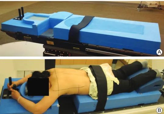
Each patient underwent two computed tomography (CT) simulations for whole breast irradiation: the first in supine position and the second in prone position. First, CT images were obtained in conventional supine and arm-up position on a breast board (CIVCO Medical Solutions, Kalona, IA). After that, second CT images were acquired in prone posi-tion using a custom-made prone breast board made of polyurethane foam with an aperture positioning left breast and a wedge to set contralateral breast away from the RT fields. Patient’s face was turned to the left to limit the rotation of her body in prone position (Fig. 1). For both setups, radio-opaque wires were placed to indicate palpable breast mass and midline of chest of the patients. On the prone breast board, radio-opaque markers were attached to point out the position of aperture and the top of board in order to provide accurate information for RT plan. Both CT images were obtained in 3.75-mm thick slices without contrast, and then transferred to the treatment planning system, Pinnacle3 (Philips Healthcare, Fitchburg, WI).
3. RT planning and dosimetric comparison
The whole breast as a clinical target volume (CTV), ipsilat-eral lung, heart, and left anterior descending coronary artery (LADCA) were contoured on each CT images by a radiation oncologist according to the Radiation Therapy Oncology
Group breast contouring atlas. Tangential-fields treatment plan with 6-MV photons in each position was designed with prescription dose of 50 Gy in 2 Gy fractions, and then the dose-volume histograms for CTV and organs at risks were compared between prone and supine positions. Primary end-point was the irradiated cardiac dose (heart and LADCA), and the secondary endpoints were the irradiated dose to ipsilateral lung and target coverage.
For target coverage, we compared the maximum and mean dose (Dmax, Dmean), the dose received by at least 95% of the
CTV (D95), homogeneity index (HI) [25], and conformity
index (CI) [26]. The HI was calculated from the formula: HI=(D2–D98)/Dp, where D98is the dose received by at least
98% of the target volume, D2is the dose received by at least
2% of the target volume, and Dpis the prescription dose to
the target volume. The D98and D2are considered to be the
minimum and maximum doses, respectively, and a lower HI value indicates a more homogeneous dose administered to the target volume. The CI was calculated from the formula: CI=(TV95/TV)×(TV95/V95), where V95 is the target volume
receiving 95% of the prescription dose, TV is the target vol-ume, and V95is the volume receiving 95% of the prescription
dose. The closer the CI is to 1 is indicative of optimal confor-mation. For normal organs, we compared Dmax, Dmean, and the
percentage of the volume that receives more than 5, 10, 20, 30, 40, and 50 Gy (V5, V10, V20, V30, V40, and V50).
4. Treatment position designation
According to study protocol, the treatment position was designated to deliver less radiation dose to the heart and LADCA, considering the maximum dose and irradiated vol-ume, while maintaining the appropriate dose (95%-107%) of the prescribed dose to CTV. The LADCA dose was consid-ered to be the determining factor of the highest priority.
As a process of designation, we evaluated the parameters between prone- and supine-position–beneficial groups for each position. Age and chest and bust sizes, volumes of CTV, heart, and LADCA in both prone and supine positions were comparable between the two groups. Lung volume in prone position for prone-position–beneficial group was smaller than for supine-position–beneficial group (1,162.1±201.1 mL vs. 1,296.4±224.7 mL, p=0.035), whereas lung volume in supine position between the two groups was not statistically different (1,081.4±213.7 mL vs. 1,197.1±232.7 mL, p=0.081). Target dose parameters for each position were all compara-ble between the two groups. Lung, heart, and LADCA dose in prone position was significantly lower for prone-position– beneficial group. In supine position, the lung dose was com-parable between the two groups but heart and LADCA dose was significantly lower for supine-position–beneficial group than for prone-position–beneficial group.
Fig. 1. Prone breast board (A) and patient setup (B).
A
5. Treatment and verification
The treatment was performed using a Varian Clinac 6Ex machine (Varian Medical Systems, Palo Alto, CA). For the patient setup verification, a weekly portal image using an electronic portal imaging device (EPID) was obtained. Intrafractional motion was assessed with images acquired at a rate of two images per second using an EPID in cine mode during one of the tangential beam delivery. To assess quan-titatively, an external marker was placed on the left breast tattoo during imaging time. The displacement of marker was compared in portal images with that of the first fraction for interfractional setup verification, and the maximum move-ment of marker during beam delivery was analyzed in the cine images for intrafractional motion.
6. Statistical analysis
Dosimetric parameters were examined by paired t test or Wilcoxon signed-rank test. The correlation between dosimet-ric benefit and treatment position were analyzed by paired t test or non-parametric statistical test including Kruskal-Wallis test and Mann-Whitney U test. Inter-/intra-fractional motions of patients were evaluated by independent samples t test. Statistical analyses were conducted using SPSS ver. 20.0.0 (IBM Corp., Armonk, NY). We considered a p-value of less than 0.05 to be statistically significant.
7. Ethical statement
This prospective study received approval from internal eview boards of Samsung Medical Center (IRB No. 2014-06-138-002) and performed in accordance with the principles of the Declaration of Helsinki and Good Clinical Practice guide-lines. All patients provided written informed consent before enrollment in this study. The trial was registered at Clinical-Trials.gov (NCT02231112).
Results
1. Total 50 enrolled patients
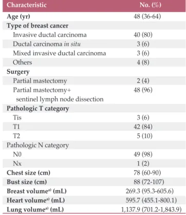
The characteristics of total 50 enrolled patients are listed in Table 1. The median age of total 50 patients was 48 years (range, 36 to 64 years). The median chest size was 78 cm (range, 60 to 90 cm) and the median bust size was 88 cm (range, 72 to 107 cm). The median breast, heart, and lung vol-umes measured in supine position was 269.3 mL (range, 95.3
mL (701.2-1,843.9 mL), respectively.
2. Dosimetric beneficial group
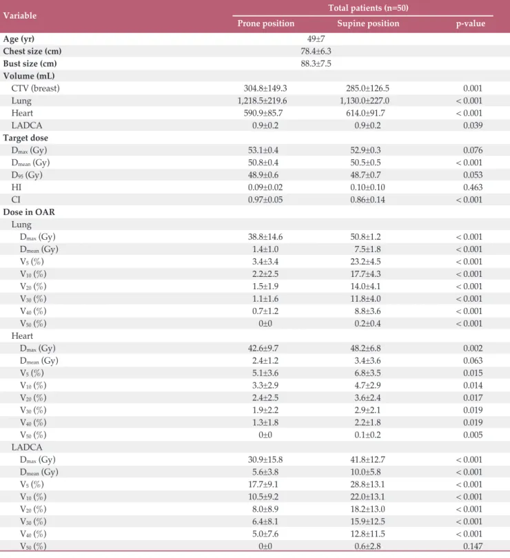
We performed the comparisons between prone and supine positions for the entire patients and the comparisons of parameters are summarized in Table 2. Prone position sig-nificantly increased the average volume of breast (CTV) (304.8±149.3 mL vs. 285.0±126.5 mL, p=0.001). Ipsilateral lung volume was also significantly increased while heart volume was decreased in prone position (1,218.5±219.6 mL vs. 1,130± 227 mL, p < 0.001 and 590.9±85.7 mL vs. 614.0±91.7 mL, p < 0.001, respectively). The volume of LADCA was equiva-lent in both positions. The radiation dose to the CTV was similar when comparing Dmaxand D95of prone and supine
positions. The Dmeanof CTV was statistically larger in prone
position but the absolute difference of the mean value bet-ween two positions was only 0.3 Gy (50.8±0.4 Gy vs. 50.5±0.5 Gy, p < 0.001). The CI in prone position was closer to 1 indi-cating more optimal target conformation (0.97±0.05 vs 0.86±
Table 1. Characteristics of total 50 participants
Values are presented as median (range) or number (%).
a)Presented volumes are measured in supine position
which is a conventional setup for patients.
Characteristic No. (%)
Age (yr) 48 (36-64)
Type of breast cancer
Invasive ductal carcinoma 40 (80) Ductal carcinoma in situ 3 (6) Mixed invasive ductal carcinoma 3 (6)
Others 4 (8)
Surgery
Partial mastectomy 2 (4)
Partial mastectomy+ 48 (96)
sentinel lymph node dissection Pathologic T category Tis 3 (6) T1 42 (84) T2 5 (10) Pathologic N category N0 49 (98) Nx 1 (2) Chest size (cm) 78 (60-90) Bust size (cm) 88 (72-107) Breast volumea)(mL) 269.3 (95.3-605.6) Heart volumea)(mL) 595.7 (455.1-800.1) Lung volumea)(mL) 1,137.9 (701.2-1,843.9)
Variable Total patients (n=50)
Prone position Supine position p-value
Age (yr) 49±7 Chest size (cm) 78.4±6.3 Bust size (cm) 88.3±7.5 Volume (mL) CTV (breast) 304.8±149.3 285.0±126.5 0.001 Lung 1,218.5±219.6 1,130.0±227.0 < 0.001 Heart 590.9±85.7 614.0±91.7 < 0.001 LADCA 0.9±0.2 0.9±0.2 0.039 Target dose Dmax(Gy) 53.1±0.4 52.9±0.3 0.076 Dmean(Gy) 50.8±0.4 50.5±0.5 < 0.001 D95(Gy) 48.9±0.6 48.7±0.7 0.053 HI 0.09±0.02 0.10±0.10 0.463 CI 0.97±0.05 0.86±0.14 < 0.001 Dose in OAR Lung Dmax(Gy) 38.8±14.6 50.8±1.2 < 0.001 Dmean(Gy) 1.4±1.0 7.5±1.8 < 0.001 V5(%) 3.4±3.4 23.2±4.5 < 0.001 V10(%) 2.2±2.5 17.7±4.3 < 0.001 V20(%) 1.5±1.9 14.0±4.1 < 0.001 V30(%) 1.1±1.6 11.8±4.0 < 0.001 V40(%) 0.7±1.2 8.8±3.6 < 0.001 V50(%) 0±0 0.2±0.4 < 0.001 Heart Dmax(Gy) 42.6±9.7 48.2±6.8 0.002 Dmean(Gy) 2.4±1.2 3.4±3.6 0.063 V5(%) 5.1±3.6 6.8±3.5 0.015 V10(%) 3.3±2.9 4.7±2.9 0.014 V20(%) 2.4±2.5 3.6±2.4 0.017 V30(%) 1.9±2.2 2.9±2.1 0.019 V40(%) 1.3±1.8 2.2±1.8 0.019 V50(%) 0±0 0.1±0.2 0.005 LADCA Dmax(Gy) 30.9±15.8 41.8±12.7 < 0.001 Dmean(Gy) 5.6±3.8 10.0±5.8 < 0.001 V5(%) 17.7±9.1 28.8±13.1 < 0.001 V10(%) 10.5±9.2 22.0±13.1 < 0.001 V20(%) 8.0±8.9 18.2±13.0 < 0.001 V30(%) 6.4±8.1 15.9±12.5 < 0.001 V40(%) 5.0±7.6 12.8±11.5 < 0.001 V50(%) 0±0 0.6±2.8 0.147
Values are presented as mean±standard deviation. CTV, clinical target volume; LADCA, left anterior descending coronary artery; Dmax, maximum dose; Dmean, mean dose; D95, dose received by at least 95% of the clinical target volume; HI,
homo-geneity index; CI, conformity index; OAR, organs at risk; VX, percentage of the volume that receives more than X Gy.
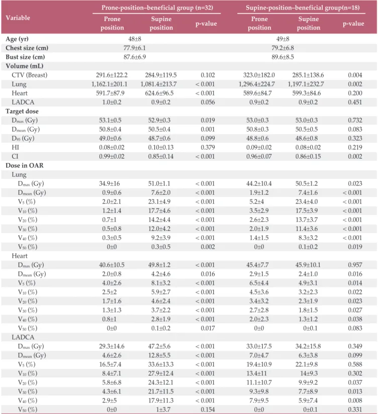
Prone-position–beneficial group (n=32) Supine-position–beneficial group(n=18)
Variable Prone Supine
p-value Prone Supine p-value
position position position position
Age (yr) 48±8 49±8 Chest size (cm) 77.9±6.1 79.2±6.8 Bust size (cm) 87.6±6.9 89.6±8.5 Volume (mL) CTV (Breast) 291.6±122.2 284.9±119.5 0.102 323.0±182.0 285.1±138.6 0.004 Lung 1,162.1±201.1 1,081.4±213.7 < 0.001 1,296.4±224.7 1,197.1±232.7 0.002 Heart 591.7±87.9 624.6±96.5 < 0.001 589.6±84.7 599.3±84.6 0.200 LADCA 1.0±0.2 0.9±0.2 0.056 0.9±0.2 0.9±0.2 0.451 Target dose Dmax(Gy) 53.1±0.5 52.9±0.3 0.019 53.0±0.3 53.0±0.3 0.732 Dmean(Gy) 50.8±0.4 50.5±0.4 0.001 50.8±0.3 50.5±0.5 0.083 D95(Gy) 49.0±0.6 48.7±0.6 0.099 48.8±0.6 48.6±0.8 0.323 HI 0.08±0.02 0.10±0.13 0.379 0.09±0.02 0.08±0.02 0.219 CI 0.99±0.02 0.85±0.14 < 0.001 0.96±0.07 0.86±0.15 0.002 Dose in OAR Lung Dmax(Gy) 34.9±16 51.0±1.1 < 0.001 44.2±10.4 50.5±1.2 0.023 Dmean(Gy) 0.9±0.6 7.6±2.0 < 0.001 1.9±1.2 7.4±1.6 < 0.001 V5(%) 2.0±2.1 23.1±4.9 < 0.001 5.2±4 23.4±4.0 < 0.001 V10(%) 1.2±1.4 17.7±4.6 < 0.001 3.5±2.9 17.5±3.9 < 0.001 V20(%) 0.7±1 14.2±4.4 < 0.001 2.6±2.3 13.7±3.7 < 0.001 V30(%) 0.5±0.8 12.0±4.2 < 0.001 2.0±1.9 11.4±3.6 < 0.001 V40(%) 0.3±0.5 9.2±3.9 < 0.001 1.4±1.5 8.3±3.2 < 0.001 V50(%) 0±0 0.3±0.5 0.002 0±0 0.1±0.2 0.019 Heart Dmax(Gy) 40.6±10.5 49.8±1.2 < 0.001 45.4±7.7 45.9±10.1 0.957 Dmean(Gy) 2.0±0.8 4.2±4.6 0.016 2.9±1.5 2.4±1.0 0.016 V5(%) 4.0±2.6 8.1±3.2 < 0.001 6.5±4.4 4.9±3.1 0.014 V10(%) 2.5±2 5.9±2.7 < 0.001 4.5±3.6 3.2±2.3 0.022 V20(%) 1.7±1.6 4.6±2.4 < 0.001 3.4±3.2 2.3±1.9 0.023 V30(%) 1.3±1.3 3.7±2.2 < 0.001 2.7±2.8 1.8±1.5 0.027 V40(%) 0.8±1 2.8±1.9 < 0.001 2.0±2.3 1.3±1.2 0.038 V50(%) 0±0 0.1±0.2 0.017 0±0 0±0.1 0.083 LADCA Dmax(Gy) 29.3±14.6 47.2±5.6 < 0.001 33.0±17.5 34.2±15.8 0.349 Dmean(Gy) 4.6±2.6 12.8±5.5 < 0.001 7.0±4.7 6.3±3.8 0.099 V5(%) 16.5±7.4 33.6±13.3 < 0.001 19.4±10.9 22.1±9.8 0.588 V10(%) 8.4±7.1 27.9±12.4 < 0.001 13.4±11 14±9.3 0.302 V20(%) 5.8±6.8 24.3±12.1 < 0.001 11.1±10.7 9.9±9.2 0.037 V30(%) 4.3±6.1 21.7±11.5 < 0.001 9.3±9.8 7.7±8.9 0.013 V40(%) 2.9±5 17.9±11.3 < 0.001 7.9±9.5 5.9±7.4 0.008 V50(%) 0±0 1±3.7 0.154 0±0 0±0.1 0.331
Values are presented as mean±standard deviation. CTV, clinical target volume; LADCA, left anterior descending coronary artery; Dmax, maximum dose; Dmean, mean dose; D95, dose received by at least 95% of the clinical target volume; HI,
homo-geneity index; CI, conformity index; OAR, organs at risk; VX, percentage of the volume that receives more than X Gy.
All dose values (Dmax, Dmean, and V5-V50) of ipsilateral lung,
heart, and LADCA were lower in prone position than in supine position (Table 2).
Among these 50 patients, 32 cases were decided as prone-position–beneficial group and 18 cases as supine-position– beneficial group in terms of dosimetric advantage after the dosimetric results of treatment plans for both setups were thoroughly compared according to study protocol. The com-parisons between prone and supine positions for each prone-and supine-position–beneficial group are described in Table 3. Age, chest size, and bust size between prone-position–ben-eficial and supine-position–benprone-position–ben-eficial groups were compara-ble (p=0.723, p=0.356, and p=0.550, respectively). For prone-position–beneficial group, prone position significantly increased lung volume (1,162.1±201.1 mL vs. 1,081.4±213.7 mL, p < 0.001) but decreased heart volume (591.7±87.9 mL vs. 624.6±96.5 mL, p < 0.001), while CTV was not statistically different between the two positions. In contrast, for supine-position–beneficial group, prone position also increased lung volume (1,296.4±224.7 mL vs. 1,197.1±232.7 mL, p=0.002) as well as CTV (323.0±182.0 mL vs. 285.1±138.6 mL, p=0.004),
but heart volume was comparable between two setups (589.6±84.7 mL vs. 599.3±84.6 mL, p=0.200). LADCA volume was comparable between the two setups for both groups.
3. Target coverage
Target coverage dose was comparable between two setups for supine-position–beneficial group in terms of Dmax, Dmean,
and D95. Target dose (Dmaxand Dmean) was higher in prone
position than in supine position for prone-position–beneficial group, but the absolute differences of those values were very minimal (Table 3). For both groups, HI was comparable between prone and supine positions, but CI was more optimal in prone position than in supine position (prone-position– beneficial group, 0.99±0.02 vs. 0.85±0.14, p < 0.001; supine-position–beneficial group, 0.96±0.07 vs. 0.86±0.15, p=0.002) (Table 3).
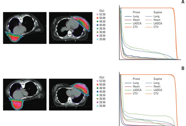
Fig. 2. Dosimetric comparison of organs at risk between supine and prone positioning with isodose lines and dose volume
histogram for a case with chest size of 84 cm and breast volume in prone position of 302 mL (A) and a case with chest size of 68 cm and breast volume in prone position of 462 mL (B). The lung (blue), heart (red), and left anterior descending coronary artery (LADCA, green) are delineated as organs at risk. CTV, clinical target volume.
A
Lung Heart LADCA CTV Prone Supine Lung Heart LADCA CTV 52.50 50.00 48.50 40.00 38.50 30.00 28.50 20.00 (Gy) (Gy)B
Lung Heart LADCA CTV Prone Supine Lung Heart LADCA CTV 52.50 50.00 48.50 40.00 38.50 30.00 28.50 20.004. Normal organ sparing
Lung dose (Dmax, Dmean, and V5-V50) was substantially
decreased in prone position than in supine position for both prone and supine-position–beneficial groups, along with the fact that absolute dose differences were considerably larger in prone-position–beneficial group than in supine-position– beneficial group (Table 3). Prone position for prone-position– beneficial group significantly decreased the dose to heart and LADCA (Dmax, Dmean, and V5-V50). On the other hand, prone
position for supine-position–beneficial group significantly increased the heart dose (Dmeanand V5-V50) (Table 3). For
supine-position–beneficial group, prone position increased V20, V30, and V40of LADCA considerably, while Dmax, Dmean,
V5, V10, and V50of LADCA were similar (Table 3). Fig. 2
showed dosimetric comparison of normal organs between supine and prone position with isodose lines and dose vol-ume histo-gram for a case with chest size of 84 cm and breast volume in prone position of 302 mL (Fig. 2A) and a case with chest size of 68 cm and breast volume in prone position of 462 mL (Fig. 2B).
5. Patient motion
Comparisons of inter- and intra-fractional motions bet-ween patients treated in supine and prone positions are depicted in Fig. 3. For interfractional movement, a total of 116 EPID images of prone position treatments and 74 images of supine position treatments were reviewed. The average of setup variation was 3.57±3.56 mm in superior-inferior (SI) direction and 2.61±2.02 mm in anterior-posterior (AP) direc-tion for prone posidirec-tion treatment and 1.35±1.05 mm in SI direction and 1.46±1.14 mm in AP direction for supine posi-tion treatment. Intrafracposi-tional moposi-tion was assessed by 0.19±0.13 mm in SI direction and 0.48±0.32 mm in AP direc-tion from 140 EPID cine images of prone posidirec-tion treatment
and 0.72±0.40 mm in SI direction and 0.86±0.49 mm in AP direction from 99 EPID images of supine position treatment. Interfractional setup variation was larger in prone position treatment (p < 0.001 for both SI and AP direction), whereas motion during treatment was lager in supine position treat-ment (p < 0.001 for both SI and AP direction).
Discussion
To save the heart for left breast cancer patients, several techniques that displace the heart from the irradiation field have been utilized, which include respiratory motion man-agement (breath hold) or patient positioning in prone or lat-eral decubitus. Breath-hold technique usually requires more than twice as much treatment time as conventional RT, and patients must control their own breathing, which is not fea-sible in elderly patients or those with poor performance sta-tus [14]. In case of lateral decubista-tus position, due to relatively unstable posture, the reproducibility, which is one of the most important challenges in RT, may be seriously degraded. Even though the prone position may also be discomfort dur-ing the treatment compared to the supine position, it is more advantageous than the two methods mentioned above [13]. Prone breast RT has been known as effective for patients with large pendulous breast, such as breast volume larger than 750 mL or 1,000 mL [19,20]. As Asian women usually have a smaller breast size, prone position was considered not having dosimetric advantage to Asian breast cancer patients. There were studies about the breast size, but they have not invol-ved such small breasts [22] or prone position did not reduce in-field heart volume statistically significantly in women with breast size smaller than 750 mL [17]. Even in
Av er ag e in tra -fr ac tio na l m ot io n (m m ) 1.5 0 0.5 Sup-Inf Ant-Post 1.0 Prone Supine Av er ag e in te r-fra ct io na l m ot io n (m m ) 8 0 2 4 Sup-Inf Ant-Post 6 Prone Supine
A
B
Fig. 3. Comparison of inter- (A) and intra-fractional (B) motion between patients treated in supine and prone positions.
evaluated for prone positioning [23,27]. However, this pro-spective study enrolled by 50 patients with mean breast size of 305 mL (in prone position) revealed that such small breast size could also gain dosimetric benefit on heart and lung by prone position while sustaining or even improving target coverage.
CTV breast volume of total 50 participants ranged 94.2 to 786.5 mL (median, 294.1 mL) in prone position and 95.3 to 605.6 mL (median, 269.3 mL) in supine position. Prone posi-tion significantly increased the average CTV volume (304.8± 149.3 mL vs. 285.0±126.5 mL, p=0.001) in total patients, but the average CTV volume was comparable between prone and supine-position–beneficial groups (prone 291.6±122.2 mL vs. supine 323.0±182.0 mL, p=0.470). Prone-position–ben-eficial group had CTV volume in prone position ranging from 103.2 to 638.4 mL. Prone position increased the volume of ipsilateral lung, and the relative lung volume change by prone position was similar between prone and supine-posi-tion–beneficial groups. Yet, the heart volume decreased in prone position for total patients, but the relative heart vol-ume change by prone position was greater in prone-position– beneficial group than in supine-position–beneficial group (but not statistically significant). However, we could not find the relationship between dosimetric gain and patient char-acteristics (breast or chest size) or volume change by prone positioning. The certain optimal subgroup having definite benefit by prone position was difficult to find probably due to a small number of participant in this Phase II study and small range of patients’ breast size. Nevertheless, we could find dosimetric advantage in prone breast RT for patients having such small breast size even around 100 ml. A further study with a larger number of patients will be needed to find the ideal patient group who will have definite dosimetric gain. This further study will allow us to identify the patient for prone breast RT without two CT simulations for the com-parison between supine and prone positions so as to avoid unnecessary radiation exposure to the patient.
Similar to earlier studies about prone breast RT [28,29], our study also found that the treatment in prone position showed larger interfractional motion but smaller intrafractional motion than the treatment in supine position. The patient's breathing was restricted due to the compression of the chest
wall by lying on the prone breast board, so the intrafractional motion was limited than the supine position. However, there is no correlation between patient characteristics and the inter-or intra-fractional motion. Among 32 patients having dosi-metric benefit in prone position, three patients showed extremely unstable setup during the process for the localiza-tion of treatment center before starting treatment, so physi-cians reviewed their treatment plans whether there was a critical clinical decision point and decided to treat them in supine position. Due to the lack of resource in our institution, we could utilize only EPID not cone-beam CT for these treat-ments. So, we were able to check the patient setup in SI and AP directions but not in lateral direction. The lateral setup of the patient was confirmed by localizing laser and patient tat-too and by checking the position of a marker atta-ched to the midline of the patient. We found that a few patients treated in prone position, who enrolled in early-phase of this study, experienced contralateral breast exposure, especially with collimator rotation in the treatment parameter. So, we che-cked whether radiation fields extended to the contralateral breast with Gafchromic EBT3 film placed under the con-tralateral breast of the patient in the first few sessions of ment. Also, we tried not to rotate the collimator in the treat-ment plan for prone position. With these efforts, the con-tralateral breast exposure problem was resolved.
In conclusion, prone breast RT could be beneficial to a sub-set of small breast patients since it substantially spared nor-mal organs while achieving adequate coverage of breast tissue. We observed patient-specific and considerable inter-fractional setup error, but it was not the extent to negatively affect treatment quality of whole breast irradiation. Further prospective study is required to validate the potential benefit and the optimal patient group with prone breast RT.
Conflicts of Interest
Conflict of interest relevant to this article was not reported.
Acknowledgments
This research has been supported by Korean Breast Cancer Foun-dation 14-03.
1. Torre LA, Islami F, Siegel RL, Ward EM, Jemal A. Global can-cer in women: burden and trends. Cancan-cer Epidemiol Biomark-ers Prev. 2017;26:444-57.
2. Jung KW, Won YJ, Kong HJ, Lee ES; Community of
Popula-tion-Based Regional Cancer Registries. Cancer statistics in Korea: incidence, mortality, survival, and prevalence in 2015. Cancer Res Treat. 2018;50:303-16.
3. Early Breast Cancer Trialists' Collaborative Group; Darby S,
References
McGale P, Correa C, Taylor C, Arriagada R, et al. Effect of radiotherapy after breast-conserving surgery on 10-year recur-rence and 15-year breast cancer death: meta-analysis of indi-vidual patient data for 10,801 women in 17 randomised trials. Lancet. 2011;378:1707-16.
4. Darby SC, McGale P, Taylor CW, Peto R. Long-term mortality from heart disease and lung cancer after radiotherapy for early breast cancer: prospective cohort study of about 300,000 women in US SEER cancer registries. Lancet Oncol. 2005;6:557-65.
5. Marks LB, Yu X, Prosnitz RG, Zhou SM, Hardenbergh PH, Blazing M, et al. The incidence and functional consequences of RT-associated cardiac perfusion defects. Int J Radiat Oncol Biol Phys. 2005;63:214-23.
6. Correa CR, Litt HI, Hwang WT, Ferrari VA, Solin LJ, Harris EE. Coronary artery findings after left-sided compared with right-sided radiation treatment for early-stage breast cancer. J Clin Oncol. 2007;25:3031-7.
7. Darby SC, Ewertz M, McGale P, Bennet AM, Blom-Goldman U, Bronnum D, et al. Risk of ischemic heart disease in women after radiotherapy for breast cancer. N Engl J Med. 2013;368: 987-98.
8. Paul Wright G, Drinane JJ, Sobel HL, Chung MH. Left-sided breast irradiation does not result in increased long-term car-diac-related mortality among women treated with breast-con-serving surgery. Ann Surg Oncol. 2016;23:1117-22.
9. Yu TK, Whitman GJ, Thames HD, Buzdar AU, Strom EA, Perkins GH, et al. Clinically relevant pneumonitis after sequ-ential paclitaxel-based chemotherapy and radiotherapy in breast cancer patients. J Natl Cancer Inst. 2004;96:1676-81. 10. Borst GR, De Jaeger K, Belderbos JS, Burgers SA, Lebesque JV.
Pulmonary function changes after radiotherapy in non-small-cell lung cancer patients with long-term disease-free survival. Int J Radiat Oncol Biol Phys. 2005;62:639-44.
11. Campana F, Kirova YM, Rosenwald JC, Dendale R, Vilcoq JR, Dreyfus H, et al. Breast radiotherapy in the lateral decubitus position: a technique to prevent lung and heart irradiation. Int J Radiat Oncol Biol Phys. 2005;61:1348-54.
12. Kirova YM, Hijal T, Campana F, Fournier-Bidoz N, Stilhart A, Dendale R, et al. Whole breast radiotherapy in the lateral decubitus position: a dosimetric and clinical solution to decrease the doses to the organs at risk (OAR). Radiother Oncol. 2014;110:477-81.
13. Huppert N, Jozsef G, Dewyngaert K, Formenti SC. The role of a prone setup in breast radiation therapy. Front Oncol. 2011; 1:31.
14. Remouchamps VM, Letts N, Vicini FA, Sharpe MB, Kestin LL, Chen PY, et al. Initial clinical experience with moderate deep-inspiration breath hold using an active breathing control device in the treatment of patients with left-sided breast cancer using external beam radiation therapy. Int J Radiat Oncol Biol Phys. 2003;56:704-15.
15. Shah C, Badiyan S, Berry S, Khan AJ, Goyal S, Schulte K, et al. Cardiac dose sparing and avoidance techniques in breast can-cer radiotherapy. Radiother Oncol. 2014;112:9-16.
16. Mulliez T, Veldeman L, van Greveling A, Speleers B, Sadeghi
S, Berwouts D, et al. Hypofractionated whole breast irradia-tion for patients with large breasts: a randomized trial com-paring prone and supine positions. Radiother Oncol. 2013;108: 203-8.
17. Formenti SC, DeWyngaert JK, Jozsef G, Goldberg JD. Prone vs supine positioning for breast cancer radiotherapy. JAMA. 2012;308:861-3.
18. Lymberis SC, deWyngaert JK, Parhar P, Chhabra AM, Fenton-Kerimian M, Chang J, et al. Prospective assessment of optimal individual position (prone versus supine) for breast radiother-apy: volumetric and dosimetric correlations in 100 patients. Int J Radiat Oncol Biol Phys. 2012;84:902-9.
19. Kirby AM, Evans PM, Donovan EM, Convery HM, Haviland JS, Yarnold JR. Prone versus supine positioning for whole and partial-breast radiotherapy: a comparison of non-target tissue dosimetry. Radiother Oncol. 2010;96:178-84.
20. Fernandez-Lizarbe E, Montero A, Polo A, Hernanz R, Moris R, Formenti S, et al. Pilot study of feasibility and dosimetric comparison of prone versus supine breast radiotherapy. Clin Transl Oncol. 2013;15:450-9.
21. Bartlett FR, Colgan RM, Donovan EM, McNair HA, Carr K, Evans PM, et al. The UK HeartSpare Study (Stage IB): ran-domised comparison of a voluntary breath-hold technique and prone radiotherapy after breast conserving surgery. Radiother Oncol. 2015;114:66-72.
22. Hannan R, Thompson RF, Chen Y, Bernstein K, Kabarriti R, Skinner W, et al. Hypofractionated whole-breast radiation therapy: does breast size matter? Int J Radiat Oncol Biol Phys. 2012;84:894-901.
23. Chen JL, Cheng JC, Kuo SH, Chan HM, Huang YS, Chen YH. Prone breast forward intensity-modulated radiotherapy for Asian women with early left breast cancer: factors for cardiac sparing and clinical outcomes. J Radiat Res. 2013;54:899-908. 24. Lee HY, Chang JS, Lee IJ, Park K, Kim YB, Suh CO, et al. The
deep inspiration breath hold technique using Abches reduces cardiac dose in patients undergoing left-sided breast irradia-tion. Radiat Oncol J. 2013;31:239-46.
25. Yoon M, Park SY, Shin D, Lee SB, Pyo HR, Kim DY, et al. A new homogeneity index based on statistical analysis of the dose-volume histogram. J Appl Clin Med Phys. 2007;8:9-17. 26. Feuvret L, Noel G, Mazeron JJ, Bey P. Conformity index: a
review. Int J Radiat Oncol Biol Phys. 2006;64:333-42.
27. Takahashi K, Morota M, Kagami Y, Okamoto H, Sekii S, Inaba K, et al. Prospective study of postoperative whole breast radiotherapy for Japanese large-breasted women: a clinical and dosimetric comparisons between supine and prone posi-tions and a dose measurement using a breast phantom. BMC Cancer. 2016;16:757.
28. Morrow NV, Stepaniak C, White J, Wilson JF, Li XA. Intra- and interfractional variations for prone breast irradiation: an indi-cation for image-guided radiotherapy. Int J Radiat Oncol Biol Phys. 2007;69:910-7.
29. Mitchell J, Formenti SC, DeWyngaert JK. Interfraction and intrafraction setup variability for prone breast radiation ther-apy. Int J Radiat Oncol Biol Phys. 2010;76:1571-7.