비조영증강
MDCT
에서 관전압과 절편 두께에 따른 요로결석의
HU
측정
동경래1· 정명진2,3· 전재두2· 김미현1,4· 권대철5· 정운관6,*1광주보건대학교 방사선과, 2조선대학교병원 영상의학과, 3조선대학교대학원 보건학과, 4전북대학교대학원 방사선과학기술학과, 5신한대학교 방사선학과, 6조선대학교 원자력공학과
HU Measurement of Urinary Stone according to the Tube
Voltage and Slice Thickness in Non-enhancement MDCT
Kyung-Rae Dong
1, Myung-Jin Jung
2,3, Jae-Doo Jeon
2, Mi-Hyun Kim
1,4,
Dae-Cheol Kweon
5and Woon-Kwan Chung
6,*
1Department of Radiological Technology, Gwangju Health University,
73, Bungmun-daero 419 beon-gil, Gwangsan-gu, Gwangju 62271, Republic of Korea
2Department of Diagnostic Radiology, Chosun University Hospital, 365, Pilmun-daero,
Dong-gu, Gwangju 61453, Republic of Korea
3Department of Health Science, Graduate School of Chosun University,
309, Pilmun-daero, Dong-gu, Gwangju 61452, Republic of Korea
4Department of Radiation Science & Technology, Graduate School of Chonbuk National University,
567 Baekje-daero, Deokjin-gu, Jeonju-si, Jeollabuk-do 54896, Republic of Korea
5Department of Radiological Science, Shinhan University, 95, Hoam-ro, Uijeongbu-si,
Gyeonggi-do 11644, Republic of Korea
6Department of Nuclear Engineering, Chosun University, 375, Seosuk-dong, Dong-gu,
Gwangju 61452, Republic of Korea
Abstract - To distinguish urinary stones, we tried to identify the components of urinary stone using HU of nonenhancement MDCT. HU was measured according to the tube voltage and the slice thickness of the section of urinary stone. After injecting urinary stone into a small plastic bottle, experimental phantom was prepared using pork similar to human body. 640 slice MDCT. HU of urinary stone was measured according to each tube voltage and slice thickness in the obtained images. HU of urinary stones according to tube voltage were 135kVp(373.68±30.20 HU), 100kVp(525.94±44.93HU) and 80kVp(691±80.80HU), and the slice thickness was 0.5mm (752±25.71HU) minimum(545.96±29.89HU), 1.5mm(462.68±46.35HU), 2mm(360.43±66.19 HU). It was found that the HU of urinary stone was increased with decreasing tube voltage, and the HU of urinary stone was decreased with increasing thickness of the section. Non-enhancement MDCT for urinary stone is thought to be useful for the diagnosis of urinary stone using thin slice thickness using low tube voltage.
Key words : HU, MDCT, Slice thickness, Tube voltage, Urinary stone
─ 119 ─ Technical Paper
* Corresponding author: Woon-Kwan Chung, Tel. +82-62-230-7166, Fax. +82-62-232-9218, E-mail. [email protected]
서 론
요로결석은 거주 지역 및 환경에 따라 발생빈도의 발병 률이 다르게 분포하지만, 최근 우리나라에서도 식생활의 서 구화로 인해 과체중, 고지혈증, 동물성 단백질의 과다 섭 취로 인하여 요로결석의 환자가 증가추세에 있다(Lee et
al. 1996; Borghi et al. 2002). 요로결석은 인구의 약 12%에
서 발생하는 질환이며 10년 이내의 재발률은 50%에 달하 며(Sierakowski et al. 1978; Leusmann et al. 1995), 요로결 석 환자의 약 87%에서 급성측복통을 호소하고, 이외의 오 심, 구토, 육안적 혈뇨 등의 증상으로 병원에 내원하게 된다 (Kim et al. 2003). 또한 요석은 흔하게 나타나는 비뇨기계 의 질환 중의 하나이며 비뇨기계통에 이물로 작용하여 요로 감염을 일으키고 배뇨의 장애를 초래하기도 하며, 드물게는 신부전증을 일으키는 질환으로 임상적으로 보고되고 있다 (Lee et al. 2006). 과거의 요로결석의 진단은 전통적으로 배설성 요로조영 술(Intravenous pyelography; IVP)을 시행하여 요로결석의 유무를 평가하였는데 배설성 요로조영술은 조영제의 사용 과 장내 가스 제거를 위한 검사의 전처치로 인하여 환자의 불편을 초래하여 신속한 검사가 힘들어 임상적인 진단에 어 려움을 수반하고 있다. 이에 비해 비조영증강 전산화단층촬 영술(Multidetector computed tomography; MDCT)은 요석 의 진단에 있어서 조영술이나 초음파에 비해 더욱 신속하고 전처치 조영제에 대한 부작용의 반응 위험이 적어 안전하 고 높은 정확성을 보이고 있으며(Levine et al. 1997) 방사선 투과성 요석을 진단할 수 있고, 요로결석 이외의 측복통의 원인도 동시에 발견할 수 있어 많은 정보를 획득할 수 있는 부가적 장점을 가지고 있다. 비뇨기계의 요로결석의 치료는 체외충격파쇄석술 (Extra-corporeal shock wave lithotripsy; ESWL)과 비뇨기과학의 발달로 괄목할 만한 발전을 이루었지만 요로결석의 원인과 발생기전에 대해서는 아직도 명확하게 밝혀지지 않았으나 (Uribarri et al. 1989) 요로결석 환자의 치료방법을 결정하는 데 있어 중요한 인자로는 환자의 증상 정도, 요로결석의 크 기 및 성분 등 치료방법의 결정에 영향을 미치는 주요인자 들 중 요로결석의 성분에 대한 정보는 효과적인 치료방법에 중요한 역할을 한다(Kakinuma et al. 1999).
Mitcheson 등(Metcheson et al. 1983)은 실험적 연구에 서 CT를 이용하여 HU(Hounsfield unit) 차이를 측정함으로 써 요산석을 다른 성분의 요석과 구분할 수 있다고 보고하 였다. 이후 다양한 연구 결과들이 보고되었으나 대부분 물, 공기 및 동물의 장기를 이용한 실험실 연구로 보고하였다 는 점이었고, HU 사이에 통계적 차이만 있을 뿐 중복되는 영역으로 인해 명확한 구분이 힘들다는 점 등에서 실제 임 상적용의 한계를 보고하였다(Nakada et al. 2000; Saw et al.
2000). 지금 임상에서는 MDCT의 단면 간격을 실험적 연구처럼 연구하기 어려움이 있어 실험적으로 관전압 및 절편 두께 에 따른 비조영증강의 MDCT를 시행하여 요로결석의 HU 측정 연구 필요성이 있다. 이에 연구의 목적은 실제적으로 요로결석의 성분분석 및 임상적 진단에 있어 비조영증강 MDCT를 이용하여 관전압과 절편의 두께에 따른 요로결석 의 HU 측정을 실험적으로 연구 보고하여 요로결석의 진단 및 치료에 일조하고자 한다.
재료 및 방법
1. 팬텀 제작팬텀 제작은 총 9개의 요로결석의 요산석(uric acid stone) 을 작은 플라스틱 병(plastic bottle) 안에 각각 삽입하여 인 체와 비슷한 돈육을 이용하여 실험팬텀을 제작하였다(Fig. 1).
2. 스캔 및 통계분석
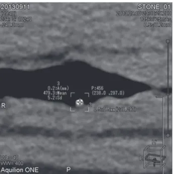
640 Slice-MDCT(Aquilion ONE, Toshiba Medical Sys-tems Corporation, Otowara, Japan)를 이용하여 rotation time 0.5sec, D-FOV 99.8(SS), 관전압은 135kVp, 100kVp, 80 kVp로 설정하여 volume scan 방식을 사용하여 스캔하였고, 관전압과 절편 두께에 따라 HU을 각각 측정하였다(Fig. 2).
본 연구에서 수집된 자료의 통계분석은 Microsoft Excel software(Microsoft Office 2007, Microsoft Corporation, Redmond WA, USA)를 사용하여 각각의 관전압과 절편 두 께에 따른 HU 차이의 유의성을 알아보기 위해 대응표본 T-검정을 통계처리하였고, p<0.05인 경우에 통계학적으로 유 의한 것으로 간주하였다.
결 과
1. 관전압에 따른 요로결석의 HU 관전압에 따른 요로결석의 HU는 관전압 135kVp에서는 373.69±30.19HU, 100kVp에서는 525.94±44.93HU, 80 kVp에서는 691.47±50.98HU이었다(Fig. 3). MDCT를 이용한 요로결석의 HU측정에서 관전압에 따른 HU값의 변화는 통계적으로 유의한 것으로 나타났다(p< 0.05)(Table 1). 2. 절편 두께에 따른 요로결석의 HU 절편 두께에 따른 요로결석의 HU는 0.5mm에서는 752.38 ±25.72HU, 1mm에서는 545.96±29.89HU, 1.5mm에서는 462.68±46.35HU, 2mm에서는 360.43±66.19HU로 측정 되었다(Fig. 4). 절편 두께에 따른 요로결석의 HU의 변화는 통계적으로 유의한 것으로 나타났다(p<0.05)(Table 2).고 찰
CT는 일반적으로 높은 관전압의 고에너지를 사용하기 때 문에 콤프톤 산란이 주로 상호작용으로 발생하고 있으며, 엑스선의 감약은 인체조직의 밀도와 비례하고 있다. 이 투 과된 엑스선의 HU는 엑스선 흡수에 따른 선감약계수(linear attenuation coefficient)를 계산하여 HU로 표현한다. 비뇨기 계의 요로결석에 대한 비조영증강의 MDCT는 기존의 영상Fig. 2. Measurement of HU using the pork inserted urinary stone
phantom.
Table 1. HU measurement of urinary stones according to the tube
voltage Tube voltage (kVp)
Maximum
(HU) Minimum(HU) Mean±SD(HU) p
135 1031.30 -73.33 373.69±30.19
100 1342.00 26.67 525.94±44.93 0.0023
80 1660.67 17.33 691.47±50.98 0.0020
Table 2. HU measurement of urinary stones for slice thickness
Slice thickness
(mm)
Maximum
(HU) Minimum(HU) Mean±SD(HU) p
0.5 958.19 551.93 752.38±25.72
1 705.67 389.44 545.96±29.89 0.0155
1.5 609.96 319.30 462.68±46.35 0.0078
2 492.07 234.07 360.43±66.19 0.0084
Fig. 3. HU distributions of the urinary stones according to the tube
voltage. Mean Maximum Minimum 2000 1500 1000 500 0 - 500 HU Tube voltage(kV) 135 100 80
Fig. 4. HU distributions of the urinary stones according to the slice
thickness. 1200 1000 800 600 400 200 0 HU Slice thickness(mm) 0.5 1 1.5 2 Mean Maximum Minimum
의학 검사에 비해 빠르고 안전하게 검사가 가능하며 정확 한 진단법으로 임상적으로 보고가 되고 있다(Yilmaz et al. 1998). 이러한 비조영증강 MDCT의 임상적 검사에서는 몇 가지 단점을 동반하고 있다. 경정맥요로조영술에 비해 상대 적인 신기능 평가가 어려우며, 전체 요관의 주행을 한눈에 파악하기 힘들고, 요석의 완전폐색 여부를 알 수 없어 요로 결석제거의 응급여부를 상대적으로 알 수 없다. 그리고 외 국과는 달리 우리나라에서 연구에 따르면 환자의 비용 부 담이 경정맥요로조영술에 비하여 4.7배 이상 증가한다고 보 고하였다(Kim et al. 2003). 하지만 요로결석이 의심되는 환 자에 있어서 비조영증강 MDCT는 비교적 짧은 시간에 데 이터를 얻을 수 있다. 한 번에 정지된 호흡기간에 많은 양의 정보를 얻을 수 있고 한 번의 정지된 호흡기간에 시행하여 환자의 움직임이나 호흡에 따른 오차를 줄일 수 있으며, 조 영제를 사용하지 않아 조영제로 인한 부작용이 없다. 요로 결석 이외의 질환을 진단할 수 있는 장점이 있어, 이 때문에 요로결석의 진단에 있어서 유용한 진단 방법이 된다.
Mitcheson 등(Metcheson et al. 1983)은 요석 성분을 구분 하기 위해 MDCT를 이용한 이래 지금까지 많은 연구에서 비조영증강 MDCT의 HU를 이용하여 요석의 성분을 구분 하고자 하였다. 실험실 연구에서 요석에 대하여 1mm의 단 면 간격으로 비조영증강 MDCT을 스캔하였을 때 요산석은 칼슘석, 시스틴석, brushite, struvite 등과 쉽게 감별된다고 보고하였다(Mostafavi et al. 1998). Newhouse 등(Newhouse
et al. 1984)은 2mm 절편 간격으로 실험실 연구를 진행하여 요산석(uric acid) 및 시스틴석(cystine stone)을 다른 요로결 석 성분과 감별할 수 있다고 하였다. 결과에서 관전압에 따른 HU의 변화는 관전압 135kVp를 기준으로 100kVp, 80kVp 모두 통계적으로 p<0.05로 유 의하게 차이가 나타남을 알 수 있었으며, 관전압 135kVp에 서는 373.69±30.19HU, 100kVp에서는 525.94±44.93HU, 80kVp에서는 691.47±50.98HU로 관전압이 감소하면 요 로결석의 HU가 커짐을 알 수 있었다. 절편 두께에 다른 HU의 변화 또한 0.5mm를 기준으로 1 mm, 1.5mm, 2mm 모두 통계적으로 p<0.05로 유의하게 차 이가 나타남을 알 수 있었다. 그러므로 관전압이 낮게 조사 될수록 요로결석의 HU가 증가하였고, 절편의 두께가 두꺼 울수록 HU가 감소함을 실험적 연구로 알 수 있었다. 임상에서 MDCT을 이용하여 절편 두께에 대한 실험적 연구는 어려움을 동반한다. 실험적 연구로는 Saw 등(Saw et al. 2000)은 127개의 요석에 대하여 1mm, 3mm, 10mm 절 편 두께로 비조영증강 MDCT를 시행하였을 때 1mm 간격 으로 스캔에서 요석 성분이 가장 명확히 구분되었다고 보 고하였으며, 이 연구를 바탕으로 요석에 대하여 비조영증강 MDCT를 검사할 때 단면 간격이 좁을수록 요석 성분의 감 별에 유리하다고 보고하였다. 이에 본 연구에서도 절편 두 께가 0.5mm에서 평균 752HU로 가장 높게 측정되었고, 절 편 두께가 얇을수록 요로결석의 HU가 낮게 측정되었다. 이 는 요로결석 주위에 위치한 연부조직(soft tissue)의 일부가 비조영증강 MDCT 절편 두께에 포함되어 HU가 낮게 측정 되는 현상을 부분용적효과(volume averaging effect) 형상으 로 보고되고 있다(Nakada et al. 2000; Zarse et al. 2004).
연구의 제한점으로는 첫째, 순수한 요산석을 주성분의 단 일성분을 대상으로 분석하였다. 혼합 및 다양한 성분의 요 로결석으로 실험을 진행하지 못해 추후 연구에서는 다양 한 성분의 요로결석을 이용하여 관전압과 절편 두께의 다양 한 파라미터를 이용하여 전향적인 연구가 시행된다면 본 연 구가 도움이 될 수 있을 것으로 생각한다. 둘째, 통계분석을 위한 자료가 부족하여 다양한 파라미터를 이용하여 분석한 다면 임상적 지침으로 적용 가능한 자료의 연구가 될 수 있 을 것으로 생각된다.
결 론
MDCT의 관전압이 감소할수록 요로결석의 HU가 높게 측정되었고, 절편 두께가 두꺼워질수록 요로결석의 HU가 감소함을 알 수 있었다. 이에 요로결석을 진단하고 측정하 기 위해서는 비조영증강 MDCT에서 낮은 관전압을 이용한 얇은 절편 두께를 사용하여 임상에서 요로결석의 진단에 도 움이 될 것으로 생각된다.사 사
This study was supported by the research fund of Chosun University, 2014.
참 고 문 헌
Borghi L, Schianchi T, Meschi T, Guerra A, Allegri F, Maggio-re U and Novarini A. 2002. Comparison of two diets for the prevention of recurrent stones in idiopathic hypercalci-uria. N. Engl. J. Med. 346(2):77-84.
Kakinuma R, Ohmatsu H, Kaneko M, Eguchi K, Naruke T, Nagai K, Nishiwaki Y, Suzuki A and Moriyama N. 1999. Detection failures in spiral CT screening for lung cancer: analysis of CT findings. Radiology. 212(1):61-66.
Kim HS, Jang SW, Jeong YB, Kim YG and Kim JS. 2003. The usefulness of unenhanced helical computerized to-mography in patients with urinary calculi. Korean J. Urol.
Lee MW, Jeong YB and Kim YG. 2006. Biochemical charac-teristics of serum and urine in the patients with uric acid stone. Korean J. Urol. 47(7):712-716.
Lee SJ, Kim DK, Rho SK, Huh JS, Lee HL, Lee CH, Chang SG and Kim JI. 1996. Clinical observations in 4468 cases of patients with urinary stones. Korean J. Urol. 37(8):877-887.
Leusmann DB, Niggemann H, Roth S and Ahlen HV. 1995. Recurrence rates and severity of urinary calculi. Scand. J.
Urol. Nephrol. 29(3):279-283.
Levine JA, Neithlich J, Verga M, Dalrymple NK and Smith RC. 1997. Ureteral calculi in patients with flank pain cor-relation of plain radiography with unenhanced helical CT.
Radiology. 204(1):27-31.
Mitcheson HD, Zamenhof RG, Bankoff MS and Prien EL. 1983. Determination of the chemical composition of urinary calculi by computerized tomography. J. Urol.
130(4):814-9.
Mostafavi MR, Ernst RD and Saltzman B. 1998. Accurate de-termination of chemical composition of urinary calculi by spiral computerized tomography. J. Urol. 159(3):673-675. Nakada SY, Hoff DG, Attai S, Heisey D, Blankenbaker D and
Pozniak M. 2000. Determination of stone composition by noncontrast spiral computed tomography in the clinical set-ting. Urology. 55(6):816-819.
Newhouse JH, Prien EL, Amis ES Jr, Dretler SP and Pfister RC. 1984. Computed tomographic analysis of urinary
cal-culi. Am. J. Roentgenol. 142(3):545-548.
Saw KC, McAteer JA, Monga AG, Chua GT, Lingeman JE and Williams JC Jr. 2000. Helical CT of urinary calculi: effect of stone composition, stone size, and scan collimation. Am.
J. Roentgenol. 175(2):329-332.
Sierakowski R, Finlayson B, Landes RR, finlayson CD and Sierakowski N. 1978. The frequency of urolithiasis inhos-pital discharge diagnosis in the United States. Invest. Urol.
15(6):438-441.
Uribarri J, Oh MS and Carroll HJ. 1989. The first kidney stone.
Ann. Intern. Med. 111(12):1006-1009.
Yilmaz S, Sindel T, Arslan G, Ozkaynak C, Karaali K, Kabaa-lioglu A and Lueleci E. 1998. Renal colic: comparison of spiral CT, US and IVU in the detection of ureteral calculi.
Eur. Radiol. 8(2):212-217.
Zarse CA, McAteer JA, Tann M, Sommer AJ, Kim SC, Pater-son RF, Halt EK. Lingeman JE and Willimams JC Jr. 2004. Helical computed tomography accurately reports urinary stone composition using attenuation values: in vitro verifi-cation using high-resolution micro-computed tomography calibrated to fourier transform infrared microspectroscopy.
Urology. 63(5):828-833.
Received: 26 February 2018 Revised: 21 March 2018 Revision accepted: 30 May 2018