Introduction
Medication-related osteonecrosis of the jaw
(MRONJ)
is a well-known complication of treatment with
bisphos-phonates, denosumab, and other drugs, such as
anti-angio-genic agents and novel anti-cancer drugs. The diagnosis of
MRONJ is currently based on clinical parameters alone.
1Osteoradionecrosis
(ORN) is a pernicious complication of
radiotherapy in head and neck cancer. The mechanism of
pathogenesis is still under investigation, although the most
frequently reported cause is radiation arteritis.
2Histological
analysis and radiographic features are not considered
man-datory for these diagnoses, and most patients do not undergo
a biopsy from the exposed necrotic bone.
3Multimodal imaging, such as panoramic radiography,
4scintigraphy, multidetector computed tomography
(MDCT),
magnetic resonance imaging,
5-8and single-photon
emis-sion computed tomography-computed tomography
9,10are
useful for detecting MRONJ and ORN. Furthermore,
cone-CBCT imaging and histopathological characteristics of osteoradionecrosis and
medication-related osteonecrosis of the jaw
Ichiro Ogura
1,*, Yoshiyuki Minami
1, Junya Ono
2, Yoriaki Kanri
2, Yasuo Okada
2,
Kensuke Igarashi
3, Maiko Haga-Tsujimura
4,5, Ken Nakahara
5, Eizaburo Kobayashi
61Department of Oral and Maxillofacial Radiology, The Nippon Dental University School of Life Dentistry at Niigata, Niigata, Japan 2Department of Pathology, The Nippon Dental University School of Life Dentistry at Niigata, Niigata, Japan
3Department of Dental Materials Science, The Nippon Dental University School of Life Dentistry at Niigata, Niigata, Japan 4Department of Histology, The Nippon Dental University School of Life Dentistry at Niigata, Niigata, Japan
5Advanced Research Center, The Nippon Dental University School of Life Dentistry at Niigata, Niigata, Japan
6Department of Oral and Maxillofacial Surgery, The Nippon Dental University School of Life Dentistry at Niigata, Niigata, Japan
ABSTRACT
Purpose: The purpose of this study was to evaluate the cone-beam computed tomographic(CBCT) imaging and
histopathological characteristics of osteoradionecrosis(ORN) and medication-related osteonecrosis of the jaw(MRONJ).
Materials and Methods: Ten surgical specimens from segmental mandibulectomy(3 ORN and 7 MRONJ) were
analyzed using CBCT. The CBCT parameters were as follows: high-resolution mode(tube voltage, 90.0kV; tube
current, 4.00mA; rotation time, 16.8s; field of view, 56mm×56mm; thickness, 0.099mm). Histopathological
characteristics were evaluated using histological slides of the surgical specimens. The Pearson chi-square test was used
to compare ORN and MRONJ in terms of CBCT findings(internal texture, sequestrum, periosteal reaction and cortical
perforation) and histopathological characteristics(necrotic bone, inflammatory cells, reactive bone formation, bacteria,
Actinomyces, and osteoclasts). A P value less than 0.05 was considered to indicate statistical significance.
Results: MRONJ showed periosteal reaction on CBCT more frequently than ORN(7 of 7[100%] vs. 0 of 3[0%],
P<0.05). Regarding histopathological characteristics, MRONJ showed osteoclasts more frequently than ORN(6 of 7
[85.7%] vs. 0 of 3[0%], P<0.05).
Conclusion: This study evaluated the CBCT imaging and histopathological characteristics of ORN and MRONJ, and
the findings suggest that CBCT could be useful for the evaluation of ORN and MRONJ.(Imaging Sci Dent 2021; 51:
73-80)
KEY WORDS: Bisphosphonate-Associated Osteonecrosis of the Jaw; Osteoradionecrosis; Cone-Beam Computed Tomography
Copyright ⓒ 2021 by Korean Academy of Oral and Maxillofacial Radiology
This is an Open Access article distributed under the terms of the Creative Commons Attribution Non-Commercial License(http://creativecommons.org/licenses/by-nc/3.0) which permits unrestricted non-commercial use, distribution, and reproduction in any medium, provided the original work is properly cited.
Imaging Science in Dentistry·pISSN 2233-7822 eISSN 2233-7830
This work was supported by JSPS KAKENHI Grant Number JP 18K09754. Received August 31, 2020; Revised October 14, 2020; Accepted October 28, 2020 *Correspondence to : Prof. Ichiro Ogura
Department of Oral and Maxillofacial Radiology, The Nippon Dental University School of Life Dentistry at Niigata, 1-8 Hamaura-cho, Chuo-ku, Niigata, Niigata 951-8580, Japan
beam computed tomography
(CBCT) provides accurate
anatomical details in 3-dimensional and multiplanar
refor-mation images for diagnosis and treatment planning.
11-15Evaluating surgical specimens of MRONJ and ORN with
CBCT is important because it assists in preparing
patho-logical specimens and reassessing the surgical margin.
Fur ther more, compared with MDCT, CBCT is easy to
perform, with short acquisition scan times and high
resolu-tion.
16However, to the best of the authors’ knowledge, little
has been published in the literature on the usefulness of
CBCT for the evaluation of surgical specimens of ORN and
MRONJ.
17,18The purpose of this study was to evaluate the
CBCT imaging and histopathological characteristics of
ORN and MRONJ.
Materials and Methods
Subjects
This study was approved by the ethics committee of
our university
(approval no. ECNG-R-318). After patients
provided written informed consent, 10 surgical specimens
of segmental mandibulectomy
(3 ORN; 3 men; mean age,
71.0 years; range, 54-85 years and 7 MRONJ
[all stage 3]:
2 men and 5 women; mean age, 77.1 years; range 56-86
years) were analyzed using CBCT at the authors’ university
hospital from September 2017 to January 2020. Patients
were diagnosed with MRONJ based on the criteria presented
in the 2014 American Association of Oral and Maxillofacial
Surgeons position paper.
1CBCT studies
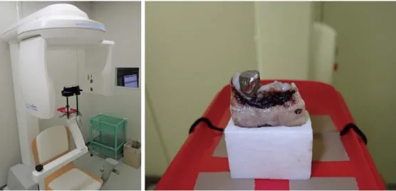
CBCT imaging of surgical specimens was performed
with a CBCT unit
(Fine Cube; Yoshida, Tokyo, Japan).
Surgical specimens in 10% formaldehyde neutral buffer
solution were fixed on the chinrest for CBCT
(Fig. 1) and
examined following the hospital’s protocol.
18The CBCT
parameters were as follows: high-resolution mode
(tube
voltage, 90.0
kV; tube current, 4.00
mA; rotation time, 16.8
s;
field of view, 56
mm×56
mm; thickness, 0.099
mm).
One oral and maxillofacial radiologist, with over 20 years
of experience, reviewed all images. The following CBCT
findings were evaluated: internal texture
(normal, sclerotic,
lytic, and sclerotic), the presence of a sequestrum,
perio-steal reaction, and cortical perforation
(buccal, lingual, and
inferior).
Histopathological studies
Three oral and maxillofacial pathologists reviewed all
histological slides. All archival slides were stained with
hematoxylin and eosin. In all cases in which bacteria were
evident on hematoxylin and eosin staining, periodic acid-
Schiff
(PAS), Gram, and Grocott stains were added.
The set of parameters evaluated in the histomorphometric
analysis included the presence or absence of necrotic bone,
inflammation, reactive bone formation, bacteria
(based on
PAS, Gram, and Grocott stains) and osteoclasts. To measure
inflammation, the percentage of the bone circumference
surrounded by inflammatory cells was evaluated in a semi-
quantitative manner on the 5-tier scale from 0 to 4 proposed
by Shuster et al.,
3as follows: 0=absence of inflammatory
cells; 1=inflammatory cells surrounding up to 10% of the
bone circumference; 2=inflammatory cells surrounding
10%-25% of the bone circumference; 3=inflammatory
cells surrounding 25%-50% of the bone circumference;
4=inflammatory cells surrounding
>50% of the bone
cir-cumference. The inflammatory infiltrate was examined
under ×10 magnification. Each slide was searched for the
area with the highest density of inflammation and the score
was assigned accordingly.
Statistical analysis
The Pearson chi-square test was used to compare ORN
and MRONJ in terms of the CBCT findings and
histopatho-logical characteristics. The statistical analysis was
per-formed with SPSS version 26
(IBM Japan, Tokyo, Japan). A
P value less than 0.05 was considered to indicate statistical
significance.
Results
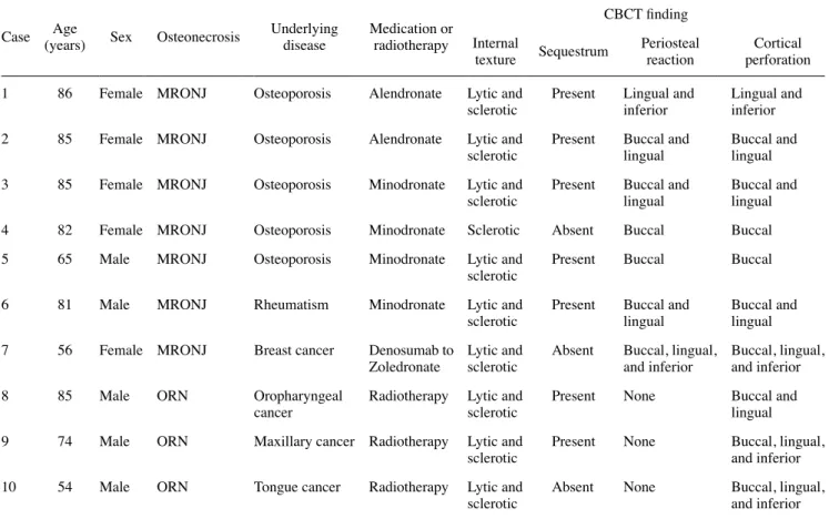
Table 1 presents data on the patients with osteonecrosis
and the CBCT findings of the surgical specimens. For
MRONJ
(case 3, Fig. 2), the CBCT findings included lytic
and sclerotic internal texture, presence of a sequestrum, and
buccal and lingual periosteal reaction and cortical
perfora-tion. The CBCT findings of ORN
(case 9, Fig. 3) included
lytic and sclerotic internal texture, presence of a sequestrum,
no periosteal reaction, and buccal, lingual, and inferior
cortical perforation.
The presence of a sequestrum was observed more
fre-quently in MRONJ than in ORN
(5 of 7
[71.4%] vs. 2 of
3
[66.7%], P
>0.05). MRONJ showed periosteal reaction
more frequently than ORN
(7 of 7
[100%] vs. 0 of 3
[0%],
P
<0.05). Buccal, lingual, and inferior cortical perforation
was found more frequently in ORN than in MRONJ
(2 of 3
[66.7%] vs. 1 of 7
[14.3%], P
>0.05).
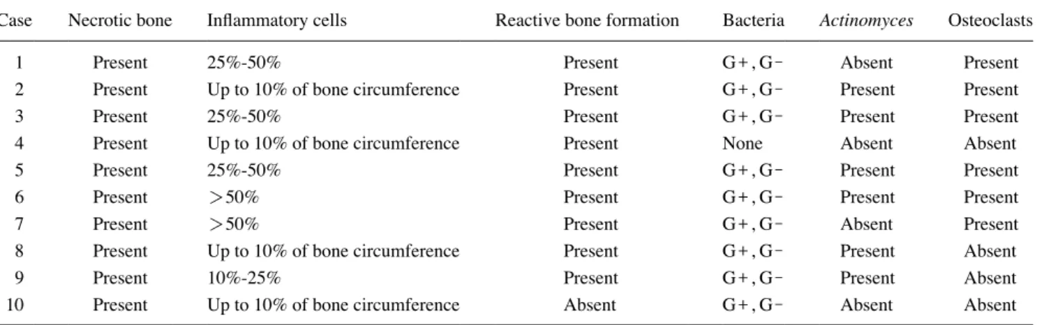
Table 2 shows the histopathological characteristics of
osteonecrosis in the surgical specimens. The
histopatho-logical characteristics of MRONJ
(case 3, Figs. 4 and 5)
included necrotic bone and granulation tissue with the bone
circumference surrounded by inflammatory cells. Six cases
of MRONJ
(6 of 7, 85.7%) revealed bacteria
(mostly Gram-
positive, but some Gram-negative). In MRONJ, osteoclasts
were rarely found around necrotic bone; however,
osteo-clasts were found on the bone surface in contact with
gran-ulation tissue. Reactive bone formation was observed on
destroyed and perforated bone, and on the outside of the
cortical bone. In ORN
(case 9, Figs. 6 and 7), the
histo-pathological findings showed necrotic bone and granulation
tissue with the bone circumference surrounded by abscess
Table 1. Data on patients with osteonecrosis and CBCT findings of the surgical specimens
Case (years)Age Sex Osteonecrosis Underlying disease Medication or radiotherapy
CBCT finding Internal
texture Sequestrum Periosteal reaction perforationCortical 1 86 Female MRONJ Osteoporosis Alendronate Lytic and
sclerotic Present Lingual and inferior Lingual and inferior 2 85 Female MRONJ Osteoporosis Alendronate Lytic and
sclerotic Present Buccal and lingual Buccal and lingual 3 85 Female MRONJ Osteoporosis Minodronate Lytic and
sclerotic Present Buccal and lingual Buccal and lingual 4 82 Female MRONJ Osteoporosis Minodronate Sclerotic Absent Buccal Buccal 5 65 Male MRONJ Osteoporosis Minodronate Lytic and
sclerotic Present Buccal Buccal 6 81 Male MRONJ Rheumatism Minodronate Lytic and
sclerotic Present Buccal and lingual Buccal and lingual 7 56 Female MRONJ Breast cancer Denosumab to
Zoledronate Lytic and sclerotic Absent Buccal, lingual, and inferior Buccal, lingual, and inferior
8 85 Male ORN Oropharyngeal
cancer Radiotherapy Lytic and sclerotic Present None Buccal and lingual 9 74 Male ORN Maxillary cancer Radiotherapy Lytic and
sclerotic Present None Buccal, lingual, and inferior 10 54 Male ORN Tongue cancer Radiotherapy Lytic and
sclerotic Absent None Buccal, lingual, and inferior
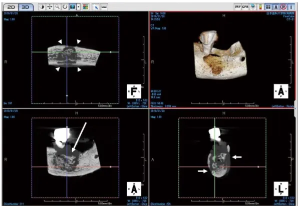
Fig. 2. Cone-beam computed tomographic(CBCT) images of surgical specimen medication-related osteonecrosis of the jaw(case 3). The CBCT findings show lytic and sclerotic internal texture, the presence of a sequestrum(long arrow), and buccal and lingual periosteal reac-tion(arrowheads) and cortical perforation(short arrows).
Fig. 3. Cone-beam computed tomographic(CBCT) images of a surgical specimen of osteoradionecrosis of the jaw(case 9). The CBCT findings show lytic and sclerotic internal texture, the presence of a sequestrum(long arrow), no periosteal reaction, and buccal, lingual, and inferior cortical perforation(short arrows).
and inflammatory cells. All cases of ORN
(3 of 3, 100%)
revealed bacteria
(mostly Gram-positive, with some Gram-
negative) and fibrosis of granulation tissue. In ORN,
osteo-clasts were rarely found around necrotic bone.
MRONJ showed reactive bone formation more frequently
than ORN
(7 of 7
[100%] vs. 2 of 3
[66.7%], P
>0.05),
whereas ORN showed Actinomyces more frequently than
MRONJ
(2 of 3
[66.7%] vs. 4 of 7
[57.1%], P
>0.05). Osteo-
clasts were observed more frequently in MRONJ than in
ORN
(6 of 7
[85.7%] vs. 0 of 3
[0%], P
<0.05).
Discussion
Radiological examinations, especially using computed
tomography, make it possible to estimate the extent of
MRONJ more accurately.
16Ogura et al.
5reported the
charac-teristics of multimodal imaging of MRONJ, and found that
periosteal bone proliferation on MDCT
(0.5-mm-thick
sec-tions, 1-mm reconstruction) was present in 56.3% of cases
(9 of 16). Baba et al.
19evaluated the CT imaging features
of bisphosphonate-related osteonecrosis of the jaw
(BRONJ)
Table 2. Histopathological characteristics of osteonecrosis in the surgical specimens
Case Necrotic bone Inflammatory cells Reactive bone formation Bacteria Actinomyces Osteoclasts
1 Present 25%-50% Present G+, G- Absent Present
2 Present Up to 10% of bone circumference Present G+, G- Present Present
3 Present 25%-50% Present G+, G- Present Present
4 Present Up to 10% of bone circumference Present None Absent Absent
5 Present 25%-50% Present G+, G- Present Present
6 Present >50% Present G+, G- Present Present
7 Present >50% Present G+, G- Absent Present
8 Present Up to 10% of bone circumference Present G+, G- Present Absent
9 Present 10%-25% Present G+, G- Present Absent
10 Present Up to 10% of bone circumference Absent G+, G- Absent Absent
G+: Gram-positive, G-: Gram-negative
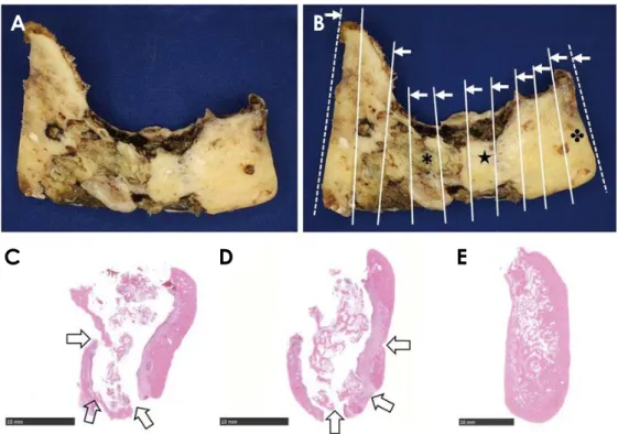
Fig. 4. Histopathological characteristics of a surgical specimen of medication-re-lated osteonecrosis of the jaw(case 3). A. Surgical specimen(buccal side) shows cortical perforation. B. Preparation of the pathological specimen based on the cone-beam computed tomographic (CBCT) findings(arrows, view side). C. Histopathological finding(B*, H&E stain, bar=5mm). Lingual side(arrow) shows cortical perforation. D. Histo-pathological findings(B★, H&E stain, bar=2.5mm). The space( ) is the tooth root. E. Histopathological findings(B , H&E stain, bar=5mm) corresponding to the coronal CBCT image in Figure 2. The buccal side(arrow) shows cortical perforation.
A B
Fig. 5. Histopathological characteristics of a surgical specimen of medication-related osteonecrosis of the jaw(case 3). A. Necrotic bone
shows no osteoclasts in resorption lacunae(H&E stain, bar=100μm). B. Lateral cortical bone(*) shows reactive bone formation(H&E stain, bar=500μm). C. Around the area of necrotic bone, granulation tissue with inflammatory cells is shown(H&E stain, bar=250μm). D. Osteoclasts(arrows) are found on the bone surface in contact with the granulation tissue(H&E stain, bar=100μm). E. Around the area of necrotic bone(★), bacteria are found(mostly Gram-positive, with some Gram-negative)(Gram stain, bar=100μm). F. Bacterial mass shows Actinomyces with methenamine silver staining(Grocott stain, bar=50μm).
A B C
D E F
Fig. 6. Histopathological characteristics of a surgical specimen of osteoradionecrosis of the jaw(case 9). A. Surgical specimen(buccal side)
shows multiple areas of destroyed and perforated cortical bone. B. Preparation of the pathological specimen based on cone-beam comput-ed tomographic findings(arrows, viewing side). C. Histopathological finding(B*, H&E stain, bar=10mm). The buccal and inferior side (arrows) shows cortical destroyed and perforated bone. D. Histopathological finding(B★, H&E stain, bar=10mm) corresponding to the coronal CBCT image in Figure 3. Lingual and inferior sides(arrows) show destroyed and perforated cortical bone. E. Histopathological finding of the surgical margin(B , H&E stain, bar=10mm) shows no necrotic bone or granulation tissue.
A B
and denosumab-related osteonecrosis of the jaw
(DRONJ),
and reported that DRONJ
(4 of 10, 40%) showed periosteal
reaction more frequently than BRONJ
(7 of 65, 10.1%) on
MDCT
(0.5-mm-thick sections, 2-mm reconstruction). In
this study, all cases of BRONJ
(6 of 6, 100%) and DRONJ
(1 of 1, 100%) showed periosteal reaction on CBCT
(0.099
mm thickness). Furthermore, this study found that MRONJ
showed periosteal reaction more frequently than ORN on
CBCT
(7 of 7
(100%) vs. 0 of 3
(0%), P
<0.05). We
con-sider that these results can be explained by the higher
res-olution of CBCT than MDCT, and therefore suggest that
evaluations using CBCT are important to assess periosteal
reaction as a parameter that may help distinguish between
MRONJ and ORN.
Regarding the histopathological characteristics of MRONJ
and ORN, Marx et al.
20reported that BRONJ involves non-
inflammatory drug toxicity to bone, with osteoclastic death
leading to over-suppression of bone renewal, whereas ORN
is another non-inflammatory condition caused by a high
lin-ear energy transfer that impairs or kills numerous cell types
in the field of radiation, including the periosteum, bone, and
all soft tissue. Shuster et al.
3compared the histopathological
characteristics of MRONJ and ORN. They reported that
necrotic bone, inflammation, and reactive bone formation
were present in both diagnoses, and that osteoclasts were
scarce in MRONJ and non-existent in ORN. In the
histo-pathological examinations in this study, MRONJ showed
osteoclasts more frequently than ORN
(6 of 7
[85.7%] vs.
0 of 3
[0%], P
<0.05). Furthermore, six cases of MRONJ
(6 of 7, 85.7%) showed bacteria, which were mostly Gram-
positive
(although some were Gram-negative), while bacteria
were not observed in 1 case of MRONJ
(case 4). A possible
explanation for this finding is that most of the lesion was
covered with mucous membranes in the surgical operation,
eliminating the exposure of necrotic bone. Osteoclasts were
found on the bone surface in contact with granulation tissue,
which may have been caused by a vital reaction.
Van Dessel et al.
17compared the CBCT and micro-CT
characteristics of trabecular bone structures in the human
mandible, and demonstrated the potential of high-resolution
CBCT imaging for in vivo applications to quantitative bone
morphometry and bone quality assessment. Ogura et al.
18showed that CBCT, especially the high-resolution mode, is
useful for the evaluation of surgical specimens of the jaw.
Furthermore, compared with MDCT, CBCT is relatively
easy to use, with short acquisition scan times and high
res-olution. The authors therefore suggest that CBCT could be
useful for evaluating surgical specimens in patients with
MRONJ and ORN.
There are several limitations of this study. The number of
Fig. 7. Histopathological characteristics of a surgical specimen of osteoradionecrosis of the jaw(case 9). A. Necrotic bone shows no
osteo-clasts in resorption lacunae(H&E stain, bar=100μm). B. Reactive bone formation is shown on the lateral cortical bone(★) and fibrosis in the bone marrow(*)(H&E stain, bar=250μm). C. Around the area of necrotic bone, abscess and granulation tissue is shown without osteoclasts(H&E stain, bar=50μm). D. Inflammatory cells and a bacterial mass(*) are found in the abscess(H&E stain, bar=100μm). E. Around the area of necrotic bone(★), bacteria are shown(mostly Gram-positive, with some Gram-negative)(Gram stain, bar=100μm). F. The bacterial mass shows Actinomyces with methenamine silver staining(Grocott stain, bar=100μm).
A B C
surgical specimens was small, because few patients
under-went segmental mandibulectomy for MRONJ and ORN.
However, Zirk et al.
21showed that CBCT image
analy-ses and volumetric measurements of osteolytic lesions in
MRONJ patients were helpful tools for further
understand-ing the clinical appearance of this condition and identifyunderstand-ing
compromised anatomical landmarks. The authors suggest
that evaluating surgical specimens of MRONJ and ORN
with CBCT is important because it is helpful for preparing
the pathological specimen, reassessing the surgical margin,
and predicting the prognosis.
In conclusion, this study evaluated the CBCT imaging
and histopathological characteristics of ORN and MRONJ,
and found that CBCT could be useful for the evaluation of
ORN and MRONJ.
Conflicts of Interest: None
References
1. Ruggiero SL, Dodson TB, Fantasia J, Goodday R, Aghaloo T, Mehrotra B, et al. American Association of Oral and Maxillofa-cial Surgeons position paper on medication-related osteonecrosis of the jaw - 2014 update. J Oral Maxillofac Surg 2014; 72: 1938-56.
2. Chronopoulos A, Zarra T, Ehrenfeld M, Otto S. Osteoradione-crosis of the jaws: definition, epidemiology, staging and clinical and radiological findings. A concise review. Int Dent J 2018; 68: 22-30.
3. Shuster A, Reiser V, Trejo L, Ianculovici C, Kleinman S, Kaplan I. Comparison of the histopathological characteristics of myelitis, medication-related osteonecrosis of the jaw, and osteo-radionecrosis. Int J Oral Maxillofac Surg 2019; 48: 17-22. 4. Ogura I, Kobayashi E, Nakahara K, Haga-Tsujimura M, Igarashi
K, Katsumata A. Computer programme to assess mandibular cortex morphology in cases of medication-related osteonecrosis of the jaw with osteoporosis or bone metastases. Imaging Sci Dent 2019; 49: 281-6.
5. Ogura I, Sasaki Y, Kameta A, Sue M, Oda T. Characteristic multimodal imaging of medication-related osteonecrosis of the jaw: comparison between oral and parenteral routes of medica-tion administramedica-tion. Pol J Radiol 2017; 82: 551-60.
6. Ogura I, Sue M, Oda T, Sasaki Y, Hayama K. Comparison be-tween mandibular malignant tumors and inflammatory lesions using 67Ga scintigraphy: relationship with panoramic
radiogra-phy, CT and MRI findings. Int J Diagn Imaging 2017; 4: 67-73. 7. Ogura I, Oda T, Sue M, Sasaki Y, Hayama K. Comparison
be-tween squamous cell carcinoma and inflammatory diseases of the oral and maxillofacial region using gallium-67 scintigraphy with computed tomography and magnetic resonance imaging. Pol J Radiol 2018; 83: e452-8.
8. Ogura I, Sasaki Y, Sue M, Oda T, Kameta A, Hayama K. Tc-99m hydroxymethylene diphosphonate scintigraphy, computed tomo-graphy and magnetic resonance imaging of osteonecrosis in the
mandible: osteoradionecrosis versus medication-related osteone-crosis of the jaw. Imaging Sci Dent 2019; 49: 53-8.
9. Ogura I, Kobayashi E, Nakahara K, Igarashi K, Haga-Tsujimura M, Toshima H. Quantitative SPECT/CT imaging for medication- related osteonecrosis of the jaw: a preliminary study using vol-ume-based parameters, comparison with chronic osteomyelitis. Ann Nucl Med 2019; 33: 776-82.
10. Ogura I, Sasaki Y, Sue M, Oda T, Kameta A, Hayama K. Tc-99m hydroxymethylene diphosphonate SPECT/CT for the evaluation of osteonecrosis of the jaw: preliminary study on diagnostic ability of maximum standardized uptake value. Clin Radiol 2020; 75: 46-50.
11. Treister NS, Friedland B, Woo SB. Use of cone-beam comput-erized tomography for evaluation of bisphosphonate-associated osteonecrosis of the jaws. Oral Surg Oral Med Oral Pathol Oral Radiol Endod 2010; 109: 753-64.
12. Wilde F, Heufelder M, Lorenz K, Liese S, Liese J, Helmrich J, et al. Prevalence of cone beam computed tomography imaging findings according to the clinical stage of bisphosphonate-related osteonecrosis of the jaw. Oral Surg Oral Med Oral Pathol Oral Radiol 2012; 114: 804-11.
13. Kämmerer PW, Thiem D, Eisenbeiß C, Dau M, Schulze RK, Al-Nawas B, et al. Surgical evaluation of panoramic radiography and cone beam computed tomography for therapy planning of bisphosphonate-related osteonecrosis of the jaws. Oral Surg Oral Med Oral Pathol Oral Radiol 2016; 121: 419-24.
14. Sue M, Oda T, Sasaki Y, Ogura I. Age-related changes in the pulp chamber of maxillary and mandibular molars on cone-beam computed tomography images. Oral Radiol 2018; 34: 219-23. 15. Mizuhashi F, Ogura I, Sugawara Y, Oohashi M, Sekiguchi H,
Saegusa H. Characteristics of root fractures: image on intraoral radiography, panoramic radiography, and cone-beam computed tomography. Oral Sci Int 2020; 17: 34-8.
16. Leite AF, Ogata Fdos S, de Melo NS, Figueiredo PT. Imaging findings of bisphosphonate-related osteonecrosis of the jaws: a criti cal review of the quantitative studies. Int J Dent 2014; 2014: 784348.
17. Van Dessel V, Huang Y, Depypere M, Rubira-Bullen I, Maes F, Jacobs R. A comparative evaluation of cone beam CT and micro-CT on trabecular bone structures in the human mandible. Dentomaxillofac Radiol 2013; 42: 20130145.
18. Ogura I, Ono J, Okada Y. Use of cone-beam computed tomo-graphy for evaluation of surgical specimen of medication-related osteonecrosis of the jaw. J Oral Maxillofac Radiol 2018; 6: 17-20.
19. Baba A, Goto TK, Ojiri H, Takagiwa M, Hiraga C, Okamura M, et al. CT imaging features of antiresorptive agent-related osteo-necrosis of the jaw/medication-related osteoosteo-necrosis of the jaw. Dentomaxillofac Radiol 2018; 47: 20170323.
20. Marx RE, Tursun R. Suppurative osteomyelitis, bisphosphonate induced osteonecrosis, osteoradionecrosis: a blinded histo-pathologic comparison and its implications for the mechanism of each disease. Int J Oral Maxillofac Surg 2012; 41: 283-9. 21. Zirk M, Buller J, Zöller JE, Heneweer C, Kübler N, Lentzen
MP. Volumetric analysis of MRONJ lesions by semiautomatic segmentation of CBCT images. Oral Maxillofac Surg 2019; 23: 465-72.