JKSMRM 12:40-48(2008)
1Department of Diagnostic Radiology and Research Institute of Radiological Science, Yonsei University College of Medicine 2Department of Cardiovascular Surgery, Yonsei University College of Medicine
This work was supported by a faculty research grant of Yonsei University College of Medicine for 2003. Received; May 1, 2008, revised; May 27, 2008, accepted; June 2, 2008
Address reprint requests to : Byoung Wook Choi, M.D., Department of Diagnostic Radiology, Severance Hospital,
Yonsei University College of Medicine, 250 Seongsanno, Seodaemoon-gu, Seoul, 120-752, Korea. Tel. 82-2-2228-7400, Fax. 82-2-393-3035 E-mail: [email protected]
Early Changes of Left Ventricular Geometry and
Function after Surgical Ventricular
Restoration and Mitral Valve Annuloplasty:
Magnetic Resonance Imaging
Byoung Wook Choi1, Byung-Chul Chang2, Young Jin Kim1, Jin Hur1, Hye-Jeong Lee1, Tae Hoon Kim1, Kyu Ok Choe1
Purpose : We sought to determine the early change of ventricular geometry and function after concomitant surgeries of modified Dor procedure and mitral valve annuloplasty by using magnetic resonance imaging.
Materials and Methods : We enrolled 21 patients with dilated heart failure who underwent modified Dor procedure (n=8), mitral valve annuloplasty (n=6), or both surgeries (n=7). Cine MRI was used to assess left ventricular dimensions and function before and after surgery. We measured the left ventricular end-diastolic and end-systolic volumes and the dimensions of the left ventricular long-axis and short-axis. Left ventricular stroke volume, ejection fraction, and sphericity index were calculated from these measurements. These parameters were analyzed and compared between three different surgery groups to explain the combined effect of the concomitant surgeries.
Results : MRI was performed within average 12 ± 15 days (range 1-58 days) before and 38 ± 50 days (range 7- 231 days) after the surgery. The patients who underwent concomitant surgeries had more profound enlargement of left ventricle and decreased contractility prior to surgery than those in the patients who underwent single surgical procedure. Left ventricular diastolic volume and end-systolic volume significantly decreased in all patients regardless of surgery type after surgery. Ejection fraction significantly increased only in the patients who got modified Dor procedure without mitral valve annuloplasty (25.4% to 40.7%). Sphericity index increased in patients with modified Dor procedure but decreased in patients with mitral valve annuloplasty (0.65 to 0.78 vs. 0.75 to 0.65). In the patients who underwent concomitant surgeries showed no significant change in sphericity index after surgery.
Conclusion : The early change of the left ventricular geometry and function after the concomitant surgeries with modified Dor procedure and mitral valve
Introduction
Surgical ventricular restoration is increasingly being utilized in the treatment of heart failure in dilated heart. The surgical strategy is composed of left ventricular volume reduction and restoration of the natural elongated shape of the ventricle to reduce wall stress and to improve ventricular function. Endoventricular circular patch plasty is the most widely used technique of the ventricular restoration surgery, which is named Dor procedure to credit Dr. Vincent Dor who first introduced it in 1985.
In dilated cardiomyopathy, Dor procedure is usually performed combined with coronary artery bypass grafting if there is any significant stenosis in coronary arteries and with ring annuloplasty (1) if there is a significant functional mitral regurgitation combined with ventricular dilatation. As bypass grafting is essential and usually performed prior to the Dor procedure itself during the surgery, the effect of Dor procedure from that of bypass grafting cannot be considered separately. Otherwise, mitral valve annuloplasty in dilated cardiomyopathy is beneficial because the presence of mitral insufficiency has a negative influence on survival with ischemic or idiopathic dilated cardiomyopathy (2). Although studies have reported the Dor procedure to be safe, the effect of the isolated Dor procedure is not well known. Because Dor procedures are often performed with concomitant mitral valve annuloplasty, improvement in postoperative systolic function and change of left
ventricular geometry may be from mitral valve annuloplasty and not from the Dor procedure (3-5). Therefore we measured volume and function changes of the left ventricle in patients who underwent only the Dor procedure and in patients who underwent only mitral valve annuloplasty and compared these results with those in patients who undrewent concomitant the Dor procedure and mitral valve annuloplasty to explain the effects in combined procedures. We used magnetic resonance imaging (MRI) to evaluate the geometry and function of the left ventricle before and after the surgery.
Materials and Methods Subjects
We enrolled 21 patients with dilated heart failure who underwent modified Dor procedure or/and mitral valve annuloplasty. Eight patients underwent only modified Dor procedure, 6 patients underwent only mitral valve annuloplasty, and 7 patients underwent concomitant modified Dor procedure and mitral valve annuloplasty. Patients’ demographic data of preoperative cardiac function on each group were summarized on Table 1. MRI was used to assess left ventricular dimensions and function. Mitral regurgitation was evaluated by echocardiography. Coronary angiography was performed in all patients.
Modified Dor procedure as a left ventricular volume reduction surgery was performed in patients with NYHA functional class III or IV and left ventricular ejection fraction below 30% (6). Patient were annuloplasty in patients with dilated heart failure includes a marked reduction in
left ventricular volume and in stroke volume. The shape of the left ventricle does not change because the effect of sphericity index decrease from mitral valve annuloplasty is counteracted by the effect of sphericity index increase from modified Dor procedure. Improvement of left ventricular ejection fraction is not the early change after the concomitant surgeries.
Index words :Surgical Ventricular Restoration
Left Ventricular Volume Reduction Surgery Dor Procedure
Mitral Annuloplasty
considered suitable for left ventricular volume reduction surgery if they demonstrated an enlarged left ventricle accompanied by left ventricular dysfunction with either dyskinetic or akinetic apical segments after myocardial infarction and had symptoms of angina, congestive heart failure, ventricular tachycardia or a combination of these.
Mitral valve annuloplasty was performed if mitral regurgitation was severe greater than grade 2 or mitral annulus diameter measured more than 35 mm with any grade of mitral regurgitation. The degree of mitral insufficiency was graded from 0 to 4 in a semi-quantitative way. Mitral valve leakage was considered secondary to left ventricular enlargement if echocardiography showed central regurgitation associated with enlarged left ventricle. Mitral annulus size was measured on cine-MRI.
Modified Dor procedure and mitral valve annuloplasty were performed at once if a patient met the criteria of both surgeries.
Surgical technique
For the modified Dor procedures, the apical left ventricle was opened and a circumferential purse-string suture was placed at the base of both papillary muscles to reduce the diameter of the left ventricle. In ischemic cardiomyopathy, the transition between infarcted aneurysm and uninfarcted myocardium was determined by inspection and palpation. All dyskinetic portions of the anterior wall and septum were excluded. The endoventricular circular suture described by Dor was performed with a 2/0 prolene purse string. Other details about this surgical procedure in these patients have been described in a previous report (7). Coronary artery bypass surgery was performed for any significantly stenotic arteries. Concomitant mitral valve annuloplasty was performed if the measurement of regurgitation and annular size met the criteria for surgery through opened left ventricle. In patients who underwent only mitral valve annuloplasty, the mitral valve was exposed by a transeptal incision. The mitral valve was repaired
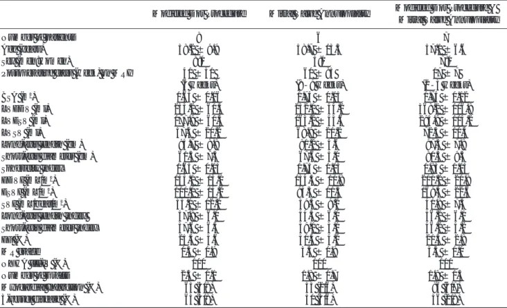
Table 1. Patients’ Demographic and Preoperative Characteristics
Modified Dor Procedure Mitral Valve Annuloplasty Modified Dor Procedure & Mitral Valve Annuloplasty
Number of patients 8 6 7
Age (years) 58.2 ± 9.9 58.7 ± 13.3 57.1 ± 6.6
Sex (men:women) 8:0 5:1 7:0
Postoperative days (week) on MRI 41 ± 31 60 ± 85 17 ± 7 (6 weeks) (8-9 weeks) (2-3 weeks)
BSA (m2) 1.65 ± 0.16 1.74 ± 0.15 1.74 ± 0.12 LVEDV (ml) 235.2 ± 61.5 232.1 ± 44.2 368.2 ± 106.8 LVESV (ml) 177.9 ± 60.3 163.2 ± 43.6 295.9 ± 114.2 LVSV (ml) 57.4 ± 20.2 68.9 ± 21.2 72.3 ± 11.4 Long-axis length (cm) 94.7 ± 8.8 91.2 ± 4.6 97.3 ± 7.8 Short-axis diameter (cm) 61.3 ± 7.5 67.6 ± 5.0 80.3 ± 9.3 Sphericity index 0.65 ± 0.03 0.75 ± 0.03 0.83 ± 0.03 EDVI (mL/m2) 145.1 ± 15.1 134.5 ± 10.8 211.2 ± 20.8 ESVI (mL/m2) 110.0 ± 14.2 95.3 ± 11.5 169.4 ± 22.6 SVI (mL/beat/m2) 35.1 ± 12.2 39.3 ± 9.2 41.8 ± 7.5 Long-axis length index 57.8 ± 6.0 54.3 ± 6.0 56.2 ± 6.1 Short-axis diameter index 37.6 ± 6.4 39.1 ± 3.1 46.2 ± 4.2 EF (%) 25.4 ± 3.6 30.4 ± 4.2 21.3 ± 2.8 MR grade 0.5 ± 0.9 3.3 ± 0.8 3.3 ± 1.0 NYHA III-IV (%) 100 100 100 Number of Grafts 2.5 ± 1.1 1.8 ± 1.7 1.9 ± 1.3 Myocardial Infarction (%) 63 (5/8) 33 (2/6) 86 (6/7) 3-vessel disease (%) 63 (5/8) 50 (3/6) 36 (2/8)
BSA: body surface area, LVEDV: left ventricular end-diastolic volume, LVESV: left ventricular end-systolic volume, LVSV: left lar stroke volume, EDVI: left ventricular end-diastolic volume index, ESVI: left ventricular end-systolic volume index, SVI: left ventricu-lar stoke volume index, EF: left ventricuventricu-lar ejection fraction, MR: mitral regurgitation, NYHA: New York Heart Association
according to the Bolling annuloplasty technique using a Cosgrove band, sutured on the posterior leaflet.
MRI protocol
Cardiac MRI was performed before and after the surgery. MRI was performed with subjects positioned supine in a 1.5-T MR unit (Gyroscan Intera; Philips medical system, Best, the Nethelands). Cine MRI was performed with balanced TFE (turbo field echo, 25 phases), which included short-axis, two-chamber and four-chamber views. Imaging parameters included the following: typical repetition time = 3.4 ms, echo time = 1.7 ms, flip angle = 50°, field of view = 360 mm, matrix 256×256, slice thickness = 10 mm, effective temporal resolution = 40 ms, number of signal average = 1, breath hold duration per slice = 14~16 s. The short-axis imaging encompassed the entire left ventricle.
Analysis of MRI
Endocardal left ventricular borders were manually traced at end-diastole and at end-systole. In left ventricular diastolic volume (LVEDV) and
end-sytolic volume (LVESV) were measured by using Simpson’s rule. We defined end-diastole as the first image of the cine-imaging set. End-systole was defined as the phase of the smallest volume in a total sum of every slice. From the measured LVEDV and EVESV, we calculated left ventricular stroke volume (LVSV) and ejection fraction (EF). We measured the dimensions of the left ventricular chamber at both end-diastole and end-systole. The dimension of the left ventricular long axis (long-axis length) was defined as a linear distance from the midpoint of the mitral annulus plane to the apex, and the dimension of the short axis (short-axis diameter) was defined as the linear distance from the anterior endocardium to the posterior endocardium at the midpapillary music plane. These dimensions were measured in end-diastole. Sphericity index was calculated as a ratio of short-axis diameter to long-axis length.
Statistical analysis
Descriptive anthropomorphic characteristics are presented as mean ± standard deviation. Analysis of variance (ANOVA) was used to assess differences
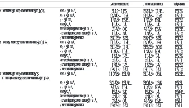
Table 2. Postsurgical Changes in Geometry and Function of Left Ventricle
Preoperative Postoperative p-value Modified Dor Procedure (n=8) EDV (mL) 235 ± 61.5 170.3 ± 36.1 0.007
ESV (mL) 177.9 ± 60.3 102.7 ± 20.9 0.001 SV (ml) 57.4 ± 20.0 67.6 ± 17.3 0.128 EF (%) 25.4 ± 3.6 40.7 ± 3.9 0.001 Long-axis length (mm) 94.7 ± 8.8 74.3 ± 10.7 0.001 Short-axis diameter (mm) 61.3 ± 7.5 57.2 ± 6.1 0.108 Sphericity Index 0.650 ± 0.03 0.779 ± 0.04 0.008 Mitral Valve Annuloplasty (n=6) EDV (ml) 232.1 ± 44.2 179.2 ± 37.4 0.005 ESV (ml) 163.2 ± 43.6 121.2 ± 29.9 0.006 SV (ml) 68.9 ± 21.2 57.9 ± 18.4 0.229 EF (%) 30.4 ± 4.2 34.3 ± 6.3 0.327 Long-axis length (mm) 91.2 ± 4.6 92.4 ± 4.4 0.321 Short-axis diameter (mm) 67.6 ± 5.0 60.0 ± 4.7 0.008 Sphericity Index 0.745 ± 0.03 0.652 ± 0.03 0.018 Modified Dor Procedure & EDV (mL) 368.2 ± 106.8 256.4 ± 92.6 0.002
Mitral Valve Annuloplasty (n=7)
ESV (mL) 295.9 ± 114.2 208.4 ± 51.7 0.003 SV (mL) 72.3 ± 11.4 48.0 ± 18.7 0.005 EF (%) 21.3 ± 2.8 20.8 ± 3.6 0.771 Long-axis length (mm) 97.3 ± 7.8 87.9 ± 8.6 0.003 Short-axis diameter (mm) 80.3 ± 9.3 74.2 ± 10.0 0.036 Sphericity Index 0.827 ± 0.03 0.848 ± 0.04 0.644 EDV: left ventricular end-diastolic volume, ESV: left ventricular end-systolic volume, SV: left ventricular stroke volume, EF: left ventricu-lar ejection fraction
among groups. Two-tailed Student test for paired data was used when appropriate. A p-value less than 0.05 was considered significant. All the analysis was done using an SPSS package (Version 12.0; Statistical Package for the Social Sciences, Chicago, IL). Left ventricular parameters were conducted after adjustment for subject body surface area (BSA).
Results
MRI was performed with an interval of average 12 ± 15 days (range 1-58 days) before and 38 ± 50 days (range 7- 231 days) after the surgery. The postsurgical changes are summarized in Table 2 and 3.
Postsurgical change in modified Dor procedure
The LVEDV and LVESV significantly decreased. The LVSV did not significantly change. The EF significant increased from 25.4% to 40.7%. The long-axis length significantly decreased from 94.7 cm to 74.3 cm, however the short-axis diameter did not change. The sphericity index increased from 0.65 to 0.78. The indices of LVEDV, LVESV, LVSV, long-axis length, and short-axis diameter showed the same tendency of changes as the unstandardized data.
Postsurgical change in mitral valve annuloplasty
The LVEDV and LVESV significantly decreased. The
LVSV did not significantly change. The EF did not significantly change. The Long-axis length significantly decreased from 67.6 cm to 60.0 cm however, the short-axis diameter did not significantly change. The sphericity index significantly decreased from 0.75 to 0.65. The LVEDV, LVESV, LVSV, long-axis length, and short-axis diameter showed the same tendency of changes as the unstandardized data.
Postsurgical change in concomitant modified Dor procedure and mitral valve annuloplasty
The LVEDV and LVESV significantly decreased. The LVSV significantly decreased from 72 ml to 48 ml. The EF did not significantly change. The Long-axis length and the short-axis diameter significantly reduced. The sphericity index showed no significant change. The LVEDV, LVESV, LVSV, long-axis length, and short-axis diameter showed the same tendency of changes as the unstandardized data.
Comparison of Different Surgeries
The LVEDV and LVESV were significant decreased in all patients regardless of surgery type done. The EF was significant increased only in the patients who got modified Dor procedure without mitral valve annuloplasty. The left ventricular short-axis diameter was significant decreased in the patients who underwent mitral valve annuloplasty with or without
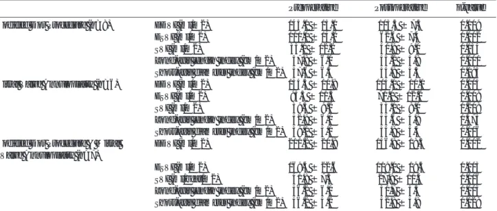
Table 3. Postsurgical Changes of Geometry and Function of Left Ventricle: Standardized with Body Surface Area
Preoperative Postoperative p-value Modified Dor Procedure (n=8) EDVI (ml/m2) 145.1 ± 15.1 103.5 ± 7.3 0.009
ESVI (ml/m2) 110.0 ± 14.2 62.6 ± 7.4 0.002 SVI (ml/m2) 35.1 ± 12.2 40.9 ± 9.0 0.134 Long-axis length index (cm/m2) 57.8 ± 6.0 45.2 ± 5.8 0.001 Short-axis diameter index (cm/m2) 37.6 ± 6.4 34.9 ± 4.5 0.095 Mitral Valve Annuloplasty (n=6) EDVI (ml/m2) 134.5 ± 10.8 104.0 ± 10.1 0.004 ESVI (ml/m2) 95.3 ± 11.5 71.0 ± 12.2 0.008 SVI (ml/m2) 39.3 ± 9.2 33.1 ± 8.0 0.208 Long-axis length index (cm/m2) 52.9 ± 6.0 53.6 ± 4.9 0.375 Short-axis diameter index (cm/m2) 39.1 ± 3.1 34.8 ± 4.4 0.006 Modified Dor Procedure & Mitral EDVI (ml/m2) 211.2 ± 20.8 146.9 ± 18.5 0.002
Valve Annuloplasty (n=7)
ESVI (ml/m2) 169.4 ± 22.6 119.0 ± 19.5 0.003 SVI (ml/beat/m2) 41.8 ± 7.5 27.9 ± 11.4 0.006 Long-axis length index (cm/m2) 56.2 ± 6.1 50.7 ± 5.3 0.003 Short-axis diameter index (cm/m2) 46.2 ± 4.2 42.8 ± 5.8 0.028 EDVI: left ventricular end-diastolic volume index, ESVI: left ventricular end-systolic volume index, SVI: left ventricular stoke volume in-dex
modified Dor procedure. The left ventricular long-axis length was significant decreased in the patients who underwent modified Dor procedure with or without mitral valve annuloplasty.
The sphericity index increased in patients with modified Dor procedure but decreased in patients with mitral valve annuloplasty. In the patients who underwent modified Dor procedure with mitral valve annuloplasty showed no significant change of the sphericity index after the surgery. These are summarized in Table 4.
Discussion
Postsurgical change in modified Dor procedure
In ischemic cardiomyopathy, coronary artery bypass grafting surgery can be an effective procedure, but the functional recovery is not sufficient and the recurrence of heart failure is high if ventricular remodeling has already occurred. Dor et al. reported another type of left ventricular volume reduction surgery, endoventricular circular patch plasty in 1985 for the dilated heart failure (8). The Dor procedure has been proved to improve pump function, clinical status, and survival, even in patients with severe cardiac dysfunction and now became one of the most widely used method among the various surgical ventricular restoration methods. Surgical ventricular restoration in patients with dilated heart failure constitutes ventricular volume reduction by elimination of dysfunctional myocardium and restoration of natural elliptical shape of left ventricle.
According to a previous animal study, after Dor procedure, left ventricular volume significantly reduces in 2 weeks then ejection fraction significantly increases
in 6 weeks (9). In a study with an angiographic follow-up in human patients, significant reduction in left ventricular volumes and an increase in EF after Dor procedure have been reported. Dor procedure shortens long-axis length but does not shorten short-axis diameter (10). In the present study, we demonstrated by using MRI that the patients who underwent modified Dor procedure without mitral valve annuloplasty showed compatible results with those of the previous reports on a mean post-operative day of 41. In the patients who underwent modified Dor procedure, surgical reduction of left ventricular volume by removing dysfunctional myocardium resulted in marked decrease of LVEDV and LVESV. However, the ratio of volume reduction was much higher in LVESV than in LVEDV (42.2%>27.5%). However, LVSV did not significantly increase while EF significantly increased. It is attributable to contractility improvement from recovered circulation by bypass grafting, elimination of uncontractile myocardium or both. While long-axis length was shortened, short-axis diameter was not shortened after surgery. Thus sphericity index rather increased post-operatively. Despite a more spherical postoperative left ventricular chamber, systolic pump function improved.
Postsurgical change in mitral valve annuloplasty
An increase in left ventricular end-systolic volume and a distortion of shape of left ventricle are associated with inadequate approximation of the mitral tetrahedron during systole, which consequently leads to functional mitral regurgitation (11). The effects of mitral valve annuloplasty in these patients are provided by two mechanisms including elimination of mitral regurgitation and reduction of short-axis diameter.
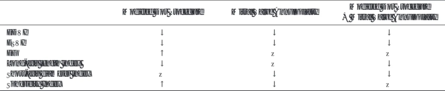
Table 4. Comparison of Early Effects on Geometry and Function of Left Ventricle after Modified Dor Procedure, Mitral Valve Annuloplasty, and Concomitant Surgery
Modified Dor Procedure Mitral Valve Annuloplasty Modified Dor Procedure + Mitral Valve Annuloplasty
EDVI ↓ ↓ ↓
ESVI ↓ ↓ ↓
EF ↑ — —
Long-axis length index ↓ — ↓
Short-axis diameter index — ↓ ↓
Sphericity Index ↑ ↓ —
EDVI: left ventricular end-diastolic volume index, ESVI: left ventricular end-systolic volume index, EF: left ventricular ejection fraction, ↑: increase, ↓: decrease, —: no change
Nicolini et al. reported that mitral valve annuloplasty improved left ventricle ejection fraction from 28.9% ± 5.2% preoperatively to 35.4% ± 8.1% (1). Left ventricular volume and sphericity index decrease after surgery (12).
In the present study, mitral valve annuloplasty eliminated mitral regurgitation resulting in a decrease of LVEDV and LVESV with similar ratio (22.8% vs. 25.8%). Total LVSV slightly decreased regardless of change of the net stroke volume ejecting into the aorta. EF is mildly increased which was not statistically significant. No significant improvement of EF can be accounted for by depressed contractility from underlying cardiomyopathy. The length of LV did not change but the short-axis diameter significantly decreased. Thus sphericity index decreased and the shape became more elliptical.
Postsurgical change in concomitant modified Dor procedure and mitral valve annuloplasty
Volume reduction surgery in patients with dilated heart failure is usually accompanied with mitral insufficiency repair. Post-infarction left ventricles are characterized by changes in shape and function frequently complicated by functional mitral regurgitation, leading to cardiac dysfunction and clinical heart failure, which heavily affect survival. Therefore mitral annuloplasty is important to reduce failure or delayed improvement rate of surgical ventricular restoration. In addition, mitral annulus size is not decreased after surgical ventricular restoration without mitral valve annuloplasty (13). In carefully selected patients with dilated cardiomyopathy with a cardiac output less than 2.5, short-axis diameter more than 7.0 cm, NYHA class exceeding III on maximal medical therapy, the Dor procedure combined with mitral valve annuloplasty achieves short-term results comparable to that after heart transplantation. In a study for the effects of the surgery, one-year post-operatively, LVSV index improved from 25.9 to 40.3 ml/m2 whereas short-axis diameter decreased from 8.4 cm to 6.6 cm (14). LVEDV and LVESV were decreased and EF improved toward 1 year, coinciding with the improvement of clinical symptoms, potentially indicating a functional remodeling after the surgery (15).
In the present study, LVEDV, LVESV, and short-axis
diameter were profoundly increased and greater than those of other groups pre-operatively. The LVEDV and LVESV significantly decreased with a similar ratio (30.3% vs. 29.6%) after the surgery. Therefore The EF was not significantly changed after the surgery. The patients with concomitant modified Dor procedure and mitral valve annuloplasty, physical reduction of LVEDV and elimination of mitral regurgitation resulted in significant LVSV reduction (33.6%) even though increased contractility after the surgery might compensate the reduction of stroke volume. However, preoperative heart failure and LV dilatation were severe in these patients, so the contractility recovery was not as good as expected. Both the long-axis length and short-axis diameter significantly decreased, which could be explained as a direct effect of each surgical procedure.
In summary, LVEDV and LVESV decreased after either surgical procedures or concormitant surgeries. However left ventricular EF in concomitant surgeries did not show a significant improvement because the patients indicated for concomitant surgeries had significantly more dilated heart and decreased contractility than the patients indicated only for either surgery. It was probably an early follow-up to evaluate the recovery of contractility in patients with concomitant surgeries. Marked reduction of stroke volume after concomitant surgeries reflected much reduction of ventricular volume and elimination of large amount of mitral regurgitation in these patients. Shortening of long-axis length from modified Dor procedure and shortening of short-axis diameter from mitral valve annuloplasty directly contributed to insignificant change of sphericity index of left ventricle after concomitant surgeries.
Limitations
Our study has some limitations. The first was that the patients were clinically heterogeneous; most of them were ischemic dilated cardiomyopathy but some were idiopathic dilated cardiomyopathy. In addition, each surgical procedure had different indication and the preoperative status was different among surgical groups. The second is that the effect of coronary bypass grafting could be a confounding factor in interpreting the results. However, it is difficult to separate the effect of bypass surgery from the surgeries we evaluated.
Conclusion
The early change of left ventricular geometry and function after concomitant modified Dor procedure and mitral valve annuloplasty in patients with dilated heart failure includes marked reduction of left ventricular volume and stroke volume. The shape of left ventricle does not change because the effect of sphericity index decrease from mitral valve annuloplasty is counteracted by the effect of sphericity index increase from modified Dor procedure. An improvement of left ventricular ejection fraction is not the early change after concomitant surgeries.
References
1.Nicolini F, Zoffoli G, Cagnoni G, Agostinelli A, Colli A, Fragnito C, et al. Mitral valve annuloplasty and myocardial revascularization in the treatment of ischemic dilated cardiomyopathy. Heart Vessels 2006; 21:28-32.
2.Grigioni F, Enriquez-Sarano M, Zehr KJ, Bailey KR, Tajik AJ. Ischemic mitral regurgitation: long-term outcome and prognostic implications with quantitative Doppler assessment. Circulation 2001; 103:1759-1764.
3.Dor V, Di Donato M, Sabatier M, Montiglio F, Civaia F. Left ventricular reconstruction by endoventricular circular patch plasty repair: a 17-year experience. Semin Thorac Cardiovasc Surg 2001; 13:435-447.
4.Popovic Z, Miric M, Gradinac S, Neskovic AN, Jovovic L, Vuk L, et al. Effects of partial left ventriculectomy on left ventricular performance in patients with nonischemic dilated cardiomyopathy. J Am Coll Cardiol 1998; 32:1801-1808. 5.McCarthy JF, McCarthy PM, Starling RC, Smedira NG, Scalia
GM, Wong J, et al. Partial left ventriculectomy and mitral valve repair for end-stage congestive heart failure. Eur J Cardiothorac Surg 1998; 13:337-343.
6.Dor V. The endoventricular circular patch plasty (“Dor
procedure”) in ischemic akinetic dilated ventricles. Heart Fail Rev 2001; 6:187-193.
7.Chang BC, Lim CY, Park PW, Park KY, Lee YT, Kim YJ. Volume reduction surgery for end-stage heart failure: experience in Korea. J Card Surg 2001; 16:159-164. 8.Dang AB, Guccione JM, Zhang P, Wallace AW, Gorman RC,
Gorman JH, 3rd, et al. Effect of ventricular size and patch stiffness in surgical anterior ventricular restoration: a finite element model study. Ann Thorac Surg 2005; 79:185-193. 9.Zhang P, Guccione JM, Nicholas SI, Walker JC, Crawford
PC, Shamal A, et al. Left ventricular volume and function after endoventricular patch plasty for dyskinetic anteroapical left ventricular aneurysm in sheep. J Thorac Cardiovasc Surg 2005; 130:1032-1038.
10.Di Donato M, Sabatier M, Dor V, Gensini GF, Toso A, Maioli M, et al. Effects of the Dor procedure on left ventricular dimension and shape and geometric correlates of mitral regurgitation one year after surgery. J Thorac Cardiovasc Surg 2001; 121:91-96.
11.Yu HY, Su MY, Liao TY, Peng HH, Lin FY, Tseng WY. Functional mitral regurgitation in chronic ischemic coronary artery disease: analysis of geometric alterations of mitral apparatus with magnetic resonance imaging. J Thorac Cardiovasc Surg 2004; 128:543-551.
12.Bolling SF, Pagani FD, Deeb GM, Bach DS. Intermediate-term outcome of mitral reconstruction in cardiomyopathy. J Thorac Cardiovasc Surg 1998; 115:381-386; discussion 387-388.
13.Menicanti L, DiDonato M, Castelvecchio S, Santambrogio C, Montericcio V, Frigiola A, et al. Functional ischemic mitral regurgitation in anterior ventricular remodeling: results of surgical ventricular restoration with and without mitral repair. Heart Fail Rev 2004; 9:317-327.
14.Kawaguchi AT, Suma H, Konertz W, Gradinac S, Bergsland J, Dowling RD, et al. Left ventricular volume reduction surgery: The 4th International Registry Report 2004. J Card Surg 2005; 20:S5-11.
15.Schafers M, Stypmann J, Wilhelm MJ, Stegger L, Kies P, Hermann S, et al. Functional changes after partial left ventriculectomy and mitral valve repair assessed by gated perfusion SPECT. J Nucl Med 2004; 45:1605-1610.
통신저자 : 최병욱, (120-752) 서울시 서대문구 성산로 250, 연세대학교 의과대학 영상의학과, 방사선의과학 연구소 Tel. (02) 2228-7400, Fax. (02) 393-3035 E-mail: [email protected]